Disturbances in glucose metabolism (insulin resistance and abnormal glucose tolerance) are frequently observed among ALS patients. In recent years, there has been growing interest in the elusive relationship between premorbid diabetes and ALS.

With regard to the risk of developing ALS, studies in European countries have demonstrated that premorbid type 2 diabetes protects people against ALS development. One exception was the study by Sun et al. , who found that premorbid type 2 diabetes increased the risk of ALS development in Asia (Taiwan, China). In addition, a 4 year delay in the onset of ALS was observed in ALS patients with premorbid type 2 diabetes.

The aforementioned literature demonstrated the distinctive effect of premorbid type 2 diabetes on ALS patients from Asia and Europe. The present study aims to provide evidence for the association between premorbid type 2 diabetes and ALS by evaluating the onset and prognostic value in our cohort of ALS patients.

Patients were recruited from the national referral Amyotrophic Lateral Sclerosis Clinic at the Department of Neurology, Peking University Third Hospital (PUTH), Beijing, between 1January 2013, and 31 December 2016. Among 1331 consecutive sporadic ALS patients, 100 (7. 5%) were labeled as ALS type 2 diabetes and 1231 were labeled as ALS control according to the presence or absence of premorbid type 2 diabetes

As in other studies, the authors found a 4. 4 year delay in the onset of ALS inpatients with premorbid type 2 diabetes after controlling for other ALS disease modifiers, including gender.

The prevalence of premorbid type 2 diabetes at base line in ALS patients was lower than the estimated rate among the Chinese population. The overall prevalence of premorbid type 2 diabetes at baseline among ALS patients was 100/1331¼7. 5%; however, the expected prevalence of type 2 diabetes in the general population (>18 years) in China is estimated to be over 11. 6% (95% CI, 11. 3% – 11. 8%). This observation also indicates a protective role of premorbid type 2 diabetes on ALS development. This result was consistent with what has been reported in other studies.

In this study, the mean age of onset of ALS symptoms was 52. 9 years (SD, 10. 2 years), which was lower than that reported in Japan (64. 8 years), Italy (64. 8 years), Europe (64. 4 years) and Germany (67. 0 years). The age of onset of Chinese patients with ALS is approximately a decade younger than that of European and Japanese patients with ALS.

It is noteworthy that the willingness to medically treat younger patients is much stronger than that of elderly patients. Since ALS is a relatively rare disease, ALS might be under recognized in older people because being weak or wasted may be regarded as a normal part of aging or because multiple medical problems may ALS diagnostic difficult. This may create bias.

A clear molecular explanation of the relationship between type 2 diabetes and ALS remains a challenge. Recently, several radiographic and molecular studies have demonstrated that structural and functional alterations in the hypothalamic melanocortin system are frequently observed at the preclinical stage of ALS patients. The essential role of melanocortins lies in the regulation of body weight and appetite. The hypothalamic melanocortin pathway is a critical center for monitoring, processing, and responding to peripheral signals, including hormones, such as ghrelin, leptin, and insulin.

In ALS patients, these alterations in the structure and function of the hypothalamic melanocortin system provide a mechanistic explanation for the abnormalities in food intake and metabolism observed inpatients with ALS. Increasingly, behavior related changes have been associated with improved survival. Similarly, these preclinical alterations in the hypothalamic melanocortin pathway, as compensatory changes for ALS development, could also be a potential explanation for ALS patients with insulin resistance and type 2 diabetes. High levels of glucose could reduce the damage to motoneurons caused by hypermetabolism in the preclinical and early stages of ALS, while in the middle and late stages of ALS, when the melanocortin system is in a decompensatory period, insulin resistance and type 2 diabetes have no beneficial effect on the progression or survival of ALS patients.

In Alzheimer disease, for example, clinical and epidemiological studies have shown that the risk of Alzheimer disease was almost doubled in type 2 diabetes patients compared to the general population. Insulin deficiency and insulin resistance, core features of type 1 diabetes and type 2 diabetes, were also observed in the brains of Alzheimer disease patients. Thus, researchers proposed the term “type 3 diabetes”, which reflects the fact that Alzheimer disease represents a form of diabetes that selectively involves the brain and has molecular and biochemical features that overlap with both type 1 diabetes and type 2 diabetes. However, the question of whether the observed insulin resistance is a cause or consequence of neuro-degeneration in Alzheimer disease remains open.

Finally, the anti type 2 diabetes drugs metformin and pioglitazone, which have been proven to be neuroprotective in Alzheimer disease and other neurodegenerative diseases, have unexpectedly failed to show beneficial effects on the progression or survival of ALS patients. It was found that, at later time points, the metformin induced trophic effects may have been overshadowed by advancing and aggressive SOD1G93A pathology and potentially negative drug effects. The cause of pioglitazone’s failed clinical translation remains elusive. It was even suggested that suboptimal glycemic control may be beneficial in ALS.

Most neurodegenerative diseases are proteopathies. Alleviating ER stress is a promising approach for treating a range of diseases. Heren researchers aimed to identify a potent chemical chaperone through High-Throughput Screening of a small molecule chemical library.

The endoplasmic reticulum (ER) is responsible for folding secretory and membrane proteins, but disturbed ER proteostasis may lead to protein aggregation and subsequent cellular and clinical pathologies. For example Tudca (see AMX00035 trial) has some efficiency in this area. Tudca is a chemical chaperone.

Chemical chaperones have recently emerged as a potential therapeutic approach for ER stress-related diseases. Scientists have identified 2-phenylimidazo[2,1-b]benzothiazole derivatives (IBTs) as chemical chaperones in a cell-based high-throughput screen.

https://elifesciences.org/articles/43302

The accumulation of unfolded or misfolded proteins in the endoplasmic reticulum (ER) is called ER stress. The cell has an adaptive system against ER stress called the unfolded protein response (UPR), which is the coordinated transcriptional upregulation of ER chaperones and folding enzymes that prevents the aggregation of unfolded and incompletely folded protein.

To overcome ER stress-related diseases, the following two pharmacological strategies can be applied: the modulation of ER protein folding environments (by UPR modulators) and a reduction in the accumulation of unfolded or misfolded ER proteins (by chemical chaperones).

In contrast, several chemical chaperones that have shown therapeutic benefits in mouse disease models have also been developed. 4-Phenylbutyrate (4PBA) and taurourso-deoxycholic acid (TUDCA) have been reported to function as chemical chaperones and have shown therapeutic benefits for a wide variety of diseases, such as diabetes, ALS and Alzheimer's disease.

Biochemical and chemical biology approaches revealed that IBT21 directly binds to unfolded or misfolded proteins and inhibits protein aggregation. IBT21 prevented cell death caused by chemically induced ER stress and by a proteotoxin, an aggression-prone prion protein. Taken together, their data show the promise of IBTs as potent chemical chaperones that can ameliorate diseases resulting from protein aggregation under ER stress.

Advertisement


This book retraces the main achievements of ALS research over the last 30 years, presents the drugs under clinical trial, as well as ongoing research on future treatments likely to be able stop the disease in a few years and to provide a complete cure in a decade or two.

Amylyx Pharmaceuticals, Inc., a pharmaceutical company focused on developing new treatments for amyotrophic lateral sclerosis (ALS) and other neurodegenerative diseases, announced today that AMX0035 has demonstrated significant treatment advantage for people with of ALS in the CENTAUR study. In the study, participants taking AMX0035 had a statistically significant slowdown in the progression of ALS disease, as measured by the revised ALS functional rating scale (ALSFRS-R) compared to placebo.

AMX0035 is an orally available candidate therapy designed to minimize the mechanisms associated with nerve cell death. It is made up of two small molecules - tauroursodeoxycholic acid (TUDCA) and sodium phenylbutyrate (PB) - which target signals in the mitochondria and endoplasmic reticulum of a cell, two compartments strongly involved in cellular stress and death of nerve cells.

TUDCA and PB have been shown to prevent cell death and damage neuroinflammation in preclinical models of ALS.

According to Justin Klee, co-founder and president of Amylyx, the therapeutic strategy followed is somewhat unique in that it does not try to prevent the root cause of ALS, the process of which, in most cases, has occurred since long before a person is diagnosed; rather, it aims to preserve motor neurons.

"What ultimately causes the clinical decline of ALS is that the motor neurons in the brain and spine degenerate and die," said Klee. "What we designed was that if we could identify or develop a therapy that could intervene in cell death and degeneration, then maybe we could have a therapy that would work for ALS, as well as neurodegeneration as a whole. "

CENTAUR participants had the opportunity, after the trial, to enroll in an open-label extension study to receive treatment with AMX0035. Almost 90 percent of participants who have completed CENTAUR have chosen to enroll in the extension study. Intermediate data from the ongoing extension study will be presented in 2020.

Sabrina Paganoni, MD, PhD, Harvard professor leading the trial, explained that CENTAUR was designed to maximize the data that could be obtained using the least number of participants and in the least possible time. This involved the use of stringent enrollment criteria - essentially, only those individuals who were expected to have the most severe ALS with the fastest disease progression, were enrolled. "In other words, when we test the drug in the most severe patients - those who need it most - if we can stop or slow the disease in these patients, we expect to do the same in all patients . "

Paganoni added that if AMX0035 reaches the point of being approved by regulatory authorities for the treatment of ALS, it should be approved for all patients with ALS.

In addition, the company will provide an update on regulatory plans and more details on expanded access plans in early 2020.

Dr. Rudolph Tanzi, Ph.D., Professor Kennedy of Neurology, Massachusetts General Hospital, Chairman of the Cure Alzheimer's Fund research leadership group and Chairman of Amylyx SAB, shared: "The positive results of the CENTAUR study ALS demonstrate that the mechanism of AMX0035 could represent a new therapeutic approach not only for ALS, but for Alzheimer's disease. I am very excited about the proven benefits of AMX0035 in people with ALS and look forward to the results of the ongoing PEGASUS trial for people with Alzheimer's disease. "

Advertisement


This book retraces the main achievements of ALS research over the last 30 years, presents the drugs under clinical trial, as well as ongoing research on future treatments likely to be able stop the disease in a few years and to provide a complete cure in a decade or two.

Amylyx Pharmaceuticals, Inc., une société pharmaceutique axée sur le développement de nouveaux traitements pour la sclérose latérale amyotrophique (SLA) et d'autres maladies neurodégénératives, a annoncé aujourd'hui que l'AMX0035 avait démontré un avantage de traitement significatif pour personnes atteintes de SLA dans l'étude CENTAUR. Dans l'étude, les participants prenant AMX0035 ont eu un ralentissement statistiquement significatif de la progression de la maladie SLA, tel que mesuré par l'échelle de notation fonctionnelle révisée SLA (ALSFRS-R) par rapport au placebo.

AMX0035 est une thérapie candidate disponible par voie orale conçue pour minimiser les mécanismes liés à la mort des cellules nerveuses. Il se compose de deux petites molécules - l'acide tauroursodésoxycholique (TUDCA) et le phénylbutyrate de sodium (PB) - qui ciblent les signaux dans les mitochondries et le réticulum endoplasmique d'une cellule, deux compartiments fortement impliqués dans le stress cellulaire et la mort des cellules nerveuses.

TUDCA et PB se sont avérés efficaces pour prévenir la mort cellulaire et endommager la neuroinflammation dans des modèles précliniques de SLA.

Selon Justin Klee, cofondateur et président d'Amylyx, la stratégie thérapeutique suivie est quelque peu unique en ce qu'elle n'essaie pas de prévenir la cause profonde de la SLA, dont le processus, dans la plupart des cas, se produit depuis longtemps avant qu'une personne ne soit diagnostiquée; il vise plutôt à préserver les motoneurones.

"Ce qui provoque finalement le déclin clinique de la SLA, c'est que les motoneurones du cerveau et de la colonne vertébrale dégénèrent et meurent", a déclaré Klee. "Ce que nous avons conçu c’est que si nous pouvions identifier ou développer une thérapie qui pourrait intervenir au niveau de la mort cellulaire et de la dégénérescence, alors peut-être pourrions-nous avoir une thérapie qui fonctionnerait pour la SLA, ainsi que la neurodégénérescence dans son ensemble."

Les participants à CENTAUR ont eu la possibilité, après l'essai, de s'inscrire à une étude d'extension en ouvert pour recevoir un traitement avec AMX0035. Près de 90 pour cent des participants qui ont terminé CENTAUR ont choisi de s'inscrire à l'étude d'extension. Les données intermédiaires de l'étude d'extension en cours seront présentées en 2020.

Sabrina Paganoni, MD, PhD, professeur à Harvard qui dirige l'essai, a expliqué que CENTAUR a été conçu pour maximiser les données qui pourraient être obtenues en utilisant le moins de participants et dans le moins de temps possible. Cela impliquait l'utilisation de critères d'inscription rigoureux - essentiellement, seules les personnes dont on prévoyait qu'elles souffraient de la SLA la plus sévère avec la progression de la maladie la plus rapide, étaient inscrites. «En d'autres termes, lorsque nous testons le médicament chez les patients les plus sévères - ceux qui en ont le plus besoin - si nous pouvons arrêter ou ralentir la maladie chez ces patients, nous nous attendons à faire de même chez tous les patients.»

Paganoni a ajouté que si l'AMX0035 atteint le point d'être approuvé par les organismes de réglementation pour le traitement de la SLA, il devrait être approuvé pour tous les patients atteints de SLA.

En outre, la société fournira une mise à jour sur les plans réglementaires et de plus amples détails sur les plans d'accès élargis au début de 2020.

Le Dr Rudolph Tanzi, Ph.D., professeur Kennedy de neurologie, Hôpital général du Massachusetts, président du groupe de leadership de recherche Cure Alzheimer's Fund et président de l'Amylyx SAB, a partagé: «Les résultats positifs de l'étude CENTAUR ALS démontrent que le mécanisme de l'AMX0035 pourrait représenter une nouvelle approche thérapeutique non seulement pour la SLA, mais pour la maladie d'Alzheimer. Je suis très enthousiasmé par les avantages démontrés de l’AMX0035 chez les personnes atteintes de SLA et j’attends avec impatience les résultats de l’essai PEGASUS en cours pour les personnes atteintes de la maladie d’Alzheimer. »

Publicité


Ce livre retrace les principales réalisations de la recherche sur la SLA au cours des 30 dernières années. Il présente les médicaments en cours d’essai clinique ainsi que les recherches en cours sur les futurs traitements susceptibles d’ici quelques années, d’arrêter la maladie et de fournir un traitement complet en une décennie ou deux.

Similarities in Parkinson and ALS proteopathies

- Posted by admin in English

You remember that in 2016, a group of scientists of Case Western Reserve University, Cleveland, found that in ALS, TDP-43 is mislocated in the mitochondria. TDP-43 is a protein which is involved in 95% of ALS cases, 1/3 of Alzheimer cases and in several other neurodegenerative diseases.

But not all neurodegenerative diseases are TDP-43 proteopathies, for example Parkinson is mainly a α-synuclein proteopathy. In Parkinson the intracellular inclusions, that contain aggregates of the intrinsically disordered protein α-synuclein, are called Lewy bodies.

Now a group of scientists in Switzerland and Sweden, have found that if the action of some chaperones (molecules that process proteins, including for protein folding) is impaired, then α-synuclein tended to accumulate in the mitochondria where it aggregated, forming clumps of protein that bear a striking resemblance to Lewy bodies.

The reseachers found that six highly divergent molecular chaperones commonly recognize a canonical motif in α-synuclein, consisting of the N terminus and a segment around Tyr39, and hinder the aggregation of α-synuclein.

Specific inhibition of the interactions between α-synuclein and the chaperone HSC70 and members of the HSP90 family, including HSP90β, results in transient membrane binding and triggers a remarkable re-localization of α-synuclein to the mitochondria and concomitant formation of aggregates.

Phosphorylation of α-synuclein at Tyr39 directly impairs the interaction of α-synuclein with chaperones, thus providing a functional explanation for the role of Abelson kinase in Parkinson’s disease. One mechanism that was invoked in Parkinson is apoptotic pathway activation via Abelson (c-Abl), so Tyrosine kinase inhibitors were envisaged to treat Parkinson. This mirrors their usage to treat ALS.

The results of the European scientists establish a master regulatory mechanism of α-synuclein function and aggregation in mammalian cells, extending the functional repertoire of molecular chaperones and highlighting new perspectives for therapeutic interventions for Parkinson’s disease.

If we return to ALS and the Case Western scientists, they similarly thought that chaperones were involved in the mislocalisation of TDP-43 in mitochondria:

Considering that molecular chaperones are usually needed to unfold protein before and during import, perhaps the final localization of TDP-43 in mitochondria depends on multiple factors, including, but not limited to, TOM and TIM22 complexes, ∆ψ and chaperones.

The article about α-synuclein : https://www.nature.com/articles/s41586-019-1808-9

Advertisement


This book retraces the main achievements of ALS research over the last 30 years, presents the drugs under clinical trial, as well as ongoing research on future treatments likely to be able stop the disease in a few years and to provide a complete cure in a decade or two.

Do breaks in the blood-brain barrier cause cognitive loss?

- Posted by admin in English

Blood-brain barrier (BBB) ​​dysfunction is increasingly emerging as an early and important mechanism that may underlie some of the cognitive changes seen in the aging process and in the development of neurodegenerative diseases, including including Alzheimer's disease.

enter image description here Source: Ben Brahim Mohammed, wikimedia.org/w/index.php?curid=12263975

A common denominator in studies on this subject is that the dysfunctional blood-brain barrier releases toxic products derived from blood in the brain, such as fibrinogen, thrombin, plasminogen, iron-containing proteins, albumin, etc., which disrupt normal neuronal function, possibly leading to neuronal and synaptic loss and/or cognitive decline in Alzheimer's disease.

In a series of experiments on mice and humans, Senatorov et al. show that BBB dysfunction occurs with aging and leads to hyperactivation of TGFβ signaling in astrocytes, which in turn leads to dysfunction of the neural network, particularly in the hippocampus. These dysfunctions could be corrected by intraperitoneal infusions of a small molecule TGFβR1 kinase inhibitor (IPW).

In a separate but related study, Milikovsky et al. show a link between electrographic anomalies - paroxysmal slow wave events (PSWE) - detected using EEG, and cognitive disorders. They show that PSWE are observed in a number of human and murine models of blood-brain barrier dysfunction, and that PSWE can be induced by exposing the mouse brain to albumin.

These two studies provide convincing evidence for a causal link between a deficiency of the blood-brain barrier and neuronal dysfunction. They suggest that this interaction may be mediated by specific astrocytic pathways, leading to electrographic dysfunction that can be quantified using EEG, and especially suggest that this pathological pathway may be a treatable therapeutic target.

Further studies are needed to determine the extent to which protein infusion (Aβ, tau, α-synuclein, TDP43) in the central nervous system contributes to the cognitive changes observed in aging and neurodegenerative diseases in humans, and what strategies to block astrocyte TGFβ signaling (eg using IPW or losartan) are feasible, safe and therapeutically useful for patients.

Advertisement


This book retraces the main achievements of ALS research over the last 30 years, presents the drugs under clinical trial, as well as ongoing research on future treatments likely to be able stop the disease in a few years and to provide a complete cure in a decade or two.

Le dysfonctionnement de la barrière hémato-encéphalique (BBB) ​​apparaît de plus en plus comme un mécanisme précoce et important qui pourrait sous-tendre certains des changements cognitifs observés dans le cadre du processus de vieillissement et dans le développement de maladies neurodégénératives, y compris la maladie d'Alzheimer. Les mécanismes sous-jacents à ces associations et leur potentiel de modification des médicaments sont moins clairs.

enter image description here Source: Ben Brahim Mohammed, wikimedia.org/w/index.php?curid=12263975

Un dénominateur commun dans les études à ce sujet est que la barrière hémato-encéphalique dysfonctionnelle laisse échapper des produits toxiques dérivés du sang dans le cerveau, tels que le fibrinogène, la thrombine, le plasminogène, les protéines contenant du fer, l'albumine, etc., qui perturbent la fonction neuronale normale , entraînant éventuellement une perte neuronale et synaptique et / ou un déclin cognitif en cas de maladie d'Alzheimer.

Dans une série d'expériences sur des souris et des humains, Senatorov et al. montrent que le dysfonctionnement BBB se produit avec le vieillissement et conduit à une hyperactivation de la signalisation TGFβ dans les astrocytes, qui à son tour conduit à un dysfonctionnement du réseau neuronal, en particulier dans l'hippocampe. Ces dysfonctionnement pourraient être corrigées par des perfusions intrapéritonéales d'un inhibiteur de kinase TGFβR1 à petite molécule (IPW).

Dans une étude distincte mais connexe, Milikovsky et al. montrent un lien entre d'une part les anomalies électrographiques - événements paroxystiques à ondes lentes (PSWE) - détectées à l'aide de l'EEG, et d'autre part les troubles cognitifs. Ils montrent que les PSWE sont observés dans un certain nombre de modèles humains et murins de dysfonctionnement de la barrière hémato-encéphalique , et que les PSWE peuvent être induits en exposant le cerveau de la souris à l'albumine.

Ces deux études fournissent des éléments convaincant pour un lien causal entre une déficience de la barrière hémato-encéphalique et la dysfonction neuronale. Ils suggèrent que cette interaction peut être médiée par des voies astrocytaires spécifiques, conduisant à un dysfonctionnement électrographique qui peut être quantifié en utilisant l'EEG, et proposent surtout que cette voie pathologique puisse être une cible thérapeutique traitable.

Des études supplémentaires sont nécessaires pour déterminer dans quelle mesure la perfusion de protéines ( Aβ, tau, α-synuclein, TDP43) dans le système nerveux central contribue aux changements cognitifs observés dans le vieillissement et les maladies neurodégénératives chez l'homme, et quelles stratégies pour bloquer la signalisation astrocytaire du TGFβ (par exemple en utilisant l'IPW ou le losartan) sont réalisables, sûres et thérapeutiquement utiles pour les patients.

Publicité


Ce livre retrace les principales réalisations de la recherche sur la SLA au cours des 30 dernières années. Il présente les médicaments en cours d’essai clinique ainsi que les recherches en cours sur les futurs traitements susceptibles d’ici quelques années, d’arrêter la maladie et de fournir un traitement complet en une décennie ou deux.

Evolutionary Connection Between Pregnancy and Cancer Metastasis

- Posted by admin in English

Placental invasion into the maternal endometrium of the uterus has substantial similarities with the early spread of cancer in the stroma (the part of a tissue or organ with a structural or connective role). These similarities have inspired the hypothesis that trophoblasts (the continuous cell layer of fibroblasts that limit the egg, which became blastocyst at day 6 after fertilization) have developed the ability to invade the endometrium, leading to invasive placentation. Invasion of a specific type of trophoblast (extravillous trophoblast) in the maternal uterus is a vital step in the establishment of pregnancy. * Zephyris Source CC BY-SA 3.0, * https://commons.wikimedia.org/w/index.php?curid=10811330
These mechanisms can be reactivated in cancer cells, leading to a predisposition to metastasis. It had been hypothesized (the ELI hypothesis) that cancer malignancy should be limited to placental mammals where invasive placentation first evolved. But there are several counterexamples.

In a recent article, the authors explore an alternative scenario in which stromal cells of the uterus evolved to resist or allow invasion, determining the outcome of placental invasiveness. The likelihood that changes in the stromal environment will lead to changes in cancer malignancy is reinforced by the fact that the molecular mechanisms used by cancer cells to metastasize are shared with other biological processes.

For example, mechanisms regulating gastrulation, wound healing, leukocyte extravasation, etc., are shared with both trophoblast and cancer invasion. This implies that invasive cancer cells use mechanisms that have evolved much earlier than placental invasion and, therefore, the evolution of invasive placentation per se can not be responsible for the origin of malignant cancer.

It is important to note, however, that the invasive nature of the placenta continued to evolve after its origin. Mammalian species differ in their tumorigenesis potential, as well as their vulnerability to cancer metastasis.

While the evolution resulted in an even higher degree of invasiveness in great apes, which includes humans, a complete loss of placental invasion has evolved in hoofed mammals, such as cows and horses and their parents, and these animals have lower malignancy rates for a variety of cancers.

In a recent review, Constanzo et al. presented compelling arguments for a model in which cancer progression in humans includes reactivation of the expression of embryonic genes normally controlling placental development and the development of the placenta. immune evasion.

For example, melanoma occurs in cattle and equines but remains largely benign; while it is very malign in the human. This correlates with the phenotype of the fetal-maternal interface (the degree of placental invasion during pregnancy). In particular, these results support that TGF-β secretion and high non-canonical WNT signaling in stromal cells are causal factors accounting for the high vulnerability of human stromal tissues to cancer invasion, at least in the case of melanoma .

Their data support the ELI hypothesis, suggesting that differences in stromal gene expression between species are critical in determining the degree of embryo implantation as well as stromal resistance to early cancer dissemination.

Advertisement


This book retraces the main achievements of ALS research over the last 30 years, presents the drugs under clinical trial, as well as ongoing research on future treatments likely to be able stop the disease in a few years and to provide a complete cure in a decade or two.

L'invasion placentaire dans l'endomètre maternel de l'utérus présente des similitudes substantielles avec la dissémination précoce du cancer dans le stroma (la partie d'un tissu ou d'un organe ayant un rôle structurel ou conjonctif).

Ces similitudes ont inspiré l'hypothèse que les trophoblastes (la couche cellulaire continue formée de fibroblastes qui limite l'œuf, devenu blastocyste au 6e jour après la fécondation) ont développé la capacité d'envahir l'endomètre, conduisant à une placentation invasive. L'invasion d'un type spécifique de trophoblaste (trophoblaste extravilleux) dans l'utérus maternel est une étape vitale dans l'établissement de la grossesse. Source Zephyris CC BY-SA 3.0, https://commons.wikimedia.org/w/index.php?curid=10811330

Ces mécanismes peuvent se réactiver dans les cellules cancéreuses, entraînant une prédisposition aux métastases. Il avait donc été fait l'hypothèse (nommée ELI) que la malignité cancéreuse devait être limitée aux mammifères placentaires où la placentation invasive a d'abord évolué. Mais il y a plusieurs contre-exemples.

Dans un article récent, les auteurs explorent un scénario alternatif dans lequel les cellules stromales de l'utérus ont évolué pour résister ou permettre l'invasion, déterminant le résultat de l'invasivité placentaire. La probabilité que l'évolution de l'environnement stromal entraîne l'évolution de la malignité cancéreuse est renforcée par le fait que les mécanismes moléculaires utilisés par les cellules cancéreuses pour métastaser sont partagés avec d'autres processus biologiques.

Par exemple, les mécanismes régulant la gastrulation, la cicatrisation des plaies, l'extravasation par les leucocytes, etc., sont partagés à la fois avec le trophoblaste et l'invasion du cancer. Cela implique que les cellules cancéreuses envahissantes utilisent des mécanismes qui ont évolué beaucoup plus tôt que l'invasion placentaire et, par conséquent, l'évolution de la placentation invasive en soi ne peut pas être responsable de l'origine du cancer malin.

Il est important de noter, cependant, que la nature invasive du placenta a continué d'évoluer après son origine. Les espèces de mammifères diffèrent par leur potentiel de tumorigenèse, ainsi que leur vulnérabilité aux métastases cancéreuses.

Alors que l'évolution a entraîné un degré d'envahissement encore plus élevé chez les grands singes, qui comprend les humains, une perte complète de l'invasion placentaire a évolué chez les mammifères à sabots, tels que les vaches et les chevaux et leurs parents, et ces animaux ont des taux de malignité inférieurs pour une variété des cancers.

Dans une revue récente, Constanzo et ses collaborateurs ont présenté des arguments convaincants pour un modèle où la progression du cancer chez l'homme comprend la réactivation de l'expression des gènes embryonnaires contrôlant normalement le développement du placenta et l'évasion immunitaire.

Par exemple, le mélanome survient chez les bovins et les équidés mais reste largement bénin; alors qu'il est très malin chez l'homme. Ceci est en corrélation avec le phénotype de l'interface fœtale-maternelle (le degré d'invasion placentaire pendant la grossesse).

En particulier, ces résultats soutiennent que la sécrétion de TGF-β et la signalisation WNT non canonique élevée dans les cellules stromales sont des facteurs causaux expliquant la forte vulnérabilité des tissus stromaux humains à l'invasion du cancer, au moins dans le cas du mélanome.

Leurs données soutiennent l'hypothèse ELI, suggérant que les différences d'expression génique stromale entre les espèces sont déterminantes pour déterminer le degré d'implantation de l'embryon ainsi que la résistance stromale à la dissémination précoce du cancer.

Publicité


Ce livre retrace les principales réalisations de la recherche sur la SLA au cours des 30 dernières années. Il présente les médicaments en cours d’essai clinique ainsi que les recherches en cours sur les futurs traitements susceptibles d’ici quelques années, d’arrêter la maladie et de fournir un traitement complet en une décennie ou deux.

Senescent cells feed on neighboring cells

- Posted by admin in English

Cells that are damaged or dysfunctional and pose a threat are either eliminated by cell death or undergo a generally irreversible state called senescence. Senescent cells usually do not divide, but they can persist in tissues and contribute to aging and cancer. Senescent cells often become larger and may have higher energy requirements than healthy cells.

In the "Journal of Cell Biology", Tonnessen-Murray and colleagues describe an activity that underlies the persistence of senescent cells - they can find extra energy by cannibalizing neighboring cells.

enter image description here

Chemotherapy that damages the DNA of cancer cells can lead cells to death or senescence. Cellular entry into senescence benefits an organism by inhibiting the development of cancer by preventing the division of cells that have accumulated significant DNA damage.

Tonnessen-Murray and colleagues investigated the effects of chemotherapy-induced senescence in breast cancer cells in mice treated with doxorubicin, a chemotherapeutic drug.

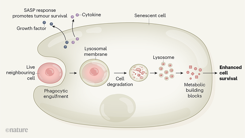
These senescent cells absorb and digest neighboring living cells. The cells are engulfed by a process that has the molecular characteristics of phagocytosis, a process used by immune cells. Once ingested, the cells are enveloped in the membrane of an organelle called lysosome and digested.

This degradation provides metabolic building blocks for the cell. Senescent cells that have ingested their neighbors survive longer than senescent cells that have not. This suggests that the metabolic building blocks recovered from lysosomal digestion of neighboring cells were used by senescent cells to promote their survival.

This surprising discovery highlights the complexity of the regulation of cell death in multicellular animals. Many mechanisms of cell death occur in animal tissues. These include forms of cell suicide, such as apoptosis, which leads to fragmentation of individual cells, and regulated forms of necrotic cell death that cause cell disruption. Some cases of engulfing cells occur through at least two distinct mechanisms:

  • One is a form of cell suicide called entosis, in which cells invade a neighboring cell and are phagocytized by it.
  • The other mechanism is cellular cannibalism resembling the process used by immune system cells such as macrophages to ingest and destroy dying cells.

The authors analyzed the gene expression profile of cancer cells treated with chemotherapy drugs (most of these cells were senescent) and found that the characteristic genes for phagocytosis were expressed. This gene expression culminated in a time that correlated with cell engulfment. They also observed that senescent cells engulfed dead cells added in vitro, providing a further clue that senescent cells engulf cells by phagocytosis.

Cellular cannibalism in cancers has already been reported. However, Tonnessen-Murray and his colleagues specifically identified an association between cannibalism and senescence, and they show that this phenomenon could make a substantial contribution to the persistence of senescent cells in cancerous tissues. Their results suggest that cellular cannibalism may be an activity that is largely associated with the induction of senescence, rather than being related to particular types of cancer or the status of proteins such as p53. It will be important to study if cannibalism is related to senescence in other contexts, for example the proteopathies that are found in neurodegenerative diseases, which moreover was suggested more than 10 years ago by Todd E Golde, and Victor M Miller. Since 2018, it has been known that senescent astrocytes and microglia accumulate in the brain in the context of diseases associated with Tau protein.

Advertisement


This book retraces the main achievements of ALS research over the last 30 years, presents the drugs under clinical trial, as well as ongoing research on future treatments likely to be able stop the disease in a few years and to provide a complete cure in a decade or two.


Please, help us continue to provide valuable information: