A failed SOD1 genetic therapy with Bannwarth syndrome commorbidity

- Posted by admin in English

Two patients with familial amyotrophic lateral sclerosis (ALS) and mutations in the gene encoding superoxide dismutase 1 (SOD1) were treated with a single intra-thecal infusion of adeno-associated virus encoding a microRNA targeting SOD1. Unfortunately it did not went well for the patients, but there are many interesting observations in this article. One of them is that one of the patient had an infection of Borrelia burgdorferi, a tick-borne spirochete bacterium also responsible for causing Lyme disease, which was discovered during the course of the disease.

Patient 1 had transient improvement in the strength of his right leg, a measure that had been relatively stable throughout his disease course, but there was no change in his vital capacity.

Patient 2 had stable scores on a composite measure of ALS function and a stable vital capacity during a 12-month period.

Patient 1

During the month of February 2017, Patient 1, a 22-year-old man, began to notice weakness in his left leg. He had the same SOD1 missense mutation (SOD1-A5V) as his mother, who had died from ALS at the age of 45 years. In March 2017, his slow vital capacity was 100% of the predicted value, and his ALSFRS-R score was 42. The flexion strength in his left hip was MRC grade 3, which indicated that he could move the limb against gravity. He could not bear full weight on his left heel or toes though.

On July 19, 2017, he received a single intrathecal infusion of 4.2×10¹⁴ vector genomes of AAV-miR-SOD1 along with an intravenous bolus of methylprednisolone (1.0 g); the latter was repeated the following day. Oral prednisone (at a dose of 60 mg per day) was then initiated, with planned tapering during a 4-week period. At that time, there was no plantar flexion or dorsiflexion in the left ankle ; the left knee flexion and extension could move only with gravity eliminated; The strength of the right leg and both arms was normal, as were sensory function and cognition.

Three weeks after the infusion, he had transient tingling in both hands, and 1 week later, he reported having a feeling of painful “electric shocks” in his left foot. The prednisone dose, which had been tapered to 10 mg per day, was increased to 30 mg.

Twenty-four weeks after treatment (46 weeks after the onset of ALS symptoms), the patient’s ALSFRS-R score was reduced to 38 from the baseline level of 42. The loss of strength in the left leg continued.

But at 12 months after treatment (nearly 18 months after the onset of ALS symptoms), the patient had transient improvement in the strength of his right leg, and could propel himself in a wheelchair using the right leg. However vital capacity was further reduced to 21% of the predicted value. At 14 months, he regained the ability to extend and flex the fingers of the left hand, a function that had been absent for the previous 20 weeks.

Lymphocytic meningoradiculitis, also known as Bannwarth syndrome, is a neurological disease characterized as intense nerve pain radiating from the spine. The disease is caused by an infection of Borrelia burgdorferi, a spirochete bacterium.

The patient died of respiratory arrest 15.6 months after the initiation of treatment and 20.5 months after the onset of ALS symptoms. The AAV-miR-SOD1 viral genome was detected in the cervical and lumbosacral spinal cord parenchyma. As compared with a baseline SOD1 level of 120 ng per milliliter in the CSF, the level was 102 ng per milliliter at 8 weeks and 120 ng per milliliter at 41 weeks

There were a loss of motor neurons in the cervical, thoracic, and left lumbosacral spinal cord but relative sparing of motor neurons in the right lumbosacral spinal cord.

The cortical ribbon in the primary motor–sensory cortex was moderately gliotic. Glial scar formation is a reactive cellular process involving astrogliosis that occurs after injury to the central nervous system.

Pyramidal neurons had pyknotic nuclei and hypereosinophilic cytoplasm, findings that were consistent with acute hypoxic–ischemic injury. Hypoxia is a condition in which the body or a region of the body is deprived of adequate oxygen supply at the tissue level. Pyknosis, is the irreversible condensation of chromatin in the nucleus of a cell undergoing necrosis or apoptosis.

The authors hypothesized that intrathecal infusion of this viral vector can trigger an adverse inflammatory response, as has been reported in some studies after the intravenous administration of AAV9 in animals.

The scientists cannot conclude that suppression of SOD1 played a role in his clinical course, since such improvements in function may have reflected recovery from meningoradiculitis.

Patient 2

For patient 2, during the year before therapy, his functional status had been stable, with ALSFRS-R scores averaging close to 28. For a year before treatment, his slow vital capacity had ranged from 42 to 58% of the predicted value;

As meningora-diculitis developed after treatment in Patient 1 and they suspected some relation between the two events, the scientists aimed to suppress B-cell activity and T-cell function with rituximab (at a dose of 375 mg per square meter of body-surface area), which was initiated in late August 2018 in weekly intravenous infusions for 3 weeks and with intrave-nous methylprednisolone (at a dose of 125 mg before each dose of rituximab and 1 g on the day of AAV-miR-SOD1 infusion).

Beginning at the initiation of study treatment, the patient began receiving daily oral sirolimus (6 mg). The day after treatment, oral prednisone (0.5 mg per kilogram of body weight) was initiated; sirolimus and prednisone were continued for 6 months.

On September 17, 2018, the patient received an intrathecal infusion of 4.2×10¹⁴ vector genomes of AAV-miR-SOD1.

On the day after treatment and at weeks 12 and 17, he received intra-venous immune globulin (at a dose of 0.4 mg per kilogram) in response to a decrease in the serum IgG to a level of less than 700 mg per deciliter, which had been induced by rituximab.

60 weeks after treatment, the ALSFRS-R score was 24, signifying worse overall function.
At 65 weeks after therapy, the slow vital capacity score value was 62% .

In contrast to the clinical course of Patient 1, the immuno-suppressive regimen in Patient 2 blunted the generation of neutralizing antibodies, antiviral antibodies, and T-cell response to the viral capsid. As of May 18, 2020, his disease course was stable, with a functional measure of 24 at 90 weeks after treatment. A course that could be typical of the slow disease progression in patients with his SOD1 genotype, so no clinical conclusions can be made about the treatment effects.

Conclusion

It is very important for the scientific knowledge acquisition process to publish failures. This study is in this respect important, but it is a quite rare publication that describes also other aspects of the story. If intrathecal genetic therapies incur an increased risk of a Borrelia burgdorferi based disease, this is an extremely important fact.

More about infections and Alzheimer

- Posted by admin in English

Flu (influenza) and pneumonia vaccinations are associated with reduced risk of Alzheimer's disease, according to new research reported at the Alzheimer's Association International Conference® (AAIC®) 2020.

Three research studies reported at AAIC 2020 suggest:

  • At least one flu vaccination was associated with a 17% reduction in Alzheimer's incidence.
  • More frequent flu vaccination was associated with another 13% reduction in Alzheimer's incidence.
  • Vaccination against pneumonia between ages 65 and 75 reduced Alzheimer's risk by up to 40% depending on individual genes.
  • Individuals with dementia have a higher risk of dying (6-fold) after infections than those without dementia (3-fold).

"With the COVID-19 pandemic, vaccines are at the forefront of public health discussions. It is important to explore their benefit in not only protecting against viral or bacterial infection but also improving long-term health outcomes," said Maria C. Carrillo, Ph.D., Alzheimer's Association chief science officer.

"It may turn out to be as simple as if you're taking care of your health in this way — getting vaccinated — you're also taking care of yourself in other ways, and these things add up to lower risk of Alzheimer's and other dementias," Carrillo said. "This research, while early, calls for further studies in large, diverse clinical trials to inform whether vaccinations as a public health strategy decrease our risk for developing dementia as we age."

Seasonal Flu Vaccine May Reduce Incidence of Alzheimer's Dementia

Previous research has suggested vaccinations may have a protective factor against cognitive decline, but there have been no large, comprehensive studies focused on the influenza (flu) vaccine and Alzheimer's disease risk, specifically. To address this gap, Albert Amran, a medical student at McGovern Medical School at The University of Texas Health Science Center at Houston, and team, investigated a large American health record dataset (n=9,066).

Amran and team found having one flu vaccination was associated with a lower prevalence of Alzheimer's (odds ratio 0.83, p<0.0001), and among vaccinated patients receiving the flu vaccine more frequently was associated with an even lower prevalence of Alzheimer's (odds ratio 0.87, p=0.0342). Thus, people that consistently got their annual flu shot had a lower risk of Alzheimer's. This translated to an almost 6% reduced risk of Alzheimer's disease for patients between the ages of 75-84 for 16 years.

The researchers found the protective association between the flu vaccine and the risk of Alzheimer's was strongest for those who received their first vaccine at a younger age — for example, the people who received their first documented flu shot at age 60 benefited more than those who received their first flu shot at age 70.

"Our study suggests that regular use of a very accessible and relatively cheap intervention — the flu shot — may significantly reduce risk of Alzheimer's dementia," Amran said. "More research is needed to explore the biological mechanism for this effect — why and how it works in the body — which is important as we explore effective preventive therapies for Alzheimer's."

Pneumonia Vaccine May Reduce Alzheimer's Risk later in life.

Repurposing of existing vaccines may be a promising approach to Alzheimer's disease prevention. Svetlana Ukraintseva, Ph.D., Associate Research Professor in the Biodemography of Aging Research Unit (BARU) at Duke University Social Science Research Institute, and team, investigated associations between pneumococcal vaccination, with and without an accompanying seasonal flu shot, and the risk of Alzheimer's disease among 5,146 participants age 65+ from the Cardiovascular Health Study. The team also took into account a known genetic risk factor for Alzheimer's — the rs2075650 G allele in the TOMM40 gene.

The researchers found that pneumococcal vaccination between ages 65-75 reduced risk of developing Alzheimer's by 25-30% after adjusting for sex, race, birth cohort, education, smoking, and number of G alleles. The largest reduction in the risk of Alzheimer's (up to 40%) was observed among people vaccinated against pneumonia who were non-carriers of the risk gene. Total number of vaccinations against pneumonia and the flu between ages 65 and 75 was also associated with a lower risk of Alzheimer's; however, the effect was not evident for the flu shot alone.

"Vaccinations against pneumonia before age 75 may reduce Alzheimer's risk later in life, depending on individual genotype," Ukraintseva said. "These data suggest that pneumococcal vaccine may be a promising candidate for personalized Alzheimer's prevention, particularly in non-carriers of certain risk genes."

Infection Substantially Increases Mortality in People with Dementia

People living with dementia commonly experience other health conditions including viral, bacterial, and other infections. There is a growing trend in research to investigate whether infections might be worsening, more life-threatening or possibly causing dementia.

Janet Janbek, a Ph.D. student at the Danish Dementia Research Centre, Rigshospitalet and the University of Copenhagen in Denmark, and team, used data from national health registries to investigate mortality in Danish residents over age 65 (n=1,496,436) who had visited the hospital with an infection. They found that people with both dementia and such hospital visits died at a 6.5 times higher rate compared with people who had neither. Study participants with either dementia alone or infection-related contacts alone had a threefold increased rate. The rate of mortality was highest within the first 30 days following the hospital visit.

The researchers also found that for people living with dementia the mortality rates remained elevated for 10 years after the initial infection-related hospital visit, and mortality rates from all infections (including major infections like sepsis to minor ear infections) were higher compared with people without dementia or without an infection-related hospital visit.

"Our study supports the need to investigate these relations even further; to find out why infections are linked to higher mortality in people with dementia, specifically which risk factors and biological mechanisms are involved. This will help advance our understanding of the role of infections in dementia," said Janbek.

"Our study suggests that the health care system — as well as relatives of people with dementia — should have increased awareness of people with dementia who get infections, so they get the medical care they need. People with dementia require more specialized treatment even when their hospital visits are not directly due to their dementia but to what might appear to be an unrelated infection," Janbek added.

Chronic neurodegenerative disorders, such as Alzheimer's disease and cerebrovascular disease, are common conditions in older people. The treatment of these conditions is aggravated by the relatively high prevalence of systemic comorbidity, which is detected in more than half of patients over 65 years of age in several surveys.

Coexisting neuropathological markers of Alzheimer's disease and cerebrovascular disease are found at autopsy in the majority of brains of many cohorts of patients clinically diagnosed with Alzheimer's disease.

A recent study, of over two hundred participants, showed the association of image markers of cerebrovascular disease, including cortical cerebral microinfarctions, with an increased risk of cognitive decline and atrophy in patients diagnosed with Alzheimer's disease.

Notably, clinical signs of the interaction of Alzheimer's disease and cerebrovascular disease can be attributed to the archetypal study Nun. A longitudinal study of aging and Alzheimer's disease of nuns was initiated in 1986 by Snowdon. The homogeneous lifestyle of the nuns makes them an ideal study population. The convent archives were made available to the investigators as a resource on the history of the participants. The study included the administration of memory and cognitive tests to nuns (some over 100 years old) and the post-mortem examination of their brains. The results of this study showed that the coexistence of rare markers of Alzheimer's disease with minor ischemic damage was associated with exacerbated dementia compared to patients diagnosed with only one or the other of the conditions.

enter image description here

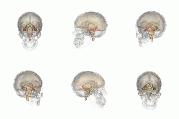
The hippocampus, one of the only cerebral structures, is one of the first structures affected in Alzheimer's disease, which explains the memory problems and disorientation that characterize the appearance of this neurodegenerative pathology. People with severe damage to the hippocampus are likely to suffer from different types of amnesia.

Scientists from the Instituto de Biofisica da UFRJ in Rio de Jaeiro, have developed an in vitro model of cerebrovascular disease co-morbid with Alzheimer's disease. This model involves organotypic cultures of the hippocampus, combining subtoxic oxygen-glucose deprivation (OGD) with exposure to subtoxic concentrations of amyloid oligomers (AβO).

This approach was designed to simulate early subclinical conditions that may precede the diagnosis of mild cognitive impairment, assuming that such subclinical conditions may be associated with minor vascular insufficiency and / or low AβO levels.

Scientists have developed an in vitro model of comorbid cerebrovascular disease / Alzheimer's disease applied to organotypic cultures of hippocampi, by combining subtoxic oxygen-glucose deprivation with exposure to also subtoxic concentrations of amyloid oligomers (AβO) Alzheimer's disease, toxins that build up in the brain and are believed to trigger synapse damage and cognitive impairment in Alzheimer's disease.

Using this experimental model, the scientists then studied in more detail the roles of glutamate receptors, which have been implicated in many approaches to cerebrovascular disease and Alzheimer's disease.

These results revealed a hitherto unappreciated synergy between subtoxic oxygen-glucose deprivation and AβO, and unveiled the complex roles of glutamate receptors on neurodegeneration in the hippocampus under conditions that mimic early and early cerebrovascular disease. comorbid / Alzheimer's disease. The results thus showed that:

  • subtoxic insults by oxygen-glucose deprivation and AβOs synergize to induce marked reductions in synaptic proteins in the absence of cell death.
  • the effects of glutamate receptor antagonists in the combined presence of subtoxic oxygen-glucose deprivation and AβO are complex and rather distinct from previous reports of their effects on experimental models of cerebrovascular disease or Alzheimer's disease alone .

Their results unveiled the complex roles of glutamate receptors on neurodegeneration in the hippocampus under conditions that mimic early and co-morbid cerebrovascular disease / Alzheimer's disease.

Advertisement


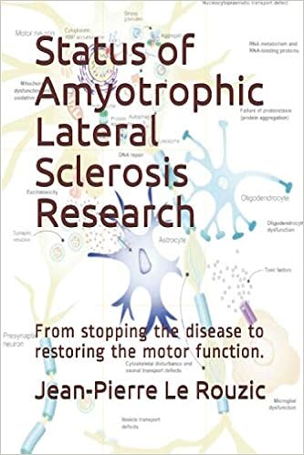
This book retraces the main achievements of ALS research over the last 30 years, presents the drugs under clinical trial, as well as ongoing research on future treatments likely to be able stop the disease in a few years and to provide a complete cure in a decade or two.

Les troubles neurodégénératifs chroniques, tels que la maladie d’Alzheimer et les maladies cérébrovasculaires, sont des pathologies courantes chez les personnes âgées. Le traitement de ces affections est aggravé par la prévalence relativement élevée de la comorbidité systémique, qui est détectée chez plus de la moitié des patients de plus de 65 ans dans plusieurs enquêtes.

Des marqueurs neuropathologiques coexistants de la maladie d’Alzheimer et des maladies cérébrovasculaires sont retrouvés à l’autopsie dans la majorité des cerveaux de nombreuses cohortes de patients diagnostiqués cliniquement avec la maladie d’Alzheimer.

Une étude récente, de plus de deux cents participants, a montré l’association des marqueurs d’image des maladies cérébrovasculaires, y compris les micro-infarctus cérébraux corticaux, avec un risque accru de déclin cognitif et d’atrophie chez les patients diagnostiqués avec la maladie d’Alzheimer.

Notamment, les signes cliniques d’interaction de la maladie d’Alzheimer et des maladies cérébrovasculaires peuvent être attribués à l’étude archétypale Nun. Une étude longitudinale du vieillissement et de la maladie d'Alzheimer de religieuses a été initiée en 1986 par Snowdon. Le style de vie homogène des religieuses en fait une population d'étude idéale. Les archives du couvent ont été mises à la disposition des enquêteurs en tant que ressource sur l'histoire des participants. L'étude comprend l'administration de la mémoire et des tests cognitifs aux religieuses (certaines de plus de 100 ans) et l'examen post mortem de leur cerveau. Les résultats de cette étude ont montré que la coexistence de marqueurs rares de la maladie d’Alzheimer avec des lésions ischémiques mineures, était associée à une démence exacerbée par rapport aux patients diagnostiqués avec seulement l’une ou l’autre des conditions.

enter image description here

L'hippocampe, une des seules structures cérébrales, est une des premières structures atteintes dans la maladie d'Alzheimer, ce qui explique les problèmes de mémoire et de désorientation qui caractérisent l'apparition de cette pathologie neurodégénérative. Les personnes subissant de graves dommages à l'hippocampe sont susceptibles de souffrir de différents types d'amnésie. L'hypoxie (la privation d'oxygène), les encéphalites et les épilepsies du lobe temporal peuvent résulter de lésions au niveau de l'hippocampe.

Des scientifiques de l’Instituto de Biofisica da UFRJ à Rio de Jaeiro, ont développé un modèle in vitro de maladies cérébrovasculaires comorbides de la maladie d’Alzheimer. Ce modèle implique des cultures organotypiques d’hippocampe, en combinant la privation subtoxique d’oxygène-glucose (OGD) avec une exposition à des concentrations subtoxiques d’oligomères amyloïdes (AβO).

Cette approche a été conçue pour simuler des conditions subcliniques précoces susceptibles de précéder le diagnostic d’une déficience cognitive légère, en supposant que de telles conditions infracliniques peuvent être associée à une insuffisance vasculaire mineure et/ou à de faibles taux d’AβO.

Les scientifiques ont développé un modèle in vitro de maladies cérébrovasculaires comorbides / maladie d’Alzheimer appliqué aux cultures organotypiques d’hippocampes, en combinant la privation subtoxique d’oxygène-glucose avec une exposition à des concentrations également subtoxiques d’oligomères amyloïdes (AβO), des toxines qui s’accumulent dans le cerveau de la maladie d’Alzheimer et sont censés déclencher des dommages aux synapses et des troubles cognitifs dans la maladie d’Alzheimer.

À l’aide de ce modèle expérimental, Les scientifiques ont alors étudié plus en détail les rôles des récepteurs du glutamate, qui ont été impliqués dans de nombreuses approches des maladies cérébrovasculaires et de la maladie d’Alzheimer.

Ces résultats ont révélé une synergie jusqu’ici inappréciée entre la privation subtoxique d’oxygène-glucose et l’AβO, et dévoilé les rôles complexes des récepteurs du glutamate sur la neurodégénérescence dans l’hippocampe dans des conditions qui imitent les maladies cérébrovasculaires précoces et comorbides / maladie d’Alzheimer. Les résultats ont ainsi montré que : * les insultes subtoxiques par privation d’oxygène-glucose et AβOs synergisent pour induire des réductions marquées des protéines synaptiques en l’absence de mort cellulaire; * les effets des antagonistes des récepteurs du glutamate en présence combinée de privation subtoxique d’oxygène-glucose et d’AβO sont complexes et plutôt distincts des rapports précédents de leurs effets sur des modèles expérimentaux de maladies cérébrovasculaires ou de la maladie d’Alzheimer seule. Leurs résultats ont dévoilé les rôles complexes des récepteurs du glutamate sur la neurodégénérescence dans l’hippocampe dans des conditions qui imitent les maladies cérébrovasculaires précoces et comorbides / maladie d’Alzheimer.

Publicité


Ce livre retrace les principales réalisations de la recherche sur la SLA au cours des 30 dernières années. Il présente les médicaments en cours d’essai clinique ainsi que les recherches en cours sur les futurs traitements susceptibles d’ici quelques années, d’arrêter la maladie et de fournir un traitement complet en une décennie ou deux.

Late-life restoration of mitochondrial function

- Posted by admin in English

The article discussed here is not related to neurodegeneration diseases, it discusses about heart failure, however it might have implications for ALS. While most ALS targeting therapies might aim at reducing TDP-43 aggregates (and similar protein aggregates in other neurodegenerative diseases), humans are indeed more than bags of identical cells, they are first living because they are composed of a multitude of physiological systems that interact to maintain homeostasis. So even if a therapy was invented that would efficiently remove TDP-43 aggregates, ALS patients would still be unable to recover health as motor neurons do not rejuvenate nor are replaced with newer cells. As this heart failure treatment improves heart muscle cells, it should also to some degree improve motor neuron cells. This article is also interesting as it mentions some drugs that are discusses ALS online internet forums, such as glutathione, N-Acetyl Cysteine (NAC) and Glycine.

This article explains precisely how some muscle cells seem to rejuvenate when a specific peptide is administrated. Very roughly: With this peptide, metabolism is rejuvenated at cellular level, so cells can use more energy, something which is clearly lacking in ALS cells which are characterized by hypermetabolism. Humans produce and consume about 65 kg of ATP every day. Because ATP cannot be stored, it is critical that the rate of ATP synthesis matches the rate of ATP consumption. The primary role of mitochondria is the generation of adenosine triphosphate (ATP) from adenosine diphosphate (ADP) using macromolecular complexes that form the electron transport chain.

enter image description here

Mitochondrial dysfunction is one of the hallmarks of aging. While mitochondria generate the bulk of cellular ATP, they are also the major source of reactive oxygen species (ROS) in most cells. ROS are sub-products inherent to ATP metabolism.

Aging is the strongest risk factor for cardiovascular diseases. It is also accompanied by a decline in cardiac function, especially diastolic dysfunction and hypertrophy of the left ventricle and left atrium. The heart is rich in mitochondria and has a high metabolic demand; therefore, it is highly susceptible to oxidative damage and the effects of mitochondrial dysfunction. Increasing evidence suggests that mitochondrial oxidative stress and mitochondrial dysfunction play critical roles in cardiovascular diseases and cardiac aging.

The mitochondrial-targeted tetrapeptide SS-31 (elamipretide), is a pharmacologic intervention that selectively concentrates in mitochondria, suppressing mitochondrial ROS and increasing skeletal muscle ATP production. Elamipretide (also named SS-31, MTP-131, Bendavia) is sold by Stealth BioTherapeutics, Newton, Massachusetts. It is a water-soluble, aromatic-cationic mitochondria-targeting tetrapeptide that readily penetrates and transiently localizes to the inner mitochondrial membrane and associates with cardiolipin to restore mitochondrial bioenergetics

it has not been established whether delivering such interventions in later life can rescue pre-existing mitochondrial and cardiac dysfunction. In this study, the authors demonstrate that mitochondrial-targeted interventions can improve mitochondrial function and reverse pre-existing cardiac dysfunction in old mice.

To determine the effects of SS-31 treatment on cardiac function in old mice, the scientists treated 24-month-old mice with the SS-31 peptide or saline control and examined cardiac function by echocardiography after 4 and 8 weeks of treatment.

SS-31 treatment was effective in aged hearts with pre-existing mitochondrial dysfunction but had little effects in young hearts with normal functioning mitochondria.

The authors acknowledge that the persistence of SS-31 induced functional benefit varied between individual mice. Other studies have reported negative effects of targeting mitochondrial ROS. In another study, suppression of mitochondrial ROS in mice resulted in impaired macrophage bactericidal activity.

However not everything is rosy, elamipretide is known since quite some time and had been tested in several different diseases. Recently it did not meet expectations stemming from promising early trial results in patients with primary mitochondrial myopathy (PMM), data from a Phase 3 (NCT03323749) trial show.

Stress granules are believed to play a critical role in modulating gene expression programs in response to environmental and nutrient stresses. However, it has been unclear how changes in cellular activity regulate stress granule formation and composition. The authors of a recent article, shown that Sam1 is recruited to yeast stress granules in response to a specific nutrient stress.

S-Adenosyl methionine is a common cosubstrate involved in methyl group transfers, transsulfuration, and aminopropylation. Although these anabolic reactions occur throughout the body, most SAM-e is produced and consumed in the liver. It is made from adenosine triphosphate (ATP) and methionine by methionine adenosyltransferase.

The scientists found that the product of Sam1, AdoMet, regulates stress granule formation in both yeast and human cells (HeLa and U2OS). This suggests that the connections between metabolism and stress granule assembly might be broader than previously believed. Their focus on the product of Sam1, AdoMet, uncovered parallels between how metabolites regulate metabolism and stress granule formation/composition. Most provocatively, AdoMet administration to iPSC-derived motor neurones cells, suppressed stress granule formation that expressed mutated forms of TDP-43 and FUS found in ALS patients. AdoMet was effective in blocking stress granule assembly in these disease models.

Cells deploy a variety of mechanisms to fine-tune biochemical processes in response to environmental stressors. One of these mechanisms is the formation of stress granules. Stress granules assemble in response to a variety of nutrient and metabolic stresses and are believed to provide a mechanism for coupling metabolic stress to post-transcriptional gene regulation.

Furthermore, stress granules act as centers to regulate cell signaling outputs and protein folding, highlighting stress granules as global integrators of the stress response. stress granules are transient and require tight regulation of both assembly and disassembly for cell function and viability. For example, disruption of stress granule formation decreases cell survival when the stress is removed.

In addition to their role in integrating the cellular stress response, stress granules have been implicated in a variety of neurodegenerative disorders. Dysregulation in stress granule dynamics in ALS patients results in accumulation of atypical cytoplasmic, stress granule-like protein aggregates in dying neurons of the brain and spinal cord. These results argue that understanding how stress granules assemble in response to metabolic or nutrient stresses is critical for both understanding the pathophysiology of ALS and FTD and developing treatment strategies focused on disrupting the formation of aberrant stress granules.

Given the linkage between stress granules and several neurodegenerative diseases, the composition of the stress granule proteome has been a subject of intense focus to identify potential therapeutic targets. Unfortunately, large-scale biochemical studies have found that stress granule composition is not only stress specific, but also organism and cell type specific. Although different techniques have helped identify which components reside within each phase, the relative role of stress granule core proteins and shell proteins in stress granule formation and pathogenesis remains unclear.

Despite the fact that stress granule formation and composition is stress specific, there has been surprisingly little exploration of the connections between metabolism and stress granule assembly. To date, only a few metabolic enzymes have been shown to be enriched or localized to stress granules via proteomic and/or targeted studies. This deficit is likely due to the limited number of stress conditions that have been used in stress granule proteomic studies. Interestingly, the localization of one metabolic enzyme, pyruvate kinase (Cdc19), to stress granules has been shown to be crucial for reactivation of growth-promoting pathways upon removal of stress. This suggests that stress granules can play a critical role in regulating metabolic enzymes in response to stress.

There are also limited examples of the reverse mode of regulation: metabolic intermediates that modulate stress granule assembly. Whereas stress granules can assemble upon the removal of glucose or amino acids, only one metabolite from intermediate metabolism, acetyl-CoA, has been implicated in regulating stress granule formation. Thus, the identification of metabolic enzymes that are recruited to stress granules in a stress-specific manner would identify new linkages between stress granule formation and metabolism as well as provide a novel set of potential therapeutic targets for ALS and FTD.

In this article the researchers have identified 17 metabolic enzymes that are recruited to yeast stress granules in a stress-specific manner. S-adenosylmethionine (AdoMet), the product of one these enzymes, is a regulator of stress granule assembly and composition.

The regulation of yeast stress granule formation by AdoMet is biphasic, with chronic changes altering stress granule composition and acute elevation of AdoMet suppressing stress granule assembly. Additionally, acute elevation of AdoMet suppresses stress granule formation in motor neurones, demonstrating conserved metabolite regulation of stress granule assembly from yeast to humans. The suppressive effect of AdoMet on stress granule formation also occurs in induced pluripotent stem cell (iPSC)-derived motor neurones from ALS patients.

AdoMet blocks the recruitment of cytoplasmic TDP-43 to remnant stress granules in this cell culture, implying that AdoMet can modify the pathogenic accumulation of stress granule material. Together, these results argue that metabolic activity controls both the composition and extent of stress granule formation and provide a framework for the identification of lead compounds that can modify or suppress stress granule formation.

Recent work on the stress granule proteome suggests that stress granule composition can vary depending on the cell type and the nature of the stress. Because many of the stresses that trigger stress granule assembly are thought to alter metabolic activity, either directly or indirectly, one might expect metabolic enzymes to be a common component of stress granules. However, few metabolic enzymes have been identified in proteomic and targeted studies of Saccharomyces cerevisiae stress granules. Their identification of 17 metabolic enzymes that are recruited to stress granules in response to physiological nutrient stresses, but are not recruited to stress granules in response to multiple acute stresses, argues that stress granule composition is tailored to the nature of the stress and that chronic stresses might require reorganization of the metabolic network.

This result also helps to explain why no metabolic enzymes have been identified in previous proteomic studies of mammalian stress granules. All of the stresses that are traditionally used to induce mammalian stress granules, such as sodium azide, do not trigger the recruitment of metabolic enzymes to yeast stress granules. Thus, one might expect to observe metabolic enzymes only in stress granules that assemble in response to the mammalian equivalent of a stationary-phase nutrient stress.

As a final thought: Whose ALS patient, have not dream of a simple drug that would alleviate their symptoms? AdoMet is interesting to investigate.


Please, help us continue to provide valuable information: