Alzheimer’s disease remains one of the hardest areas in drug development. One major reason is the behavior of amyloid-beta (Aβ), a peptide strongly associated with the disease, especially its 42-amino-acid form Aβ42.
Unlike many proteins targeted by conventional drugs, Aβ does not maintain one stable 3D structure. Instead, it behaves as an intrinsically disordered peptide, constantly shifting among many conformations. This makes it difficult to apply the usual “lock-and-key” approach of drug design, where a molecule is built to fit a well-defined binding pocket.
A recent paper by Morita, Maruyama and colleagues in Chemistry – A European Journal proposes a different strategy: rather than searching for a fixed pocket, the researchers use chirality-guided molecular recognition to bind a short sequence motif within Aβ42 and interfere with its aggregation.

Why amyloid-beta is difficult to drug
Aβ42 is a classic example of a target that resists standard medicinal chemistry.
Traditional small-molecule drugs work best when a protein has:
- a stable fold
- a persistent groove or pocket
- a clearly defined active site
Aβ42 has none of these features. Its pathological behavior comes from self-assembly into oligomers and fibrils, rather than from an enzyme-like function.
This is why many researchers describe intrinsically disordered proteins as hard to drug, even though the term “undruggable” is probably too absolute.
The challenge is therefore not simply “finding something that sticks,” but finding a molecule that selectively redirects or blocks a highly dynamic aggregation pathway.
What is actually new about the chirality idea?
The use of chirality itself is not new.
Researchers have studied D-peptides (mirror-image peptides made from D-amino acids) for years, including in Alzheimer’s research. D-peptide candidates such as RD2 already exist, and chirality has also been used to study Aβ uptake, aggregation, and membrane interactions.
So the novelty of this paper is more specific.
What appears genuinely new is the systematic design framework:
The authors first studied how short peptide sequences form stereocomplexes with their mirror images, identified the sequence features that favor this interaction, then used those rules to rationally design a D-peptide against the –FFAE– motif of Aβ42.
This moves the work beyond the older “try D-peptides and screen what works” approach.
The conceptual advance is the idea that mirror-image recognition can become a design principle, especially for intrinsically disordered proteins where fixed-structure targeting is difficult.
That broader framing may be more important than the Alzheimer’s angle alone.
Why the approach is scientifically interesting
The designed D-peptide inhibited Aβ42 fibril formation in vitro and reduced Aβ42-associated toxicity in neuronal-like cells. In the authors’ assays, it even outperformed RD2, an existing clinical-stage D-peptide comparator.
Several features make this attractive in principle:
1) It targets a sequence motif rather than a rigid structure
Because the interaction is based on sequence complementarity and stereochemistry, it may work even when the target peptide remains flexible.
That is a useful idea for intrinsically disordered proteins more broadly, including proteins implicated in Parkinson’s disease and some cancers.
2) D-peptides are protease-resistant
A practical advantage of D-amino-acid peptides is that most biological proteases evolved to degrade L-peptides.
This often gives D-peptides:
- longer half-life
- greater metabolic stability
- improved persistence in biological fluids
This is one reason mirror-image therapeutics have attracted long-standing interest.
3) It may reduce trial-and-error screening
Perhaps the most important long-term potential is methodological.
If stereocomplexation rules can be generalized, this could become a rational route to designing binders for disordered proteins, which is still a major unmet need in drug discovery.
Roadblocks before this becomes therapeutic
This remains an early proof-of-concept, and several major uncertainties remain.
Brain delivery is still a major challenge
A D-peptide that works in solution or cultured cells still needs to cross the blood–brain barrier.
This is one of the central bottlenecks in Alzheimer’s therapeutics, and the paper does not solve that translational problem.
Real Aβ biology is more complex than purified assays
Aβ42 in the brain does not exist as one clean species.
It transitions among:
- monomers
- soluble oligomers
- protofibrils
- mature fibrils
- membrane-associated forms
A binder optimized against one motif in vitro may behave differently in this far more heterogeneous environment.
Cell protection is not disease modification
The cell experiments are encouraging, but rescue of cultured neuronal-like cells is still very far from demonstrating efficacy in animals or humans.
Many anti-amyloid strategies have looked convincing at this stage and later failed in vivo.
Manufacturing and formulation remain nontrivial
D-peptides are chemically synthesizable, which is an advantage, but scaling highly pure sequences can still be expensive.
For CNS delivery, formulation requirements may further complicate development.
A broader perspective: why this may matter beyond Alzheimer’s
The most valuable part of this study may not be the immediate therapeutic claim.
Its broader significance is that it offers a general molecular-recognition strategy for intrinsically disordered proteins, a class of targets that remains difficult across neuroscience and oncology.
In that sense, the paper is less about “a near-term Alzheimer’s drug” and more about expanding the design toolbox for hard protein targets.
That is a meaningful contribution, even if the path to a medicine remains long.
Bottom line
This study should be seen as a carefully reasoned molecular design paper, not as evidence that Alzheimer’s treatment is about to change.
The real advance is not simply “using mirror-image chemistry,” since that field already exists. The more interesting step is the rule-based use of chirality to design sequence-targeting ligands for disordered proteins.
If that principle proves transferable, it could become useful well beyond amyloid-beta.
For now, it is best understood as a promising strategy at the chemistry and early cell-biology stage, with major delivery, validation, and translational hurdles still ahead.
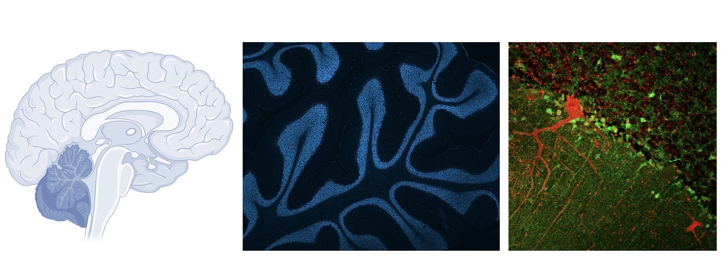 To understand this major breakthrough, it's helpful to dispel a common misconception about DNA damage. Our genetic code isn't a static library; it constantly undergoes routine damage. Every time a brain cell processes oxygen, generates metabolic energy, or emits an electrical impulse, it creates oxidative stress that naturally breaks its own DNA strands.

 By testing this approach on mouse models of both diseases and on human patient stem cells (iPSCs), the team observed several crucial improvements: