A growing number of studies are exploring the role of perineuronal nets (PNNs) in Parkinson’s disease. PNNs are specialized structures in the brain’s extracellular matrix that limit synaptic plasticity, like a stabilizing "glue" between neurons. While this stabilization helps maintain brain function in adulthood, it also reduces the brain’s ability to rewire or adapt, which is important for learning and recovery after injury.
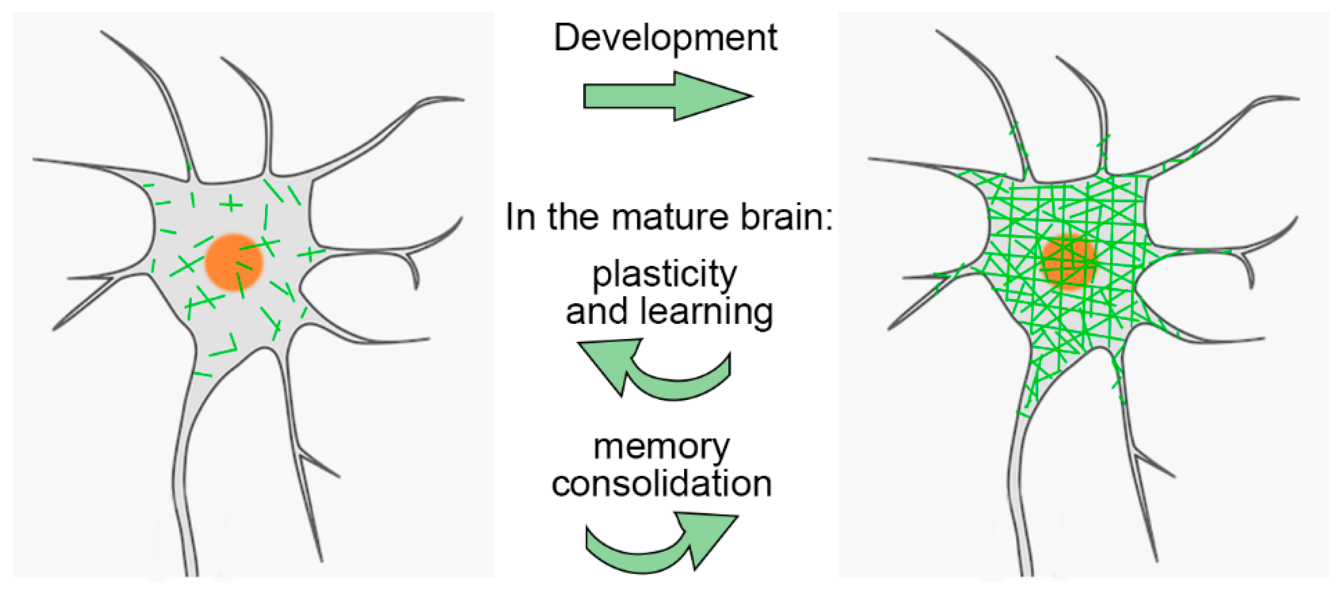 PNNs are especially important during brain development. They help close the "critical period" of heightened plasticity in childhood. Interestingly, while PNNs are degraded in adults, this plasticity can be partially restored. For example, PNN removal can promote recovery in stroke models
PNNs serve multiple functions: protecting neurons from oxidative stress and harmful molecules, regulating plasticity, and helping stabilize long-term memories. Abnormal changes in PNNs have been observed in aging and various neurological conditions, making them a promising target for therapeutic intervention.
In the cerebral cortex, PNNs are primarily found around inhibitory interneurons, particularly those that produce the protein parvalbumin. These interneurons help maintain a balance between excitatory and inhibitory signals in the brain. In Parkinson’s disease, where dopamine-producing neurons in the substantia nigra are lost, this balance is disrupted—suggesting that changes in PNNs may contribute to the disorder.
A recent study investigated the effects of temporarily reducing PNNs in the primary motor cortex (M1) of healthy adult mice using chondroitinase ABC (ChABC). This intervention caused temporary impairments in motor function, suggesting that PNNs contribute to motor stability. Using ChABC makes sense as perineuronal nets are composed of chondroitin sulfate proteoglycans, and ChABC is an enzyme that digests them. Chondroitinase treatment has been shown to allow adults' vision to be restored; moreover, there is some evidence that Chondroitinase could be used for the treatment of spinal injuries.
The researchers created a Parkinson’s disease mouse model by damaging one side of the midbrain of mice with 6-hydroxydopamine (6-OHDA). Two weeks after the lesion, PNN levels dropped in both hemispheres of the motor cortex but returned to normal within five weeks.
The researchers then applied ChABC to reduce PNNs again in the motor cortex and paired this with motor training. This combination improved motor recovery slightly in the Parkinsonian mice. The improvement was linked to an increase in parvalbumin interneurons surrounded by PNNs and a normalization of excitatory signals at their cell bodies. These findings suggest that PNNs respond dynamically—first to the injury and later to therapeutic intervention—and that manipulating them could help restore motor function.
In summary, perineuronal nets in the motor cortex appear to play a subtle but significant role in regulating movement. Modifying their structure could open new avenues for motor rehabilitation in Parkinson’s disease.