Nervous system injuries affect over 10 of thousands people every year. It is estimated that spinal cord injuries alone affect 10,000 each year. Nervous system injuries includes also effects of stroke, glaucoma and some injuries to the eye.
enter image description here

The nervous system is divided into two parts: the central nervous system, which consists of the brain and spinal cord, and the peripheral nervous system, which consists of cranial and spinal nerves along with their associated ganglia.

Nerve regeneration (the regrowth or repair of nervous tissues), a subfield of neural tissue engineering, is becoming a rapidly growing field dedicated to the discovery of new ways to recover nerve functionality after injury. However neuroregeneration differs between the peripheral nervous system (PNS) and the central nervous system (CNS). When an axon is damaged, the distal segment undergoes Wallerian degeneration, while the proximal segment can either die by apoptosis or undergo the chromatolytic reaction, which is an attempt at repair.

There is currently no treatment for recovering human nerve function after injury to the central nervous system. Unlike peripheral nervous system injury, injury to the central nervous system is limited by the inhibitory growth environment which is, in part, created by the migration of myelin-associated inhibitors, astrocytes, oligodendrocytes, oligodendrocyte precursors, and microglia.

Although neuroinflammation is often depicted as detrimental, there is growing evidence that alternatively activated, reparative leukocyte subsets and their products can be deployed to improve neurological outcomes.

Using a mouse model, researchers at Ohio State and the University of Michigan discovered a new type of immune cell that not only rescues damaged nerve cells from death, but partially reverses nerve fiber damage. The research team also identified a human immune cell line, with similar characteristics, that promotes nervous system repair.

The cell discovered by these researchers is a granulocyte, a type of white blood cell that has small granules. The most common granulocytes, neutrophils, normally help the body fight off infection. The unique cell type resembles an immature neutrophil but is distinctive in possessing neuroprotective and neuroregenerative properties. It drives central nervous system axon (nerve) regrowth in vivo, in part through the secretion of a cocktail of growth factors.

In the current study, the researchers identify a unique granulocyte subset, with characteristics of an immature neutrophil, that had neuroprotective properties and drove CNS axon regeneration in vivo, in part via secretion of a cocktail of growth factors. This pro-regenerative neutrophil promoted repair in the optic nerve and spinal cord, demonstrating its relevance across CNS compartments and neuronal populations. Their findings could lead to the development of new therapies that reverse CNS damage and restore lost neurological function across a spectrum of diseases.

The researchers demonstrated the therapeutic potency of the immature neutrophils subset by injecting them into mice with crush injury to the optic nerve or lacerated nerve fibers in the spinal cord. Mice injected with the new neutrophil subset, but not more typical mature neutrophils, grew new nerve fibers.

The next step for the Ohio State and University of Michigan researchers is to collect the neuro-enhancing granulocytes they discovered and figure out how to enhance them in the lab. Ultimately, they hope to determine whether the cells could be injected into patients to reverse damage to the central nervous system.

"Our findings could ultimately lead to the development of novel immunotherapies that reverse central nervous damage and restore lost neurological function across a spectrum of diseases," said first author Dr. Andrew Sas, an assistant professor and physician scientist in the Department of Neurology at Ohio State.

Dr. Benjamin Segal, professor and chair of the Department of Neurology at The Ohio State College of Medicine and co-director of the Ohio State Wexner Medical Center's Neurological Institute said: "In the future, this line of research might ultimately lead to the development of novel cell based therapies that restore lost neurological functions across a range of conditions."

Advertisement


This book retraces the main achievements of ALS research over the last 30 years, presents the drugs under clinical trial, as well as ongoing research on future treatments likely to be able stop the disease in a few years and to provide a complete cure in a decade or two.

Leslie P. Weiner, MD proposed in 1980 that the mechanism of motor neuron death is sometimes related to the loss of androgen receptors.

(doi:10.1001/archneur.1980.00500520027002)

A recently published article brings some substance to his hypothesis.

Weiner based his hypothesis on characteristic of ALS.

  1. Male-to-female ratio. There have been frequent reports of preponderance of male to female patients with ALS. The ratio of 1.5 to 2.5 has been reported. A 1:1 ratio has been calculated for patients over the age of 65.

  2. Age. The average age of ALS patients is 55 to 60 years.

  3. Sparing of neuronal populations. In almost all cases of ALS, the extraocular muscles are not involved. The urinary and anal sphincters are also spared. The neurons of cranial nerves III, IV, and VI and sacral spinal cord motor neurons (S-2) are left intact even in far advanced cases of ALS.

  4. Axonal changes. There are changes in ALS that suggest axonal involvement.

  5. Certain types of axonal injury and axonal repair.

Leslie P. Weiner hypothesis:

  1. Could androgen receptors, and hence, androgens themselves, be important in motor neuron function?
  2. Could the sparing of cranial nerves in ALS be due to the lack of dependence of these neurons on androgens?
  3. Could the role of androgens be important in repair processes following axonal injury?

One could postulate that normal people have insults to their nerves and muscles hundreds of times in a life time. The neuron, with the "anabolic" help of androgen, can repair its axon. In ALS, whether due to toxins, viruses, trauma, or an accelerating "aging" process, androgen receptors are lost and axonal changes result in the death of the motor neuron.

What about dihydrotestosterone in ALS?

Fast forward in 2020, dr Nishit Sawal and colleagues aimed at testing Cerebrospinal fluid (CSF) levels of free testosterone and dihydrotestosterone in 13 ALS patients [7 males, 6 females] and 22 controls [12 males, 10 females].

While testosterone is well known for its role in sexual development, it does not stop here. Androgen including testosterone enhances muscle growth. Testosterone also regulates platelet aggregation in humans. It has been correlated with health deterioration in several neurodegenerative diseases, including Alzheimer.

Adult testosterone effects are more clearly demonstrable in males than in females, but are likely important to both sexes. Some of these effects may decline as testosterone levels might decrease in the later decades of adult life.

Androgens affect the cerebral vasculature and may contribute to sex differences in cerebrovascular diseases. Men are at a greater risk for stroke and vascular contributions to cognitive impairment and dementia (VCID) compared to women throughout much of the lifespan. In men, low androgen levels have been linked to metabolic and cardiovascular diseases including hypertension, diabetes, hyperlipidemia, and obesity, which greatly increase the risk of stroke and VCID.

Dihydrotestosterone (DHT) is the most potent natural androgen in humans. There has been an increasing interest in this androgen and its role in the development of primary and secondary sexual characteristics as well as its potential roles in diseases ranging from prostate and breast cancer to Alzheimer's disease. Dihydrotestosterone is created when testosterone is converted into a new form, dihydrotestosterone. About 10% of the testosterone in the bodies of both men and women is converted into dihydrotestosterone in adults, with a much higher amount in puberty. This may be why it is so closely related to the triggering of puberty. The dihydrotestosterone hormone is much more powerful than testosterone.

What the scientists found

What they found was that CSF free testosterone levels did not show any significant differences but CSF dihydrotestosterone levels were significantly decreased in all male and female ALS patients.

What did the scientists conclude?

They concluded that dihydrotestosterone is probably integral to survival of motor neurons. In patients predisposed to develop ALS, there is possibly a sort of “testosterone resistance” at level of blood–brain barrier [BBB] existing right from birth and is likely the result of dysfunctional transport protein involved in testosterone transfer across the BBB. In these patients, lesser amount of testosterone is able to breach the BBB and enter the central neural axis.

Lesser amount of testosterone is available to dihydrotestosterone and so fewer dihydrotestosterone is generated. There is inadequate negative feedback suppression of Luteinizing hormone at the level of anterior pituitary by dihydrotestosterone. As a result of higher Luteinizing hormone levels, testosterone levels rise in the peripheral testosterone fraction [the fraction outside the BBB] and this explains the various physical attributes of ALS patients like lower Ratio of the index and ring finger lengths (2D:4D ratio), increased incidence of early onset alopecia etc.

This deficiency of dihydrotestosterone leads to motor neuron death causing ALS.

Advertisement


This book retraces the main achievements of ALS research over the last 30 years, presents the drugs under clinical trial, as well as ongoing research on future treatments likely to be able stop the disease in a few years and to provide a complete cure in a decade or two.

La maladie d'Alzheimer est une maladie neurodégénérative chronique qui commence généralement lentement et accélère progressivement. Bien qu'elle soit variable, l'espérance de vie typique après le diagnostic est de trois à neuf ans.

enter image description here Source Alzforum

Les deux principales hypothèses sur l’origine de cette maladie, affirment que des quantités anormales de protéines amyloïdes ou encore de protéines tau s’agglomèrent dans le cerveau. Les plaques issues de l’agglomération de protéines, entraîneraient une perte progressive de la fonction cérébrale.

On ne sait pas pourquoi ces dysfonctionnements protéiques se produisent. De nombreux essais cliniques ayant pour but de réduire ces plaques, n’ont jamais réussi à améliorer la condition des patients, aussi les scientifiques se tournent petit à petit vers des hypothèses concurrentes.

D’après une nouvelle étude publiée le 12 Octobre 2020 dans le Journal of Neuroscience, au cours de la maladie d'Alzheimer une altération du flux sanguin vers les régions du cerveau coïncide avec l'accumulation de protéines tau et β amyloïde. Cette relation se renforce à mesure que la cognition diminue. Les chercheurs ont aussi testé l’hypothèse selon laquelle un mauvais fonctionnement de la barrière hémato-encéphalique peut être impliqué et leurs résultats semblent la confirmer. Albrecht et ses collègues de la Keck School of Medicine of the University of Southern California ont utilisé l'IRM et la TEP pour comparer le flux sanguin et l'accumulation de protéine tau dans le cerveau des personnes âgées.

Cette corrélation concerne de plus en plus de régions du cerveau au fur et à mesure que la maladie progresse en gravité. Ces résultats suggèrent que le ciblage de la fonction vasculaire pourrait être essentiel pour prévenir et traiter la démence d'Alzheimer.

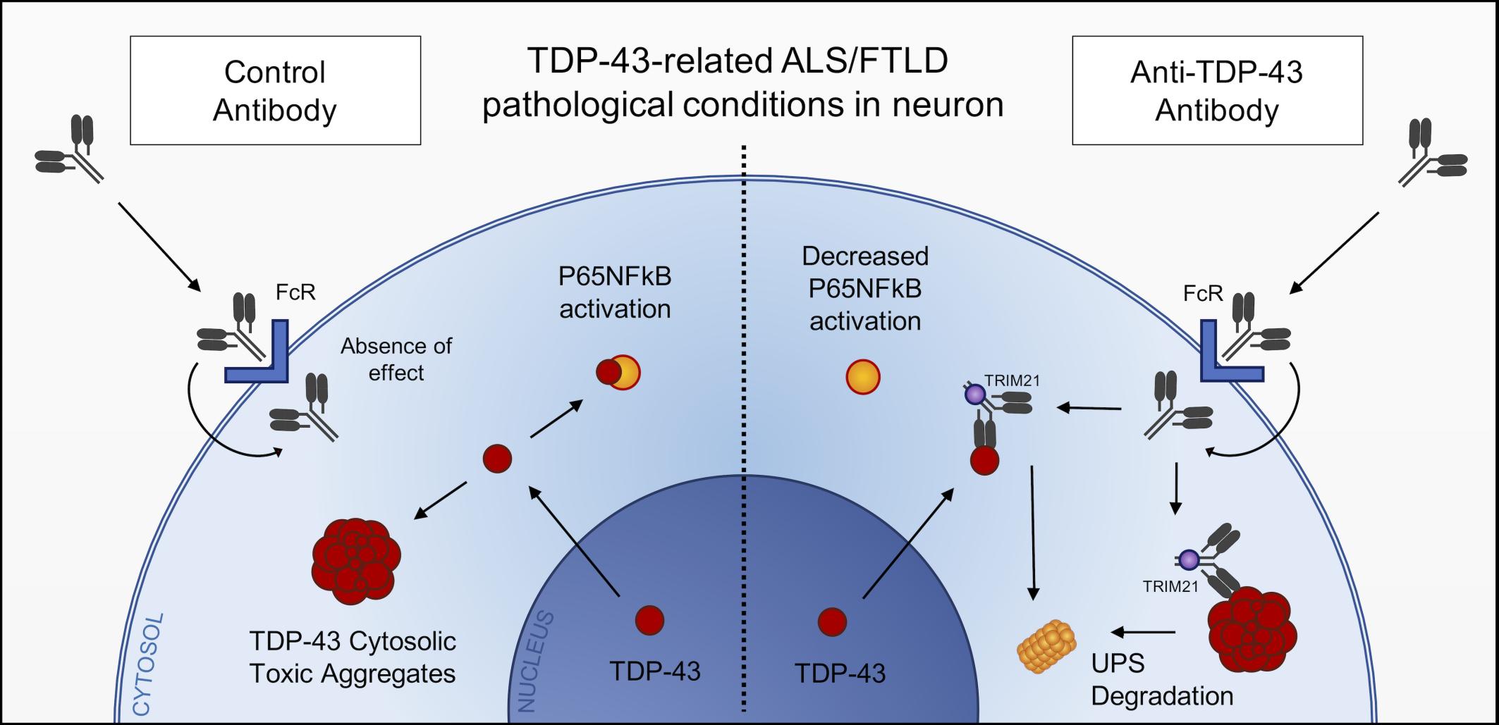
Monoclonal full-length antibody against TDP-43

- Posted by admin in English

Dr. Silvia Pozzi, Jean-Pierre Julien and colleagues have designed a second antibody therapy against misplaced TDP-43, the most obvious cellular problem in ALS. enter image description here

2020 has seen several announcements in this area, which could create a vaccine against ALS. Penetrating the cell is a challenge. Promis neurosciences had announced that they designed a peptide (a tiny protein) targeting TDP-43, and JP Julien's lab worked on single chain antibodies. Single chain antibodies are miniaturized antibodies, their size is roughly half of a normal antibody.

Now JP Julien's team now announced that they successfully designed and tested a monoclonal antibody on a mice model of the disease. Some signs that they have hope it may progress towards clinical trials is that they tested several administration modes and applied for a patent (US 15/532,909).

This is great news and a further step toward a vaccine, as monoclonal antibodies production is well controlled.

Advertisement


This book retraces the main achievements of ALS research over the last 30 years, presents the drugs under clinical trial, as well as ongoing research on future treatments likely to be able stop the disease in a few years and to provide a complete cure in a decade or two.

A hypothesis linking diet and Alzheimer's disease

- Posted by admin in English

A hypothesis describing a mechanism leading to Alzheimer's disease.

Scientists in an interdisciplinary team, have developed a hypothesis that the disease begins when we ingest excess fructose, this phenomenon would be accentuated if there is concomitant consumption of salt and /or alcohol. This excess fructose induces behavioral and metabolic changes, which ultimately lead to Alhzeimer's disease.

The study was published in Frontiers in Aging Neuroscience, it brought together an interdisciplinary team of neurologists, neuroscientists and experts on sugar metabolism

Behavioral and metabolic changes

In the wild, starving animals activate behavioral and metabolic changes to aid their survival once fat stores are depleted. This includes the development of foraging behavior, reduced energy production, and the development of insulin resistance which reduces the uptake of glucose into muscle, thereby promoting preferential uptake by the brain (Koffler and Kisch, 1996; Cahill, 2006).

Studies in humans have largely confirmed these results. The administration of fructose has unique effects on the attention and reward centers, resulting in more hunger and desire for sugary foods than glucose and which is linked to reduced cortical activity (Purnell et al., 2011).

In addition to stimulating hunger, thirst and the search for food, fructose increases the storage of fats, including in the liver, blood (triglycerides) and adipose tissue, thus providing stored energy as well as metabolic water when needed (Johnson et al., 2016).

Deleterious effects of uric acid. Uric acid, generated by fructose, increases blood pressure responses by reducing endothelial nitric oxide, stimulating oxidative stress, and activating the renin-angiotensin system. There is also a very active stimulation of innate immunity, probably mediated by uric acid. These are all protective systems to help survive extreme conditions like famine.

But it is speculated that if it lasts too long, chronic mitochondrial oxidative stress results in impaired mitophagy with the buildup of damaged mitochondria and fewer functioning mitochondria (Shefa et al., 2019), thus affecting overall energy production. and metabolism and resulting in increased dependence on glycolysis.

Generation of fructose in the body Certain foods that do not contain fructose also activate aldose reductase (AR) and stimulate endogenous fructose production, including high glycemic carbohydrates, salty foods, and alcohol (Lanaspa et al., 2013, 2018 ; Wang et al., 2020).

Aldose reductase is expressed in neurons, including the hippocampus (Picklo et al., 2001; Hwang et al., 2017). The activation of aldose reductase leading to the generation of fructose has been shown in the brain after dehydration in rats (Song et al., 2017) as well as after glucose loading in humans (Hwang et al. , 2017). Importantly, there is evidence of endogenous fructose production in patients with Alzheimer's disease, with intracerebral sorbitol and fructose levels 3-5 times higher than normal (Xu et al., 2016) .

Conclusion In one of the scenarios described by Johnson and colleagues, glucose hypometabolism increases oxidative stress and induces progressive loss of mitochondria, ultimately leading to neuronal dysfunction and death. In this scenario, the amyloid plaques and neurofibrillary tangles are part of the inflammatory response and participate in the injury, but are not the central factors of the disease.

Advertisement


This book retraces the main achievements of ALS research over the last 30 years, presents the drugs under clinical trial, as well as ongoing research on future treatments likely to be able stop the disease in a few years and to provide a complete cure in a decade or two.

Une hypothèse reliant régime alimentaire et maladie d’Alzheimer

- Posted by admin in Français

Une hypothèse décrivant un mécanisme aboutissant à la maladie d’Alzheimer.

Des scientifiques dans une équipe interdisciplinaire, ont développé une hypothèse selon laquelle la maladie commence lorsque nous ingérons un excès de fructose, ce phénomène serait accentué s’il y a une consommation concomitante de sel et/ou d’alcool. Cet excès de fructose induit des changements comportementaux et métaboliques, qui à terme induisent la maladie d’Alhzeimer.

L'étude a été publiée dans les Frontiers in Aging Neuroscience, elle a réuni une équipe interdisciplinaire de neurologues, de neuroscientifiques et d'experts sur le métabolisme du sucre

Changements comportementaux et métaboliques

Dans la nature, les animaux qui meurent de faim activent des changements comportementaux et métaboliques pour favoriser leur survie une fois que les réserves de graisse sont épuisées. Cela comprend le développement d’un comportement de recherche de nourriture, une réduction de la production d'énergie et le développement d'une résistance à l'insuline qui réduit l'absorption du glucose dans le muscle, favorisant ainsi l'absorption préférentielle par le cerveau (Koffler et Kisch, 1996; Cahill, 2006).

Pour éviter la famine, les animaux stockent de la graisse en prévision des périodes de pénurie alimentaire, comme avant l'hibernation, la migration sur de longues distances ou la nidification. Une approche utilisée par de nombreux animaux consiste à ingérer des aliments riches en fructose, tels que des fruits et du miel.

Au fil du temps, les animaux développent une résistance à la leptine qui entraîne une consommation excessive de nourriture tout en réduisant l'oxydation des graisses (Shapiro et al., 2008).

Des études chez l'homme ont largement confirmé ces résultats. L'administration de fructose a des effets uniques sur les centres d'attention et de récompense, ce qui entraîne plus de faim et de désir d'aliments sucrés que le glucose et qui est lié à une activité corticale réduite (Purnell et al., 2011).

En plus de stimuler la faim, la soif et la recherche de nourriture, le fructose augmente préférentiellement le stockage des graisses, y compris dans le foie, le sang (triglycérides) et les tissus adipeux, fournissant ainsi de l'énergie stockée ainsi que de l'eau métabolique en cas de besoin (Johnson et al. ., 2016).

Effets délétères de l'acide urique. L'acide urique, généré par le fructose, augmente les réponses de la pression artérielle en réduisant l'oxyde nitrique endothélial, en stimulant le stress oxydatif et en activant le système rénine-angiotensine. Il existe également une stimulation très active de l'immunité innée probablement médiée par l'acide urique. Ce sont tous des systèmes de protection pour aider à survivre dans des conditions extrêmes comme une famine.

Mais il est spéculé que s'il dure trop longtemps, le stress oxydatif mitochondrial chronique entraîne une mitophagie altérée avec l'accumulation de mitochondries endommagées et moins de mitochondries fonctionnelles (Shefa et al., 2019), affectant ainsi la production d'énergie globale et le métabolisme et entraînant une dépendance accrue à la glycolyse.

Génération de fructose dans l'organisme Certains aliments qui ne contiennent pas de fructose activent également l'aldose réductase (AR) et stimulent la production endogène de fructose, y compris les glucides à indice glycémique élevé, les aliments salés et l'alcool (Lanaspa et al., 2013, 2018; Wang et al., 2020).

L'aldose réductase est exprimée dans les neurones, y compris dans l'hippocampe (Picklo et al., 2001; Hwang et al., 2017). L'activation de l’aldose réductase entrainant la génération de fructose a été montrée dans le cerveau après une déshydratation chez le rat (Song et al., 2017) ainsi qu'après une charge en glucose chez l'homme (Hwang et al., 2017). Surtout, il existe des preuves d'une production endogène de fructose chez les patients atteints de la maladie d'Alzheimer, avec des niveaux intracérébraux de sorbitol et de fructose 3 à 5 fois plus élevés que la normale (Xu et al., 2016).

Conclusion Dans l'un des scénarios décrits par Johnson et ses collaborateurs, l'hypométabolisme du glucose augmente le stress oxydatif et induit une perte progressive des mitochondries , conduisant finalement à un dysfonctionnement neuronal et à la mort. Dans ce scénario, les plaques amyloïdes et les enchevêtrements neurofibrillaires font partie de la réponse inflammatoire et participent à la blessure, mais ne sont pas les facteurs centraux de la maladie.

Parkinson's disease is not one but two diseases

- Posted by admin in English

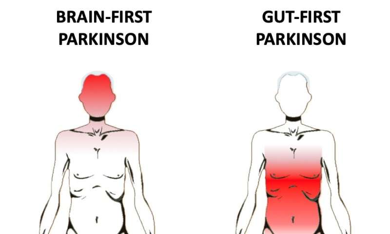
A study which has just been published in the leading neurology journal Brain, indicates that Parkinson's disease is not one but two diseases, starting either in the brain or in the intestines.

These two groups of patients displayed strikingly different profiles on multimodal imaging battery.

These profiles support the existence of a brain-first and body-first subtype of Parkinson’s disease, and furthermore, that premotor rapid eye movement sleep (REM) sleep behaviour disorder is a highly predictive marker of the body-first subtype. enter image description here

Neuropathological autopsy studies of patients with Parkinson’s disease, dementia with Lewy bodies, or incidental Lewy body disease have shown discrepant results.

Heiko Braak’s staging system was derived from a cohort of patients, who were selected based on the presence of Lewy pathology in the dorsal motor nucleus of the vagus, and all these patients conformed to a brainstem predominant type.

Other studies reported that some post-mortem cases do not harbour Lewy pathology in the dorsal motor nucleus of the vagus, and a minority of patients do not follow the Braak staging scheme.

A pathological hallmark of Parkinson’s disease is the presence of intraneuronal a-synuclein inclusions termed Lewy pathology. However, it remains unknown from where the initial a-synuclein aggregates originate.

It has been hypothesized that a-synuclein inclusions initially form in nerve terminals of the enteric nervous system, and then subsequently spread via autonomic connections to the dorsal motor nucleus of the vagus and intermediolateral cell columns of the sympathetic system (Braak et al., 2003).

In addition, Lewy pathology has been detected in gastrointestinal nerve fibres years prior to Parkinson’s disease diagnosis. There is also epidemiological evidence that complete but not partial vagotomy may protect against later Parkinson’s disease.

Autopsy studies have shown that a minority of cases with Lewy pathology do not have pathological inclusions in the dorsal motor nucleus of the vagus, and that a fraction of cases display a limbic-predominant distribution of a-synuclein inclusions with less pathology in the brainstem.

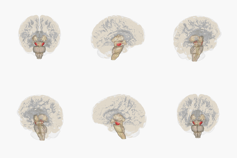
The scientists hypothesized that the appearance of isolated REM sleep behaviour disorder well before parkinsonism is a strong marker of the body-first subtype. The presence of REM sleep behaviour disorder in the premotor phase is a marker of the body-first type, probably a reflection of ascending a-synuclein pathology reaching the pons before the substantia nigra. enter image description here The substantia nigra in the brain. Source Wikipedia
To test this hypothesis, they recruited de novo patients with Parkinson’s disease and performed video-polysomnography to divide patients into de novo Parkinson’s disease without REM sleep behaviour disorder and de novo Parkinson’s disease with pre-motor REM sleep behaviour disorder. The study was conducted between August 2018 and January 2020.

The scientists have shown that prodromal and de novo patients with Parkinson’s disease can be categorized by means of multimodal imaging into distinct clusters, which are compatible with a brain-first and body-first Parkinson’s disease subtype.

Their conclusion is that the Parkinson’s disease comprises two subtypes: (i) a body-first (bottom-up) subtype, where the pathology originates in the enteric or peripheral autonomic nervous system, and then ascends via the vagus nerve and sympathetic connectome to the CNS;

(ii) a brain-first (top-down) subtype, in which the a-synuclein pathology initially arises in the brain itself or sometimes enters via the ol- factory bulb, and subsequently descends to the peripheral autonomic nervous system.

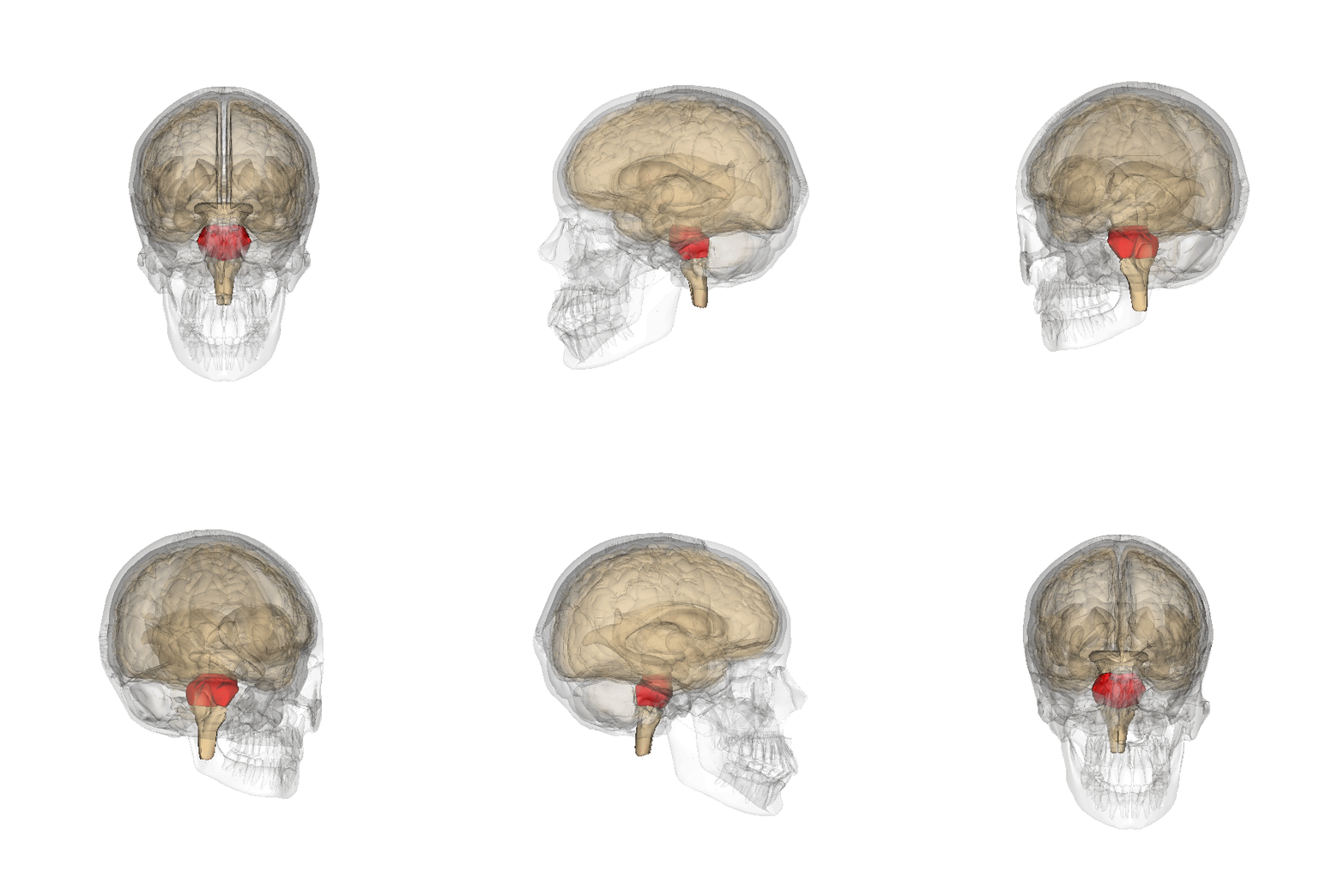
Body-first (bottom-up) subtype: The initial a-synuclein pathology appears in the enteric or peripheral autonomic nervous system. It then propagates via the sympathetic connectome to the heart, and via the vagus nerve to the dorsal motor nucleus of the vagus. enter image description here The pons area in the brain. Source Wikipedia

Ascending pathology affects pontine structures giving rise to REM sleep behaviour disorder before the substantia nigra shows substantial involvement. When parkinsonism appears, signifying a loss of 450% of nigrostriatal dopamine terminals, all lower Braak stage structures show marked damage on relevant imaging markers.

Brain-first (top-down) subtype: In the brain-first type, the initial a-synuclein pathology appears in the CNS. The most likely site of origin seems to be the amygdala or connected structures, or the pathology may in some cases enter via the olfactory bulb. Rarely, the pathology arises in the upper brainstem (substantia nigra or locus coeruleus). The pathology spreads from the site of origin to the brainstem and cortex. When parkinsonism appears, the brainstem shows a rostro-caudal gradient of pathology with marked involvement of the substantia nigra, moderate involvement of the neighbouring pons, but little involvement of the medulla and autonomic nervous system.

Citation: Jacob Horsager et al, Brain-first versus body-first Parkinson's disease: a multimodal imaging case-control study, Brain (2020). DOI: 10.1093/brain/awaa238


Please, help us continue to provide valuable information: