Metastasis as a metabolic disease

- Posted by admin in English

Metastase as a metabolic disease

The risk of cancer and associated mortality increases substantially in humans from the age of 65 years onward. Nonetheless, our understanding of the complex relationship between age and cancer is still in its infancy. For decades, this link has largely been attributed to increased exposure time to mutagens in older individuals. However, more and more publications point toward metabolic aging as an important factor in cancer etiology and Ana Gomes, John Blenis, at Weill Cornell Medicine in New York, and their colleagues have made significant progress in this direction.

It is well known that many physiological processes are degraded or even severely altered in aging:
* lack of normal hepatic synthesis, excess of ammonia in the blood is a dangerous condition that may lead to brain injury and death. * gut microbiome dysbiosis, * development of insulin resistance, * impaired immune processes with persistent chronic neuro-inflammation, and persistent infectious.

Metabolic deregulation of the aged host may play a central role in the acquisition of aggressive properties that contribute to tumor progression.

Considering the growing body of evidence that cancer cell-extrinsic factors are key in modulating tumor progression, the scientists hypothesized that aging might produce a systemic environment that supports tumor progression and aggressiveness. To test this hypothesis, they cultured human cancer cells from 30 young and 30 old healthy donors.

Cells into young serum

Whereas the majority (25 out of 30) of cells treated with young donor serum (plasma from which the clotting proteins have been removed) maintained their epithelial morphology, cells treated with 25 out of the 30 old donors sera became mesenchymal, losing their polarity and displaying a spindle-shaped morphology. These phenotypes were independent of donor ethnicity, and resembled the epithelial-to-mesenchymal transition (EMT), a developmental process that is hijacked by cancer cells to acquire pro-metastatic properties.

Cells into old blood serum

Cells cultured with aged-donor serum displayed a pronounced loss of the epithelial marker E-cadherin and gain of the mesenchymal markers fibronectin and vimentin, in addition to increased expression of serpine1 and MMP2 (proteins associated with aggressive phenotypes). Moreover, the aged sera promoted resistance to two distinct and widely used chemotherapeutic drugs, carboplatin and paclitaxel.

Cancer cells into mice

To determine whether the cells treated with the old donor sera would also show heightened metastatic potential, the scientists treated breast cancer cells with human serum before injecting them into the tail veins of athymic mice. In contrast to the young sera, the aged sera potentiated the ability of the cells to colonize the lungs and form metastatic lesions.

Assessment of metabolites

Out of the 179 circulatory metabolites detected by targeted metabolomics, only 10 were altered at a statistically significant level. A pronounced decline in levels of glutathione, spermidine, glutamine and α-ketoglutarate was expected, considering their known or suggested roles in the ageing process. Notably, only three metabolites were consistently increased in the sera of aged donors: phosphoenolpyruvate, quinolinate and methylmalonic acid (MMA). * phosphoenolpyruvate is involved in glycolysis and gluconeogenesis * Quinolinic acid has a potent neurotoxic effect. * Methylmalonic acid (MMA), is converted into succinyl-CoA by methylmalonyl-CoA mutase, in a reaction that requires vitamin B12 as a cofactor. In this way, it enters the Krebs cycle (a series of chemical reactions used by all aerobic organisms to release stored energy through the oxidation of acetyl-CoA derived from carbohydrates, fats, and proteins). 20–25% of patients over the age of 70 have elevated levels of MMA, but 25–33% of them do not have B12 deficiency. For this reason, MMA test is not routinely recommended in the elderly.

Which metabolite is responsible for the cells metastase-like behavior?

To test whether any of these three metabolites was responsible for inducing the pro-aggressive effects, the scientists treated cancer cells with each metabolite. Only MMA induced a complete pro-aggressive EMT-like phenotype with a decline in E-cadherin and a concurrent increase in fibronectin and vimentin. Loss of E-cadherin function or expression has been implicated in cancer progression and metastasis. Fibronectin may promote lung tumor growth/survival and resistance to therapy

Focus on MMA

The scientists measured the absolute concentration of MMA in the sera from all 60 donors. This analysis revealed that MMA levels were higher in the sera of the old donors (15–80 μM) than in that of the young donors (0.1–1.5 μM). Moreover, in the case of the ten outlier samples (five samples from old donors that did not induce EMT and five samples from young donors that did induce EMT), MMA levels consistently correlated with the phenotypes observed in cancer cells, supporting the idea that MMA is, at least in part, responsible for the observed age-related aggressive phenotypes.

Confirming that MMA is implicated in metastasis

To better understand the pro-aggressive properties of MMA, the scientists treated cells model for EMT studies with MMA. Concentrations of 1 mM and above were sufficient to induce an EMT-like phenotype and the expression of pro-aggressive proteins. Notably, the pro-aggressive effects of MMA were specific, as different acids with similar structures and pK a values did not induce the same complete phenotype under the specific conditions used.

MMA also induced resistance to carboplatin and paclitaxel, two common chemotherapy medication, and increased the migratory and invasive capacity of the cells, and promoted stem-like properties, as shown by an upregulation of CD44 and a decline in CD24.

Treatment of MDA-MB-231 cells in vitro with MMA was sufficient to robustly increase the ability of the cells to colonize the lungs of athymic mice in a concentration-dependent manner

MMA is not enough to induce metastasis

To assess whether another component of the serum could facilitate the entrance of MMA into cancer cells, the scientists depleted the old blood serum of lipids or of molecules larger than 3 kDa—two manipulations that should not affect the levels of polar metabolites such as MMA.

In both cases, the ability of the depleted old blood serum to induce pro-aggressive properties was abolished. Strikingly, both manipulations also caused a pronounced decrease in serum MMA levels.

MMA complexed with large lipidic structures

This suggests that the MMA has to be complexed with lipidic structures larger than 3 kDa in the serum in order to facilitate its entry into cancer cells.

To test this hypothesis, the scientists first complexed MMA with synthetic lipidic structures (lipofectamine) or with lipidic structures purified from fetal bovine serum (FBS). With both approaches, the concentration of MMA necessary to induce pro-aggressive properties was reduced to the levels similar to that of the old donor serum. Moreover, MMA complexed with lipidic structures from FBS produced a similar intracellular concentration of MMA within the same time frame as treatment with old donor serum.

MMA complexed with lipidic structures has similar properties to old blood serum

In support of this idea, treatment of cancer cells with lipidic structures isolated from old blood serum, but not from young serum, or isolated from young serum and loaded with MMA at concentrations similar to the ones found in the old blood serum, was sufficient to drive pro-aggressive properties. Conversely, depletion of lipidic structures from old blood serum resulted in a reduction in total serum MMA levels and was sufficient to abrogate the pro-aggressive phenotype. Orthotopic injections of MDA-MB-231 cells into the mammary fat pads of athymic mice with elevated circulatory MMA levels further demonstrated that circulatory MMA has a substantial role in tumor progression by promoting tumor growth and metastatic spread.

Conclusion

Aging promotes an increase in circulatory MMA, which in turn endows cancer cells with the properties necessary to migrate, invade, survive and thrive as metastatic lesions, which results in decreased cancer-associated survival. Although more in-depth studies are necessary to fully determine the scope of age-driven changes that contribute to the tumorigenic process, this study adds metabolic reprogramming to the complex relationship between aging and cancer.

Couvertures lestées et essais cliniques

- Posted by admin in Français

Pourquoi utiliser des couvertures lestées?

Les troubles du sommeil et l'anxiété représentent une comorbidité courante chez les enfants et adolescents atteints de troubles du spectre autistique (TSA), avec une prévalence allant de 50 à 80%.

Si l'on excepte leur usage sportif (handicap pour l’entraînement), les objets lestés sont souvent utilisés pour déclencher une rétroaction proprioceptive qui a un effet calmant. Cependant un essai thérapeutique en 2014 (3) n’a pas démontré un intérêt mesurable à l’usage de couvertures chez des enfants autistes. Par contre lors de cet essai, parents et enfants ont manifesté un fort intérêt pour ce type de couverture.

L’académie américaine de neurologie (4) conseille aux cliniciens de dire à leurs patients que bien qu'il n'y ait actuellement aucune preuve à l'appui de l'utilisation systématique de couvertures lestées pour améliorer le sommeil, elles peuvent cependant être une approche non pharmacologique raisonnable pour certaines personnes.

Cependant la situation a évolué en 2020 avec deux essais cliniques.

Cas du cancer

Les patients atteints de cancer peuvent ressentir de l'anxiété liée au diagnostic et aux traitements, comme les perfusions de chimiothérapie. Les couvertures pondérées ont été utilisées comme outil de pression tactile pour soulager l'anxiété chez de nombreux patients. Un essai contrôlé randomisé (1) a récemment évalué l'efficacité des couvertures pondérées thérapeutiques de qualité médicale sur l'anxiété chez des patients recevant leurs première et deuxième perfusions de chimiothérapie dans un centre de perfusion ambulatoire.

Les patients recevant une chimiothérapie ont éprouvés un soulagement lorsqu'une couverture lestée a été utilisée. Sur la base de ces résultats, une couverture pondérée thérapeutique de poids standard de qualité médicale peut être utilisée en toute sécurité pour réduire l'anxiété chez les patients.

Cas de la psychiatrie

Là aussi, un essai contrôlé randomisé visant à évaluer l'effet des couvertures pondérées sur l'insomnie et les symptômes diurnes liés au sommeil, a été réalisé chez les patients présentant un trouble dépressif majeur, un trouble bipolaire, ou un trouble d'anxiété généralisée (TAG) ou encore un trouble d'hyperactivité avec déficit de l'attention (TDAH).

Cent vingt patients ont été randomisés : soit dans une couverture lestée par chaînes métalliques, soit dans une couverture à chaîne en plastique léger pendant quatre semaines. Après ces quatre semaines, il est apparu qu’il y avait un avantage significatif à l’utilisation de couverture pondérée par rapport à une couverture légère. L’usage de couverture lestée a entraîné un meilleur maintien du sommeil, un niveau d'activité diurne plus élevé et une réduction des symptômes diurnes de fatigue, de dépression et d'anxiété. Aucun incident n’a été noté.

Au cours d'une phase de suivi ouvert de 12 mois après l’essai, les participants continuant à utiliser des couvertures pondérées ont vu cet effet positif persister sur leur sommeil, tandis que les patients passant d'une couverture légère à une couverture pondérée ont obtenu un meilleur repos.

Publicité


Pour des couvertures lestées faites main et Made in France, vous pouvez consulter la boutique de SisterCatBlankets. Léonore confectionne des couvertures lestées à la main, et accepte les commandes personnalisées. Elle-même personne autiste, elle échangera volontiers avec vous par mail sur les couvertures et leurs effets.

Références:

(1) Weighted Blankets: Anxiety Reduction in Adult Patients Receiving Chemotherapy Jaime Vinson , Jan Powers , Kelly Mosesso DOI: 10.1188/20.CJON.360-368

(2) A randomized controlled study of weighted chain blankets for insomnia in psychiatric disorders Bodil Ekholm , Stefan Spulber , Mats Adler DOI: 10.5664/jcsm.8636

(3) Weighted blankets and sleep in autistic children--a randomized controlled trial Paul Gringras et al. DOI: 10.1542/peds.2013-4285

(4) Practice guideline: Treatment for insomnia and disrupted sleep behavior in children and adolescents with autism spectrum disorder: Report of the Guideline Development, Dissemination, and Implementation Subcommittee of the American Academy of Neurology DOI: 10.1212/WNL.0000000000009033

A new tool for the design of oligonucleotide therapeutics

- Posted by admin in English

Antisense therapy

Antisense therapy is a form of treatment that uses antisense oligonucleotides (ASOs) to target messenger RNA (mRNA). ASOs are capable of altering mRNA expression through a variety of mechanisms. Several ASOs have been approved in the United States, European Union, and elsewhere.

Approved therapies

Approved therapies include Batten disease (Milasen), Cytomegalovirus retinitis (Fomivirsen), Duchenne muscular dystrophy (eteplirsen, golodirsen, viltolarsen), Familial chylomicronaemia syndrome (Volanesorsen), Familial hypercholesterolemia (mipomersen), Hereditary transthyretin-mediated amyloidosis, (Inotersen), Spinal muscular atrophy (nusinersen).

Amyotrophic lateral sclerosis

Tofersen (also known as IONIS-SOD1Rx and BIIB067) is currently being tested in a phase 3 trial for amyotrophic lateral sclerosis (ALS) due to mutations in the SOD1 gene. Results from a phase 1/2 trial have been promising. It is being developed by Biogen under a licensing agreement with Ionis Pharmaceuticals.

Design of oligonucleotide therapeutics

The development of oligonucleotide therapeutics remains challenging as they often have unintended off-target effects and cause non-specific hepatic and renal toxicity. Additionally, it is exceptionally difficult to deliver oligonucleotides to most tissue types and organs.

This can result in the need for tissue specific delivery systems and several siRNA compounds have advanced into development using this paradigm. Designing antisense oligonucleotides (ASOs) and siRNA can be logistically challenging given the many competing design criteria that can be incorporated into the selection of tool or therapeutic sequences.

Design considerations may include splice variants, cross-species targeting for validation, single nucleotide polymorphisms (SNPs), secondary structure, undesirable motifs (e.g. toxic, poly-A, or poly-G repeats), complementarity with off-target sequences, intron/exon boundaries, chemical modification pattern of nucleotides, and predicted activity.

These factors need to be weighed according to the intended use of the oligonucleotide. For example, compounds designed as in vitro tools only need to be active in a single species, whereas it is advantageous for therapeutic oligonucleotides to be active in model organisms as well as patients. A number of computational tools have been developed to address different aspects of the design process.

PFizer RNAi Enumeration and Design tool

In the present paper, the scientists describe PFRED ( PFizer RNAi Enumeration and Design tool), a client-server software system designed to assist with the entire oligonucleotide design process, starting with the specification of a target gene (Ensembl ID) and culminating in the design of siRNAs or RNase H-dependent antisense oligonucleotides. Sequences are chosen using bioinformatics algorithms built upon careful mining of the sequence-activity relationships found in public datasets as well as internal collections.

The tool provides researchers with a user-friendly interface where the only required input is an accession number for the target gene and it returns a list of properties that are believed to contribute to the efficacy of an siRNA or ASO. These properties include human transcripts and cross-species homology, GC content, SNPs, intron-exon boundary, duplex thermodynamics, efficacy prediction score and off-target matches.

An automated oligonucleotide selection procedure is available to quickly select one potential set of sequences with an appropriate property profile. The selection protocol can be customized by the user through changes of the selection cutoffs or the addition of alternate design parameters and algorithms.

Advertisement


This book retraces the main achievements of ALS research over the last 30 years, presents the drugs under clinical trial, as well as ongoing research on future treatments likely to be able stop the disease in a few years and to provide a complete cure in a decade or two.

Combination of acamprosate and baclofen (PXT864) for ALS

- Posted by admin in English

PXT864 is an example of a repurposed drug combination. It uses baclofen and acamprosate, taken twice a day. Baclophen is a derivative of γ-aminobutyric acid, aka GABA, and acts as a GABA-B receptor agonist. It is used as a muscle relaxant to treat spasticity, for example in cerebral palsy and multiple sclerosis. Acamprosate is a drug of unclear mechanism of action, which is used to treat alcohol dependence.

While no double blind phase I/II/III clinical trial has tested PXT864, two small studies tested it for Alzheimer disease in 2013 to 2015. There was also a publication in 2015 in Nature's Scientific reports about effects of PXT864 on a rat model of Parkinson disease (6-OHDA). They used stereotaxic injection rat model to assess the efficacy of the combination in vivo in 6-OHDA rats. This model offers the benefit that each animal serves as its own control.

In a new publication PXT864 activity was assessed in primary cultures of motoneurons derived from SOD1G93A rat embryos. These motoneurons presented severe maturation defects that were significantly improved by PXT864. In this model of ALS, glutamate application induced an accumulation of TDP-43 protein in the cytoplasm, a hallmark that was completely prevented by PXT864. The anti-TDP-43 aggregation effect was also confirmed in a cell line expressing TDP-43 fused to GFP. These results demonstrate the value of PXT864 as a promising therapeutic strategy for the treatment of ALS.

Testing in-vitro is cheap with respect to test with animal models, and it is well known that no animal model of neurodegenerescent diseases reflects what is happening in humans.

Typical of biotech players, Pharnext, which began in 2007, has had its struggles. It is deeply unprofitable but the driving force behind Pharnext’s strategy is its founder, Daniel Cohen, who led the team that mapped the human genome in the 1990s, and now functions as his company’s chief scientist.

Nevertheless baclofen and acamprosate are two common drugs, so this might interest many ALS patients. However both drugs have strong and indesirable side effects.

Advertisement


This book retraces the main achievements of ALS research over the last 30 years, presents the drugs under clinical trial, as well as ongoing research on future treatments likely to be able stop the disease in a few years and to provide a complete cure in a decade or two.

Widespread tissue hypoxia dysregulates cell and metabolic pathways

- Posted by admin in English

Neurodegenerative diseases are also vascular diseases. This has long been observe in Alzheimer, but it is also true for amyotrophic lateral sclerosis or multiple sclerosis. The purpose of this study by Elena Hernandez-Gerez and al was to determine the extent and role of systemic hypoxia in the pathogenesis of spinal muscular atrophy (SMA).

Necrosis at the tip of toes and fingers has been observed in SMA patients, as well as thrombotic occlusions of small vessels, together with significant reductions in vascular density in the mouse models of severe SMA, these symptoms suggest defects in blood supply. In fact SMA mouse spinal cord is hypoxic, suggesting that other tissues may also be affected. All tissues can be damaged by hypoxia, but motor neurons are particularly sensitive.

enter image description here Source: James Heilman, MD via Wikipedia
Historically, non-neuronal pathology has been of less interest, as the severe motor neuron pathology negated the need to consider additional therapies.

However, now that treatments have become available, including Nusinersen (Spinraza) and Onasemnogene abeparvovec (Zolgensma), survival is increasing, but so is the likelihood of significant non-neuronal co-morbidities. It is now clear that a number of non-neuronal tissues also show disease-related pathology, and that the cardiovascular system is particularly involved, with heart, vessels, and circulating cells all described as pathological in severe mouse models and now also increasingly in patients.

The authors were keen to understand the extent to which vascular dysfunction could contribute to and potentially exacerbate motor neuron death, having previously shown decreased vasculature associated with tissue hypoxia in spinal cord. Hypoxia was assayed in vivo in early-symptomatic (postnatal day 5) SMA-model mice.

Hypoxia is widespread in neuronal and nonneuronal tissues in pre/early-symptomatic SMA mice Here the authors show using qualitative and quantitative techniques that a wide range of tissues from brain and eye, through skeletal muscle to visceral organs are significantly hypoxic at pre/ early symptomatic time points. Importantly skin and lung, which can absorb atmospheric oxygen, showed no evidence of hypoxia.

All assays found significant levels of hypoxia in multiple organ systems in early symptomatic SMA mouse pups, except aerated tissues such as skin and lungs. This was accompanied by significantly increased glucose uptake in many affected organs, consistent with a metabolic hypoxia response. SMN protein levels were shown to vary widely between motor neuron precursors in vitro, and those with lower levels were most susceptible to cell death. In addition, SMA-model motor neurons were particularly sensitive to hypoxia, with reduced ability to transport lactate out of the cell in hypoxic culture, and a failure in normal cell cycle progression.

Not only is there widespread tissue hypoxia and multi-organ cellular hypoxic response in SMA model mice, but SMA-model motor neurons are especially susceptible to that hypoxia. The data support the hypothesis that vascular defects leading to hypoxia are a significant contributor to disease progression in SMA, and offer a route for combinatorial, non-SMN related therapy. This immediately suggests a need to assess SMA patients, and particularly those undergoing therapy, for ongoing, perhaps low-level chronic hypoxia, and also potential utilization of oxygenation as an easy to deliver, ameliorative, therapy.

Advertisement


This book retraces the main achievements of ALS research over the last 30 years, presents the drugs under clinical trial, as well as ongoing research on future treatments likely to be able stop the disease in a few years and to provide a complete cure in a decade or two.

Alzheimer's disease is a severe neurodegenerative disease characterized by progressive damage to synaptic bonds and neuronal cells mainly in the cerebral region of the hippocampus.

Optical spectroscopy methods can provide a unique means of analyzing body fluids, enabling rapid, sensitive, non-invasive and reliable diagnosis.

enter image description here * Source: Toommm via Wikipedia. *

The development of Alzheimer's disease has been associated with a dysfunctional blood-brain barrier with a reduced ability to clear Aβ and tau proteins from the brain. These two substances are biomarkers of the progression of Alzheimer's disease and are a reliable sign of a neurodegenerative process even at an early stage. These biomarkers can be transported in the vascular system across the blood-brain barrier. The blood-brain barrier plays an important role in the selective passage of several substances between the brain and the blood system and in the maintenance and integrity of the brain.

Traces of these biomarkers are present in the systemic bloodstream and can be found in the vasculature of other areas of the body. The relevance of changes occurring in the eye as a consequence of Alzheimer's disease has been reported by Lim et al. Therefore, tears are an interesting way to study the changes induced by neurodegenerative pathologies. They are easily accessible and can be collected using minimally invasive methods.

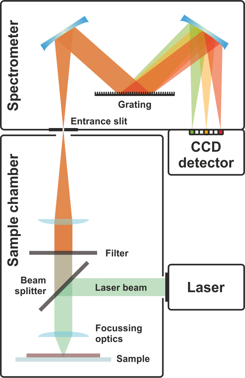
Over the past decades, a large number of studies have been conducted to analyze the composition of tears using conventional biochemical methods.These conventional techniques have been largely replaced by optical spectroscopy methods, such as those based on Raman scattering, which take less time and require only little preparation of the samples.

Raman spectroscopy has already been used to identify biomarkers of Alzheimer's disease in blood serum and saliva and to study the basic aggregation mechanisms of amyloids. However, it has some limitations regarding sensitivity, noise / signal level and repeatability.

The objective of the study which concerns us today was to demonstrate the spectral differences in the SERS spectroscopic response of human tears in subjects suffering from Alzheimer's disease, subjects suffering from mild cognitive and healthy control subjects. Human tears were characterized by SERS coupled with multivariate data analysis.

Thirty-one informed subjects (Ctr, MCI and AD) were considered. Eighteen subjects with Alzheimer's disease (7 women, 11 men and mean age 71 ± 10 years) and seven subjects with mild cognitive impairment (3 women, 4 men and mean age 73 ± 9 years) were included In this study.

While the Raman spectrum does not provide any valuable information apart from very weak characteristics, conversely, the SERS spectrum clearly demonstrated the various contributions of human tears components. SERS measurements were performed with standard microscope glasses coated with a homemade colloid of gold nanoparticles (GNP).

The data processing was implemented using software routines (wavelet toolbox). The fully processed dataset of all mean spectra was analyzed by PCA and i-PCA to describe it using a set of orthogonal eigenvectors separately accounting for different sources of variance in the original data. enter image description here

The differences between the SERS spectra of tears from subjects at different clinical states are sometimes very subtle and are mainly localized in certain specific spectral ranges. Notably, the authors found that PCA performed over the entire spectral range did not differentiate between the subtle changes that occurred in the spectra examined. To better highlight these differences and take advantage of them to classify the spectra, the authors therefore performed the PCA in intervals (interval partial component analysis (i-PCA)).

The average SERS spectra of the Ctr, MCI and AD subjects showed differences related to the protein components of lactoferrin and lysozyme. Quantitative changes were also observed by determining the intensity ratio between the selected bands. Scientists also built a classification model that discriminates between AD, MCI and Ctr subjects. This model was built using the scores obtained by performing a interval principal component analysis on specific spectral regions (i-PCA). enter image description here

Although it has not been possible to discriminate specific biomarkers of Alzheimer's disease, the overall response of SERS reflects small but interesting changes in tear composition that can be attributed to altered levels of specific pathological substances. and stimulated by disease.

The analysis of the partial interval components (i-PCA) of the spectra by the authors made it possible to distinguish subjects with Alzheimer's disease from healthy subjects with MCI. The precision of classification of the constructed method was very encouraging with interesting prospects for medical applications as a support for clinical diagnosis and discrimination of Alzheimer's disease from other forms of dementia.

Advertisement


This book retraces the main achievements of ALS research over the last 30 years, presents the drugs under clinical trial, as well as ongoing research on future treatments likely to be able stop the disease in a few years and to provide a complete cure in a decade or two.

La maladie d'Alzheimer est une pathologie neurodégénérative sévère caractérisée par une lésion progressive des liaisons synaptiques et des cellules neuronales principalement dans la région cérébrale de l'hippocampe.

Les méthodes de spectroscopie optique peuvent fournir un moyen unique d'analyser les fluides corporels, permettant un diagnostic rapide, sensible, non invasif et fiable.

enter image description here Source: Toommm via Wikipedia.

Les bêta-amyloïdes (Aβ) et la protéine tau sont impliquées dans le développement de la maladie d'Alzheimer. Ces deux substances sont des biomarqueurs de la progression de la maladie d'Alzheimer et sont un signe fiable d'un processus cérébral neurodégénératif également au stade précoce. Ces biomarqueurs peut être transportés dans le système vasculaire à travers la barrière hémato-encéphalique. La barrière hémato-encéphalique joue un rôle important dans le passage sélectif de plusieurs substances entre le cerveau et le système sanguin et dans le maintien et l'intégrité du cerveau. Le développement de la maladie d'Alzheimer a été associé à une barrière hémato-encéphalique dysfonctionnelle avec une capacité réduite à éliminer les protéines Aβ et tau du cerveau.

Des traces de ces biomarqueurs sont présentes dans la circulation sanguine systémique et peuvent rejoindre la vascularisation d'autres zones du corps, comme la rétine, le cristallin et les régions lacrymales, affectant la composition des larmes. La pertinence des changements survenant dans l'œil en tant que La conséquence de la maladie d'Alzheimer a été rapportée par Lim et al. Par conséquent, les larmes sont un moyen intéressant pour étudier les changements induits par les pathologies neurodégénératives. Elles sont facilement accessibles et peuvent être collectés à l'aide de méthodes peu invasives.

Au cours des dernières décennies, un grand nombre d'études ont été menées pour analyser la composition des larmes en utilisant des méthodes biochimiques conventionnelles.

Ces techniques conventionnelles ont été largement remplacées par les méthodes de spectroscopie optique, telles que celles basées sur la diffusion Raman, qui prennent moins de temps et ne nécessitent qu'une faible préparation préalable des échantillons.

La spectroscopie Raman a déjà été utilisée pour identifier les biomarqueurs de la maladie d'Alzheimer dans le sérum sanguin et la salive et pour étudier les mécanismes d'agrégation de base des amyloïdes. Cependant, certaines limites concernant la sensibilité, le niveau de bruit / signal et la répétabilité ont mises en évidence.

L'objectif de l'étude qui nous occupe aujourd'hui était de mettre en évidence les différences spectrales dans la réponse spectroscopique SERS des larmes humaines chez des sujets atteints de la maladie d'Alzheimer, des sujets atteints de troubles cognitifs légers et des sujets témoins sains. Les larmes humaines ont été caractérisées par SERS couplé à une analyse de données multivariée. Trente et un sujets informés (Ctr, MCI et AD) ont été considérés.

Dix-huit sujets atteints de la maladie d'Alzheimer (7 femmes, 11 hommes et âge moyen 71 ± 10 ans) et sept sujets atteints de troubles cognitifs légers (3 femmes, 4 hommes et âge moyen 73 ± 9 ans) ont été inclus dans cette étude.

Alors que le spectre Raman ne fournit aucune information précieuse en dehors de caractéristiques très faibles, à l'inverse, le spectre SERS a mis clairement en évidence les diverses contributions des composants des larmes. Les mesures SERS ont été effectuées avec des verres de microscope standard revêtus d'un colloïde fait maison de nanoparticules d'or (GNP). Le traitement des données a été mis en œuvre à l'aide de routines logicielles (boîte à outils ondelettes). L'ensemble de données entièrement traité de tous les spectres moyens a été analysé par PCA et i-PCA pour le décrire en utilisant un ensemble de vecteurs propres orthogonaux tenant compte séparément des différentes sources de variance dans les données d'origine. enter image description here

Les différences entre les spectres SERS des larmes de sujets présentant des états cliniques différents sont parfois très subtiles et sont principalement localisées dans certaines plages spectrales spécifiques. Notamment, les auteurs ont constaté que la PCA réalisée sur toute la gamme spectrale ne différenciait pas les changements subtils qui se produisaient dans les spectres examinés. Pour mieux mettre en évidence ces différences et en profiter pour classer les spectres, les auteurs ont donc réalisé la PCA dans les intervalles (interval partial component analysis (i-PCA)).

Les spectres SERS moyens des sujets Ctr, MCI et AD ont mis en évidence des différences liées aux composants protéiques de la lactoferrine et du lysozyme. Des changements quantitatifs ont également été observés en déterminant le rapport d'intensité entre les bandes sélectionnées. Les scientifiques ont également construit un modèle de classification qui discrimine les sujets AD, MCI et Ctr. Ce modèle a été construit en utilisant les scores obtenus en effectuant une analyse en composantes principales sur des régions spectrales spécifiques (i-PCA).

Même s'il n'a pas été possible de discriminer des biomarqueurs spécifiques de la maladie d'Alzheimer, la réponse globale du SERS reflète des modifications petites mais intéressantes de la composition des larmes qui peuvent être attribuées à des niveaux modifiés de substances pathologiques spécifiques et stimulées par la maladie.

L'analyse des composantes partielles d'intervalle (i-PCA) des spectres par les auteurs a permis de distinguer les sujets atteints de la maladie d'Alzheimer des sujets sains et atteints de MCI. La précision de classification de la méthode construite était très encourageante avec des perspectives intéressantes pour les applications médicales comme support du diagnostic clinique et de la discrimination de la maladie d'Alzheimer des autres formes de démence.

Publicité


Ce livre retrace les principales réalisations de la recherche sur la SLA au cours des 30 dernières années. Il présente les médicaments en cours d’essai clinique ainsi que les recherches en cours sur les futurs traitements susceptibles d’ici quelques années, d’arrêter la maladie et de fournir un traitement complet en une décennie ou deux.

Multiple studies have signaled brain atrophy in relationship with obesity, particularly in elderly cohorts. Initially, this relationship was shown in the Cardiovascular Health Study in 94 cognitively normal participants in their late 70s who remained cognitively normal 5 years after their brain MRI scan.

The findings of brain atrophy in relation to higher BMI were replicated in the separate Alzheimer's Disease Neuroimaging Cohort and then in a larger Cardiovascular Study Cohort. However, what makes the present study different from that work, is the focus on brain perfusion, that shows greater sensitivity and earlier changes related brain dysfunction than atrophy.

These changes have been demonstrated even in cognitively normal individuals, as well as persons with mild cognitive impairment and Alzheimer's disease. Regional cerebral blood flow has also been used to track obesity-related brain abnormalities. enter image description here

A SPECT scan monitors level of biological activity at each place in the 3-D region analyzed. Emissions from the radionuclide indicate amounts of blood flow in the capillaries of the imaged regions. Because blood flow in the brain is tightly coupled to local brain metabolism and energy use, the radionuclide is used to assess brain metabolism regionally, in an attempt to diagnose and differentiate the different causal pathologies of dementia. SPECT imaging is performed by using a gamma camera to acquire multiple 2-D images (also called projections), from multiple angles. A computer is then used to apply a tomographic reconstruction algorithm to the multiple projections, yielding a 3-D data set.

Recent studies have shown the accuracy of SPECT in Alzheimer's diagnosis may be as high as 88%. SPECT was superior to clinical exam and clinical criteria in being able to differentiate Alzheimer's disease from vascular dementias. This latter ability relates to SPECT's imaging of local metabolism of the brain, in which the patchy loss of cortical metabolism seen in multiple strokes differs clearly from the more even or "smooth" loss of non-occipital cortical brain function typical of Alzheimer's disease.

The authors have previously utilized SPECT functional neuroimaging to review and identify patterns of abnormality relevant to the diagnosis of traumatic brain injury, depression versus dementia classification , marijuana-related influences in the brain , omega-3 fatty acid associated improved cerebral blood flow , gender-related differences in the brain , and brain aging.

The purpose of their current article is to identify potential brain perfusion abnormalities in adults related to being overweight or obese.

A large psychiatric cohort of 35,442 brain scans across 17,721 adults were imaged with SPECT. ANOVA was done to identify patterns of perfusion abnormality in this cohort across BMI designations of underweight, normal weight, overweight, obesity, and morbid obesity.

With over 35,000 functional neuroimaging scans across more than 17,000 individuals, this study is one of the larger studies linking obesity with brain dysfunction, as evidenced here by quantifiable regional perfusion.

In particular, brain areas noted to be vulnerable to Alzheimer's disease: the temporal and parietal lobes, hippocampus, posterior cingulate gyrus, and precuneus were found to have reduced perfusion along the spectrum of weight classification from normal weight to overweight, obese, and morbidly obese.

While the work presented here focused on body tissue adiposity and cerebral perfusion in a large cohort, other studies have suggested a negative relationship between BMI, obesity, and the brain, particularly with neuroimaging as a proxy of structure or function.

Across adulthood, higher BMI correlated with decreased perfusion on both resting and concentration brain SPECT scans. These are seen in virtually all brain regions, including those influenced by AD pathology such as the hippocampus.

Perfusion is the passage of fluid through the circulatory system or lymphatic system to an organ or a tissue, it usually refers to the delivery of blood to a capillary bed in tissue. Cerebral perfusion may therefore warrant further study as a biomarker of caloric restriction in related efforts to improve brain health.

Overall, the scientists have found a strong set of relationships between being overweight and obese and brain hypoperfusion across a large adult cohort spanning young adults to late life.

The persistence of these abnormalities despite adjusting for demographic and psychiatric factors further highlights the need to address obesity as a target for interventions designed to improve brain function, be they Alzheimer's disease prevention initiatives or attempts to optimize cognition in younger populations.

Advertisement


This book retraces the main achievements of ALS research over the last 30 years, presents the drugs under clinical trial, as well as ongoing research on future treatments likely to be able stop the disease in a few years and to provide a complete cure in a decade or two.


Please, help us continue to provide valuable information: