Despite often being described as two separate neurodegenerative disorders, several independent studies confirmed that clinical symptoms and pathologies of Alzheimer's disease and Parkinson's disease can overlap.

Indeed dementia with Lewy bodies involves deposition of both extra-cellular amyloid-β plaques and intra-cellular α-synuclein containing Lewy bodies which are pathological hallmarks of Alzheimer's disease and Parkinson's disease respectively. enter image description here Robin Williams (pictured in 2011) was diagnosed during autopsy as having diffuse Lewy bodies.

Scientists correlated two interesting observations: - First, humans as well as mice with a strong α-synuclein pathology, appear to have a lower burden of amyloid-β. - Second, a common feature of neurodegenerative diseases is the occurrence of olfactory dysfunctions at a very early stage of disease progression, even years before other clinical symptoms occur

Diagnosis of Dementia with Lewy bodies is extremely difficult and often leads to a misdiagnosis of Alzheimer's disease or Parkinson disease. Until now, an accurate diagnosis can only be made by postmortem analysis. So there is an urgent need of biomarkers to improve identification of dementia subtypes.

Therefore, studies of early pathological alterations in the olfactory bulb are of great interest in order to use olfactory deficits as a biomarker for early diagnoses and disease progression.

In the present study, the scientists therefore examined the effect of α-synuclein on the formation of amyloid-β aggregates specifically in the olfactory bulb.

They shown on a mice model of dementia with Lewy bodies, that as α-synuclein increases, the amyloid-β load in the olfactory bulb of these mice diminishes and that led to a significant amelioration of olfactory performance.

Via inoculation of amyloid-β rich brain extracts in the olfactory bulb, the authors could show that the amyloid-β seeding area was significantly decreased in the olfactory bulb of aged (8 months) mice model of dementia with Lewy bodies compared to same age mice with a model of Alzheimer's disease.

Experiments with grafts of embryonic neurons supported the inhibitory role of α-synuclein as the number of plaques was significantly reduced when healthy grafts were transplanted into a mouse model of Lewy body dementia, compared to the same graft in a mice model of Alzheimer's disease.

The scientists determined that the decreased Amyloid-β plaque load in 8-month-old mice was not attributable to an increased phagocytic capacity of microglial cells. Extra-cellular α-synuclein seems to interfere with the formation of Aβ deposits.

In conclusion, this study verifies an inhibitory role of α-synuclein on amyloid-β plaque formation in the olfactory bulb of a mice model od Dementia with Lewy body mice, which further lead to the amelioration of olfactory deficits in mice harbouring both α-synuclein and amyloid-β pathologies.

Indeed this work was done on mice models of diseases, and it rarely translate in similar human findings. In addition it is not clear if these mice were standardized commercial products or in-site raised animals. Finally the authors do no explain what is the relation between the extra-cellular α-synuclein, which was the subject of their study and the intra-cellular α-synuclein found in Lewy bodies.

So anosmia (loss of the ability to detect one or more smells) might be a clinical sign helping to differentiate Alzheimer disease from Dementia with Lewy bodies

Contact the author

Advertisement


This book retraces the main achievements of ALS research over the last 30 years, presents the drugs under clinical trial, as well as ongoing research on future treatments likely to be able stop the disease in a few years and to provide a complete cure in a decade or two.

MRi is a new way to diagnose ALS. It replaces the unreliable practice of old school neurologists with instantaneous, factual information.

Yet there is a need of specialists to analyze MRi pictures. In the long term it will disrupt the way neurologists work, and they may take a back seat in their relation with neurodegenerescent patients.

MRi also reintroduces anatomy in the global picture and scientists may notice what is obvious to patients: That muscle wasting is a central feature of ALS, and not a epiphenomenon.

MRi brings also a new vocabulary, T1, T2, relaxivity, FLAIR, STIR, STRATE. enter image description here Source: KieranMaher at English Wikibooks

T relaxivity contrast imaging may serve as a potential imaging biomarker for amyotrophic lateral sclerosis by noninvasively quantifying the tissue microstructure.

In this preliminary longitudinal study, the authors investigated the Transverse Relaxivity at Tracer Equilibrium (TRATE, an MRI technique) in three muscle groups between SOD1-G93A rat and a control population at two different timepoints. The control group was time matched to the Amyotrophic Lateral Sclerosis group such that the second timepoint was the onset of disease. Other groups have as well experimented this new way to diagnose ALS with MRi on muscles.

They observed a statistically significant decrease in TRATE over time in the gastrocnemius, tibialis, and digital flexor muscles in the SOD1-G93A model, whereas TRATE did not change over time in the control group.

Immunofluorescent staining revealed a decrease in minimum fiber area and cell density in the SOD1-G93A model when compared to the control group. These microstructural changes observed from histology align with the theorized biophysical properties of TRATE.

The scientists here demonstrate that TRATE can longitudinally differentiate disease associated atrophy from healthy muscle and has potential to serve as a biomarker for disease progression and ultimately therapy response in patients with Amyotrophic Lateral Sclerosis.

Let's hope the usage of MRi whill shorten the diagnosis "black hole" that characterize old school neurologists practice.

Let's hope MRi will help to shift the paradigm of "ALS as a Motor Neuron Disease". Many signs hint a a disease striking the skeletal muscles as well, including the presence of TDP-43 aggregates.

Read the original article here

Contact the author

Advertisement


This book retraces the main achievements of ALS research over the last 30 years, presents the drugs under clinical trial, as well as ongoing research on future treatments likely to be able stop the disease in a few years and to provide a complete cure in a decade or two.

Neurodegenerative diseases are characterized by the selective degeneration of neuronal populations in different brain regions and frequently the formation of distinct protein aggregates that often overlap between diseases.

Contrary to most other cells, neurons cannot divide, and therefore cannot dilute misfolded/aggregated proteins that accumulate in their cytosol. Their removal is a complex task involving recognition proteins, chaperones, and eventually activation of degradation pathways such as the ubiquitin-proteasome system and the autophagy-lysosomal pathway.

Mechanisms to recycle proteins are complex, one of them uses lysosomes. A lysosome is a membrane-bound organelle found in many animal cells. They are spherical vesicles that contain hydrolytic enzymes that can break down many kinds of biomolecules.

Lysosomal health is crucial for the degradation of dysfunctional proteins and in particular for the clearance of autophagic vacuoles. An impaired lysosomal system contributes to autophagy stress, accumulation of damaged mitochondria, and restricts clearance of proteins aggregates.

Vacuolar protein sorting ortholog 35 (VPS35) is a protein involved in autophagy and is implicated in neurodegenerative diseases, such as Parkinson's disease (PD) and Alzheimer's disease (AD). Yet it was not commonly associated with ALS.

VPS35 is part of a complex called the retromer, which is responsible for transporting select cargo proteins between vesicular structures (e.g., endosomes, lysosomes, vacuoles) and the Golgi apparatus.

So any defects in VPS35 is associated with neurodegenerative diseases. Indeed it is certainly not the unique cause of those diseases. Mutations in the VPS35 gene have been identified to cause late-onset, autosomal dominant familial Parkinson's disease, whereas reduced VPS35 protein levels are reported in vulnerable brain regions of subjects with Alzheimer's disease, neurodegenerative tauopathies such as progressive supranuclear palsy and Pick's disease, and amyotrophic lateral sclerosis.

Here, Dorian Sargent and colleagues develop conditional knockout mice with the selective deletion of in neurons to better elucidate its role in neuronal viability and its connection to neurodegenerative diseases.

Surprisingly, the pan-neuronal deletion of induces a progressive and rapid disease with motor deficits and early post-natal lethality. Neuronal loss is accompanied and preceded by the formation of p62-positive protein inclusions and robust reactive astrogliosis.

The authors' study reveals a critical yet unappreciated role for VPS35 function in the normal maintenance and survival of motor neurons during post-natal development that has important implications for neurodegenerative diseases, particularly amyotrophic lateral sclerosis.

In 2020 another team achieved a substantial reduction of CRC proteins in motor neurons of SOD1 G93A mice. They designed, synthetize and characterize a small array of bis-guanylhydrazones. Such retromer stabilizers possess good in vivo bioavailability, potency, and stability.

Read the original article on Pubmed

Does Indiana vesiculovirus cause neurodegenerative diseases?

- Posted by admin in English

Although it is not yet universally accepted that all neurodegenerative diseases (NDs) are prion disorders, there is little disagreement that Alzheimer's disease (AD), Parkinson's disease, frontotemporal dementia (FTD), and other NDs are a consequence of protein misfolding, aggregation, and spread. The precise mechanism of extracellular aggregate transfer and induction of new aggregates is unclear.

Yet only a small fraction of released soluble or aggregated proteins are associated with extracellular vesicle, while the vast majority is freely secreted.

So there is an apparent paradox: If proteins aggregates are not usually found in extracellular vesicles, how could it be that they are causing aggregates?

The usual explanation is that extracellular vesicles are seeding protein aggregates, which might be a good enough explanation in extracellular medium. Yet in humans only Alzheimer disease has extracellular proteins aggregates but has also intracellular aggregates of Tau protein, for most other diseases, the protein aggregates are only intracellular.

Scientists from the German Center for Neurodegenerative Diseases Bonn (DZNE) and the German Centre for the Protection of Laboratory Animals (Bf3R), hypothesized that for one extracellular vesicle to penetrate in a foreign cell, it has to have ligands are present that bind to receptors on the cell surface and then cause the two membranes to fuse. https://www.nature.com/articles/s41467-021-25855-2

The researchers induced cells to produce viral proteins that mediate target cell binding and membrane fusion. Two proteins were chosen as prime examples: SARS-CoV-2 spike protein S, which stems from the virus causing COVID-19, and vesicular stomatitis virus glycoprotein VSV-G, which occurs in a pathogen that is clinically similar to the Foot-and-mouth disease but from a different family.

Moreover, cells expressing receptors for these viral proteins, and with poor aggregate-inducing activity in recipients were chosen.

They found that vesicular stomatitis virus glycoprotein and SARS-CoV-2 spike S increase extracellular aggregates of misfolded proteins in infected cells.

  • Expression of viral glycoprotein VSV-G drastically increases cell-to-cell spreading of cytosolic prions
  • Enhanced extracellular transmission of Tau aggregation upon VSV-G expression
  • VSV-G extracellular vesicle efficiently transmit scrapie prions to recipient cells

There is little about intracellular aggregates in this article, as the researchers' focus is obviously on neurodegenerative animal diseases.

Misfolded proteins are located in the cytosol, proteins fold for a reason, it is the endoplamic reticulum (ER) which folds them. There is no mention of the ER in this article. Yet it looks like that a protein which cause membranes to fuse would destroy organelles like the ER.

The German scientists worked on Tau protein and PrP protein, but there is no mention of TDP-43 and synuclein. Probably because those two proteins are found in (human) intracellular aggregates.

In another recent article, another team found that VSV-G caused marked alterations in cell's secretory trafficking, with VSV-G accumulating mainly in the Golgi complex . https://www.ncbi.nlm.nih.gov/labs/pmc/articles/PMC8059059/ The Golgi apparatus is the dispatch station of protein received from the endoplasmic reticulum (ER). ER is the place where linear proteins just produced by the ribosomes are correctly folded. enter image description here

So a protein disturbing the ER or Golgi apparatus is certainly creating proteopathies such as the one seen in human neurodegenerescence.

Yet that does not prove that VSV-G is the cause of neurodegenerescence. There is nearly nearly no publications associating VSV-G and neurodegenerescence.

Advertisement


This book retraces the main achievements of ALS research over the last 30 years, presents the drugs under clinical trial, as well as ongoing research on future treatments likely to be able stop the disease in a few years and to provide a complete cure in a decade or two.

Evolution of pure autonomic failure towards Parkinson or MSA

- Posted by admin in English

Pure autonomic failure is a rare degenerative disease of the autonomic nervous system. Symptoms include dizziness and fainting (caused by orthostatic hypotension), visual disturbances and neck pain. Chest pain, fatigue and sexual dysfunction are less common symptoms that may also occur. enter image description here 
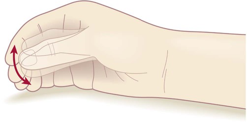
‘Pill-rolling’ rest tremor as found in Parkinson’s disease.

The autonomic nervous system is a control system that acts largely unconsciously and regulates bodily functions. It has a sequential organization: The preganglionic neuron synapse onto a postganglionic neuron before innervating the target organ. It's a bit similar to the upper/lower motor neuron/muscle assemblage.

Longitudinal studies have reported that Pure autonomic failure can phenoconvert to a central synucleinopathy with motor or cognitive involvement-i.e., to Parkinson disease, dementia with Lewy bodies, or multiple system atrophy.

Synucleinopathies are neurodegenerative diseases characterised by the abnormal accumulation of aggregates of alpha-synuclein protein in neurons, nerve fibres or glial cells.

These longitudinal studies have classified patients clinically as having Pure autonomic failure based on neurogenic orthostatic hypotension without an identified secondary cause or clinical evidence of motor or cognitive impairment due to central neurodegeneration.

This approach lumps together two neurogenic orthostatic hypotension syndromes that are pathologically and neurochemically distinct.

  • One is characterized by intraneuronal cytoplasmic alpha-synuclein aggregates and degeneration of postganglionic sympathetic neurons, as in Parkinson disease and Dementia with Lewy Bodies.
  • The other is not, as in multiple system atrophy.

Clinical and postmortem data show that the form of Pure autonomic failure that involves sympathetic intraneuronal synucleinopathy and noradrenergic deficiency can phenoconvert to Parkinson disease or Dementia with Lewy Bodies-but not to multiple system atrophy.

Conversely, Pure autonomic failure without these features leaves open the possibility of premotor multiple system atrophy.

Multiple system atrophy is a rare neurodegenerative disorder characterized by autonomic dysfunction, tremors, slow movement, muscle rigidity, and postural instability (collectively known as parkinsonism) and ataxia. This is caused by progressive degeneration of neurons in several parts of the brain including the basal ganglia, inferior olivary nucleus, and cerebellum.

MSA generally show little response to the dopamine medications used to treat Parkinson's disease and only about 9% of MSA patients with tremor exhibit a true parkinsonian pill-rolling tremor.

Read the original article on Pubmed

Disappointing results from the VALOR study on Tofersen (BIIB067)

- Posted by admin in English

Biogen announced the first results of its pivotal phase 3 VALOR study in tofersen (BIIB067), an experimental antisense oligonucleotide (ASO) under evaluation for people with amyotrophic lateral sclerosis (ALS) who carry a mutation on the SOD1 gene.

The results are depressing.

enter image description here

The VALOR study showed that there was no slowing of disease progression, as measured by the overall score of the ALSFRS-R scale.

However, improvements have appeared on specific points or technical aspects.

In particular, the therapy has achieved its technical goal, although this does not translate into clinical terms. The production of SOD1 protein was indeed reduced, as differences were observed between the Tofersen and placebo groups of 38% and 26% in the faster and slower progressing populations respectively.

Regarding the baseline value of the plasma neurofilament light chain (NfL), a potential marker of neuronal degeneration, differences were observed between the tofersen and placebo groups of 67% and 48% in the more rapidly progressing populations. and slower respectively.

In the fastest growing population, respiratory function evolved somewhat slower than expected (SVC); difference of 7.9%). This is also the case for muscle strength.

However, serious neurological events have been reported in one in twenty patients receiving Tofersen, including 2 cases of myelitis (2.0%). One death was reported in the Tofersen group in the VALOR study, which was determined to be unrelated to Tofersen.

Tofersen binds to SOD1 mRNA, allowing its degradation by RNase-H1 to reduce the synthesis of mutant SOD1 protein production.

The idea that the production of a mutated gene should be reduced is very common among biologists, but it is medically counter-intuitive. Indeed our genes would not have been constantly selected through a billion year if they were useless, and if we could put them "KO" without serious consequences. SOD1 is a gene that is essential for survival. It is present in most organisms. So it's not surprising cases of myelitis appear when SOD1 production was inhibited. It happened as well in other clinical trials.

Moreover, an ASO is only effective for a certain type of mutation, but there are more than a hundred known mutations for SOD1, some with very rapid progression, others on the contrary almost harmless.

If the problem was due to a "gain of function" of the mutated protein, then it was not enough to reduce its production, it had to be corrected, or to increase the production of SOD2. Even the key 1993 article on SOD1 involvement in ALS suggested something similar.

Hopefully this failure will trigger a global strategic reflection at Biogen, which has stopped all research on ALS in general a few years, to concentrate on ALS subsets deemed (at the time) less risky through licenses with Ionis Pharmaceuticals.

This failure is not only that of an ALS therapy, it is the overall failure of research on neurodegenerative diseases which is unable to produce results despite colossal investments (more than 500 unsuccessful clinical trials for ALS, nearly 2,500 unsuccessful clinical trials for Alzheimer's). These numbers are dizzying and insane.

Advertisement


This book retraces the main achievements of ALS research over the last 30 years, presents the drugs under clinical trial, as well as ongoing research on future treatments likely to be able stop the disease in a few years and to provide a complete cure in a decade or two.

Résultats décevants de l'étude VALOR sur le Tofersen (BIIB067)

- Posted by admin in Français

Biogen a annoncé les premiers résultats de son étude pivot de phase 3 VALOR sur le tofersen (BIIB067), un oligonucléotide antisens expérimental (ASO) en cours d'évaluation pour les personnes atteintes de sclérose latérale amyotrophique (SLA) et porteuse d'une mutation sur le gène SOD1.

Les résultats sont déprimants.

L'étude VALOR a montré qu'il n'y avait aucun ralentissement de la progression de la maladie, tel que la mesure le score global de l'échelle ALSFRS-R.

Cependant des améliorations sont apparues sur des points particuliers ou des aspects techniques.

En particulier la thérapie a bien atteint son but technique, même si cela ne se traduit pas en terme cliniques. La production de protéine SOD1 a bien été réduite, des différences ont été observées entre les groupes Tofersen et placebo de 38 % et 26 % dans les populations à progression plus rapide et plus lente respectivement.

En ce qui concerne la valeur initiale de la chaîne légère des neurofilaments plasmatiques (NfL), un marqueur potentiel de dégénérescence neuronale, des différences ont été observées entre les groupes tofersen et placebo de 67 % et de 48 % dans les populations à progression plus rapide et plus lente respectivement.

Dans la population qui progresse le plus rapidement, la fonction respiratoire a évoluée un peu moins vite que prévu (SVC) ; différence de 7,9 %). C'est aussi le cas pour la force musculaire.

Des événements neurologiques graves ont cependant été rapportés chez un patient sur vingt recevant du Tofersen, dont 2 cas de myélite (2,0 %). Un décès a été signalé dans le groupe traité au Tofersen dans l'étude VALOR, qui a été déterminé comme n'étant pas lié au Tofersen.

Tofersen se lie à l'ARNm de SOD1, permettant sa dégradation par la RNase-H1 pour réduire la synthèse de la production de protéines SOD1 mutantes.

L'idée qu'il faille réduire la production d'un gène muté est très courante chez les biologistes, elle est cependant contre-intuitive sur le plan médical. En effet nos gènes n'auraient pas été sélectionnés s'ils étaient inutiles, et qu'on pouvait les mettre "KO" sans conséquences graves. SOD1 est un gène qui est indispensable à la survie. Il est présent dans la plupart des organismes.

Par ailleurs, un ASO n'est efficace que pour un certain type de mutation, or il y a plus d'une centaine de mutations connues pour SOD1, certaines à progression très rapide, d'autres au contraire quasi inoffensives.

Si le problème était dû à un "gain de fonction" de la protéine mutée, alors il ne suffisait pas de réduire sa production, il fallait la corriger, ou augmenter la production de SOD2.

Tofersen est également à l'étude dans l'étude de phase 3 ATLAS, qui est conçue pour évaluer la capacité de Tofersen à retarder l'apparition clinique lorsqu'elle est initiée chez des individus présymptomatiques présentant une mutation génétique SOD1 et des preuves biomarqueurs de l'activité de la maladie.

Espérons que cet échec va déclencher une réflexion stratégique globale chez Biogen qui a déjà arrêté toute recherche sur la SLA en général, pour ce concentrer sur des segments jugés (à l'époque) moins risqués à travers des licenses avec Ionis Pharmaceuticals.

Cet échec n'est pas seulement celui d'une thérapie de la SLA, c'est globalement l'échec d'une recherche sur les maladies neurodégénérative qui est incapable de produire des résultats malgré des investissements colossaux (plus de 500 essais cliniques infructueux pour la SLA, près de 2500 essais cliniques infructueux pour Alzheimer). Les chiffres sont vertigineux et insensés, un sorcier Vaudou aurait statistiquement de meilleurs résultats.

Il est temps d'arrêter de recruter des "meilleurs élèves" ou des leaders d'opinion comme scientifiques, la société a besoin de personnes innovantes pas de scientifiques courants après leur carrière ou les plateaux de télévision.

Advertisement


This book retraces the main achievements of ALS research over the last 30 years, presents the drugs under clinical trial, as well as ongoing research on future treatments likely to be able stop the disease in a few years and to provide a complete cure in a decade or two.

Alzheimer may be a comorbidity of ALS

- Posted by admin in English

As observed in other neurodegenerative conditions, mixed pathologies also exist in ALS. In similar fashion to TDP-43 pathology playing a role in Alzheimer disease, Shuangwu Liu, Chuanzhu Yan and colleagues suggest here that Alzheimer disease pathology also plays a role in ALS. Indeed they found alterations at early stage in the subiculum, which is located in the temporal lobe of the brain, a region different from the frontal lobe which hosts the motor cortex which is classically implicated in ALS.


enter image description here 
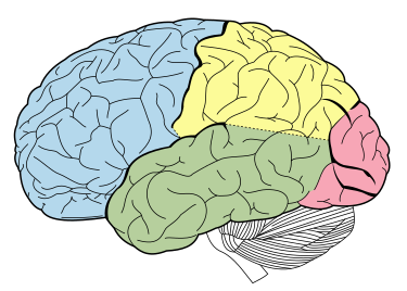
Source: Wikipedia. The temporal lobe is shown in green, while the motor area is in the frontal lobe in blue


ALS is now considered a multisystemic disorder in which almost half of patients present with varying degrees of cognitive deficits, yet unfolded TDP-43 aggregates in cytosol are found in most ALS cases.

TDP-43 pathology in ALS can be divided into four stages (Braak stages): it begins focally, and then spreads persistently in sequential and regional patterns that typically originate from the motor cortex and extend to the prefrontal cortex, thalamus and eventually, the hippocampus.

However neuroimaging studies of hippocampal volumes in patients with amyotrophic lateral sclerosis (ALS) have reported inconsistent results. The group of Chinese scientists from 10 institutions, aimed to demonstrate that such discrepancies are largely due to atrophy of different regions of the hippocampus that emerge in different disease stages of ALS and to explore the existence of co-pathology in ALS patients.

They used King’s clinical staging system for ALS to classify patients into different disease stages. The scientists then investigated in vivo hippocampal atrophy patterns across subfields and anterior-posterior segments in different King’s stages using structural MRI in 76 ALS patients and 94 health controls.

The thalamus, corticostriatal tract and perforant path were used as structural controls to compare the sequence of alterations between these structures and the hippocampal subfields.

In summary:

  • ALS patients at King’s stage 1, had lower volumes in the bilateral posterior subiculum and presubiculum;
  • ALS patients at King’s stage 2 exhibited lower volumes in the bilateral posterior subiculum, left anterior presubiculum and left global hippocampus;
  • ALS patients at King’s stage 3 showed significantly lower volumes in the bilateral posterior subiculum, dentate gyrus and global hippocampus. Thalamic atrophy emerged at King’s stage 3.

White matter tracts remained normal in a subset of ALS patients.

In the present study, the authors demonstrated that the earliest hippocampal alterations in ALS patients occurred in the posterior subiculum and presubiculum, and these alterations emerged at King’s stage 1. This indicates that subiculum atrophy occurs earlier and independent of TDP-43 pathology in ALS.


enter image description here Hagmann P, Cammoun L, Gigandet X, Meuli R, Honey CJ, et al. 


Taken together, their data suggest that patients with ALS have additional pathologies that are independent of TDP-43 pathology.

Increasingly, studies have shown that at least 20% of ALS patients present significant Alzheimer disease pathology of both Aβ and tau proteins. Recently, Gómez-Pinedo and colleagues showed that in ALS patients, the amyloid cascade of the amyloid precursor protein is activated in the hippocampus of ALS patients, and cytoplasmic Aβ peptide and pTDP-43 expression levels are moderately correlated.

Thus the motor cortex and subiculum seem to represent two independent centres of ALS during the early stages of the disease, which represent TDP-43 pathology and Alzheimer disease pathology, respectively, and these pathologies may converge as the disease progresses toward advanced stages.

If these findings are confirmed in further studies, they will have a profound effect on the understanding of the aetiology and pathogenic mechanisms underlying ALS and other neurodegenerative diseases.

Advertisement


This book retraces the main achievements of ALS research over the last 30 years, presents the drugs under clinical trial, as well as ongoing research on future treatments likely to be able stop the disease in a few years and to provide a complete cure in a decade or two.

Résultats décevants de l'étude VALOR sur le Tofersen (BIIB067)

- Posted by admin in Français

Voir notre post sur les résultats ici.

In Silico Computer Models for Deep Brain Stimulation

- Posted by admin in English

Deep brain stimulation (DBS) is an established therapy for patients with Parkinson's disease. enter image description here In silico computer models for DBS allow to pre-select a set of potentially optimal stimulation parameters. If efficacious, they could further carry insight into the mechanism of action of DBS and foster the development of more efficient stimulation approaches. In recent years, the focus has shifted towards DBS-induced firing in myelinated axons, deemed particularly relevant for the external modulation of neural activity.

The scientists in this new medRxiv publication, use the concept of pathway activation modeling, which incorporates advanced volume conductor models and anatomically authentic fiber trajectories to estimate DBS-induced action potential initiation in anatomically plausible pathways that traverse in close proximity to targeted nuclei.

Then the scientists applied the method on a retrospective dataset with the aim of providing a model-based prediction of clinical improvement following DBS (as measured by the motor part of the Unified Parkinsons Disease Rating Scale).

Based on differences in outcome and activation rates for two DBS protocols in a training cohort, the authors computed a theoretical 100% improvement profile and enhanced it by analyzing the importance of profile matching for individual pathways.

Finally, the authors validated the performance of authors' profile-based predictive model in a test cohort.

As a result, the authors demonstrated the clinical utility of pathway activation modeling in the context of motor symptom alleviation in Parkinsons patients treated with DBS.

Read the original article on medRxiv


Please, help us continue to provide valuable information: