The theory of infectious origin of the Alzheimer disease

- Posted by admin in English

The theory of infectious origin of the Alzheimer's disease

The hypothesis that Alzheimer's disease has an infectious origin, has a long and controversial history. The data at the origin of this hypothesis are contradictory and mainly associative in nature, without it being possible to demonstrate a causal link. Interest in this theory has been renewed, however, by several recently published observations. In the section Viewpoint de la revue Nature Reviews Neurology, Ben Readhead, a researcher at the Biodesign Institute's ASU-Banner Center for Research on Neurodegenerative Diseases, joined several distinguished colleagues to discuss the idea that bacteria, viruses or other infectious pathogens can play a role in Alzheimer's disease.

A hypothesis that has never been favored by researchers

This hypothesis may have been rejected too quickly. For example, microorganisms do not only cause acute illnesses, in fact certain microorganisms can hide in the body for decades in latent form, causing damage intermittently or after long periods of silence.

In addition, being infected does not necessarily mean being symptomatic. For example, out of the millions of people infected with Mycobacterium tuberculosis, only about a tenth of them will develop tuberculosis. Likewise, most people infected with HSV1 do not develop cold sores so it is possible that asymptomatic carriers of this virus were often mistakenly included in the control groups. It should also be noted that many viruses of the Herpes family (HSV1, HSV2, VZV) live preferentially in neurons.

A role for an infectious agent - in particular the herpes simplex 1 virus (HSV1) - in Alzheimer's disease (Alzheimer's disease) was proposed about 30 years ago based discovery of HSV1 DNA in the brain tissue of a large proportion of the elderly, followed by evidence that e the virus confers a high risk of disease to carriers of the ε4 allele of the gene apolipoprotein E (APOE * ε4).

Shortly after the detection of HSV1 DNA, two different species of bacteria, Borrelia burgdorferi and Chlamydia pneumoniae, were implicated in Alzheimer's disease, and a third species, Porphyromonas gingivalis, was recently added to the list.

Doubts remain, however

Nevertheless, it is known that an acute end-of-life infection, such as pneumonia, can cause a dramatic increase in the amount of microorganisms in the brain. They will then be detected post-mortem but that does not mean that these microorganisms are at the origin of the Alzheimer's disease. In addition, the issue of reverse causation is never really addressed: For example, clinical Alzheimer's disease can lead to poor dental hygiene and, therefore, damage to the oral microbiome.

Indeed, there are many challenges to prove the theory of microbial origin of Alzheimer's disease. A potential challenge is that each drug has a relatively narrow spectrum of antimicrobial activity. However, since a large number of microorganisms have been associated with Alzheimer's disease by a range of researchers, it would be difficult to interpret what a negative result in a clinical trial would mean, which would necessarily use a specific antimicrobial.

Another problem is the duration of the disease. We know that the underlying pathology of Alzheimer's disease begins 20 years or more before the onset of symptoms. So, how to prove that an infectious process that occurred decades before the onset of symptoms, really contributed to the disease process?

One may also wonder why bacteria or viruses would escape the innate innate immune defense mechanisms, which are responsible for protecting the brain against such an invasion.

Advertisement


This book retraces the main achievements of ALS research over the last 30 years, presents the drugs under clinical trial, as well as ongoing research on future treatments likely to be able stop the disease in a few years and to provide a complete cure in a decade or two.

La théorie de l'origine infectieuse de la maladie d’Alzheimer

- Posted by admin in Français

La théorie de l'origine infectieuse de la maladie d’Alzheimer

L’hypothèse que la maladie d’Alzheimerde a une origine infectieuse a une histoire longue et controversée. Les données à l’origine de cette hypothèse sont contradictoires et de nature principalement associative, sans que l'on puisse mettre en évidence un lien de causalité. Cependant, l’intérêt pour cette théorie a été renouvelé par plusieurs observations récemment publiées. Dans la section Viewpoint de la revue Nature Reviews Neurology, Ben Readhead, chercheur au Centre de recherche sur les maladies neurodégénératives ASU-Banner du Biodesign Institute, s'est joint à plusieurs collègues distingués pour discuter de l'idée que les bactéries, les virus ou d'autres agents pathogènes infectieux peuvent jouer un rôle dans la maladie d'Alzheimer.

Une hypothèse qui n'a jamais eu la faveur des chercheurs

Cette hypothèse a peut-être été rejetée trop vite. Par exemple les micro-organismes ne causent pas que des maladies aiguës, en effet certains micro-organismes peuvent se cacher des dizaines d'années dans le corps sous une forme latente, causant des dommages par intermittence ou après de longues périodes de silence.

De plus, être infecté ne signifie pas nécessairement être manifester des symptômes. Par exemple, sur les millions de personnes infectées par la bactérie Mycobacterium tuberculosis, seulement un dixième d'entre eux vont développer une tuberculose. De même, la plupart des personnes infectées par le HSV1 ne développent pas de boutons de fièvre aussi il se peut que des porteurs asymptomatiques de ce virus ont souvent été inclus par erreur dans les groupes témoins. On notera aussi que de nombreux virus de la famille Herpès (HSV1, HSV2, VZV) vivent préférentiellement dans les neurones.

Un rôle pour un agent infectieux - en particulier le virus de l’herpès simplex 1 (HSV1) - dans la maladie d’Alzheimer (maladie d’Alzheimer) a été proposé il y a environ 30 ans sur la base de la découverte d’ADN de HSV1 dans le tissu cérébral d’une forte proportion de personnes âgées, suivi par la preuve que le virus confère un risque élevé de maladie aux porteurs de l’allèle ε4 du gène de l’apolipoprotéine E (APOE * ε4).

Peu après la détection de l’ADN du HSV1, deux espèces différentes de bactéries, Borrelia burgdorferi et Chlamydia pneumoniae, ont été impliquées dans la maladie d’Alzheimer, et une troisième espèce, Porphyromonas gingivalis, a récemment été ajoutée à la liste.

Des doutes subsistent pourtant

Cependant l’on sait qu’une infection aiguë en fin de vie, comme une pneumonie, peut entraîner une augmentation conséquente de la quantité de micro-organismes dans le cerveau. Celle-ci sera alors détectée post-mortem mais cela ne signifie pas pour autant que ces micro-organismes seront la cause de la maladie d’Alzheimer. De plus, la question de la causalité inverse n’est jamais vraiment abordée : Par exemple, la maladie d’Alzheimer clinique peut entraîner une mauvaise hygiène dentaire et, par conséquent, une altération du microbiome buccal.

Il y a donc de nombreux défis à relever pour prouver la théorie de l’origine microbienne de la maladie d’Alzheimer. Un défi potentiel est que chaque médicament possède un spectre d’activité antimicrobien relativement étroit. Or comme un grand nombre de micro-organismes ont été associés à la maladie d’Alzheimer par un éventail de chercheurs, il serait difficile d’interpréter dans un essai clinique ce que signifierait un résultat négatif qui utiliserait forcément un antimicrobien spécifique.

Un autre problème est la durée de la maladie. Nous savons que la pathologie sous-jacente à la maladie d’Alzheimer commence 20 ans ou plus avant l’apparition des symptômes. Alors, comment prouver qu’un processus infectieux survenant des décennies avant l’apparition des symptômes est à l'origine de la maladie?

On peut aussi se demander pourquoi les bactéries ou les virus échapperaient-ils aux mécanismes intrinsèques de défense immunitaire innée, qui sont chargés de protèger le cerveau contre une telle invasion.

Enfin et comme l’on suspecte une large gamme d’organisme des microbes, aux organismes fongiques en passant par les virus de contribuer significativement à la maladie d’Alzheimer, pourquoi alors ne pas considérer que la caractéristique centrale pertinente serait plutôt un dysfonctionnement général du système immunitaire inné ?

Publicité


Ce livre retrace les principales réalisations de la recherche sur la SLA au cours des 30 dernières années. Il présente les médicaments en cours d’essai clinique ainsi que les recherches en cours sur les futurs traitements susceptibles d’ici quelques années, d’arrêter la maladie et de fournir un traitement complet en une décennie ou deux.

Micro-wire arrays Integrated with CMOS chips for neural recording

- Posted by admin in English

Technologies to study neural activity

Recording the activity of a sufficient number of neurons at significant time scales and spatial distributions is one of the main challenges for a better understanding of how neural ensembles work.

Optical methods are increasingly used because they allow activity over a large area to be monitored from the same layer of brain tissue. In addition, they are inherently limited to recording from superficial structures of the brain or require the use of probes or surgery to provide access to deep brain regions.

A number of innovations have been made in electrical recording using flexible materials on the surface of the brain, and microelectrodes have long been the norm, but their number of channels is limited due to connection, volumetric displacement and tissue damage.

At the same time, electronics continue to evolve at a rapid pace, but few of these technological improvements make their way to neuroscience in vivo.

A new strategy for interfacing chips with three-dimensional micro-wire arrays

In this article, the authors report a new strategy to take advantage of the scalability and power of electronic components combined with a 3D neural interface.

This neural interface consists of a bundle of isolated micro-wires coupled perpendicularly to imaging networks such as those found in camera chips. enter image description here

By organizing them into bundles, the authors control the three-dimensional structure of the distal end, with a robust parallel contact plane on the proximal side which is coupled to an array of pixels.

The density of the microwires for the proximal end and the distal end can be independently adjusted, allowing wire-to-wire spacing to be customized as required.

Design and Manufacturing

enter image description here

  • (A) Procedure for manufacturing bundles of micro-wires.
    • (i) The individual micro-wires are electrically insulated with a robust ceramic or polymer coating.
    • (ii) A sacrificial layer is applied to the wires to ensure spacing.
    • (iii) The tips of the micro-wires can be shaped with an angular tip..
    • (iv) The wires are then grouped together by winding the wire or by mechanical aggregation. The threads pile up naturally in a honeycomb network.
    • (v) The bundle is infiltrated with biomedical epoxy to hold the wires together, then the upper (proximal) end is polished to mate with the CMOS chip.
    • (vi) The proximal end is etched from 10 to 20 μm to mate with the CMOS chip and the distal end of the wires is released by etching.
    • (B) An electron microscope backscatter view of an individual microwire.
    • (C) The wires are grouped in a structure in honeycomb and epoxy is infiltrated in between to fill in the gaps.
    • (D) Proximal end of a 177 bundle wires after etching to expose the common thread.
    • (E) Expected volumetric displacement of the micro-wire bundles as a function of the wire-to-wire distance, determined by the wire size and the thickness sacrificial coating.
    • (F) The distal end of a 600 bundle 7.5 μm W wires covered with 1 μm glass after etching (G and H). The distal end can be precisely shaped to simultaneously access different depths in the tissue.

Compatibility with different imaging chips and tests on a retina and on a motor cortex

Tests with different imaging matrices

The process described is very flexible and agnostic as to the identity of the chip; the authors successfully interface with the imaging matrix chip of a Xenics Cheetah camera, with an organic light emitting diode display chip from Olightek and with a multi-electrode matrix device.

Retinal tests

To test the ability of the completed device to record neural activity on a flat surface, the authors used an ex vivo preparation of rat retina. A dialysis membrane held a small piece of isolated retina against the bundle in an infusion chamber, then a 138-thread bundle was lowered into contact with the retina. The recorded spikes exhibited typical unit signatures, i.e. a detected action potential located on a wire with smaller peaks on the adjacent wires. These retinal recordings demonstrate the system's ability to record individual units at high data acquisition rates and a high signal-to-noise ratio.

Test on a motor cortex

Next, the researchers tested whether it was possible to record neural activity in the deep cortical and subcortical areas across a large spatial region in rodents in vivo. The recordings were made within 2 hours of implantation of the bundle into deep layers of the motor and somatosensory cortexes and of the dorsal striatum. The mice were allowed to run on a spherical treadmill, in a state of head restraint during the recording. Neural activity was easily observed in most of the wires in the bundle through a horizontal layer. Approximately 100 to more than 200 putative neurons were reliably identified over a large horizontally extended area in each recording during a typical 5-minute recording session.

enter image description here

Conclusion

Conventional two-photon imaging is generally limited in time and space, so bundles of micro-wires coupled to CMOS arrays can simultaneously record the activity of hundreds of neurons. In addition, the flexibility along the length of the distal end of the beam allows precise recordings from normally inaccessible areas, such as the striatum.

Advertisement


This book retraces the main achievements of ALS research over the last 30 years, presents the drugs under clinical trial, as well as ongoing research on future treatments likely to be able stop the disease in a few years and to provide a complete cure in a decade or two.

Technologies pour étudier l'activité neuronale

L’enregistrement de l’activité d’un nombre suffisant de neurones à des échelles de temps et des distributions spatiales significatives est l’un des principaux défis pour une meilleure compréhension du fonctionnement des ensembles neuronaux.

Les méthodes optiques sont de plus en plus utilisées, car elles permettent de surveiller l’activité sur une grande surface à partir de la même couche de tissu cérébral. En outre, elles sont intrinsèquement limitées à l’enregistrement à partir de structures superficielles du cerveau ou nécessitent l’utilisation de sondes ou une intervention chirurgicale pour fournir un accès à des régions cérébrales profondes.

Un certain nombre d’innovations ont été apportées à l’enregistrement électrique à l’aide de matériaux flexibles à la surface du cerveau, et les microélectrodes sont depuis longtemps la norme, mais leur nombre de canaux est limité en raison de la connexion, du déplacement volumétrique et des dommages tissulaires.

Dans le même temps, l’électronique continue d’évoluer à un rythme rapide, mais peu de ces améliorations technologiques font leur chemin vers les neurosciences in vivo.

Une nouvelle stratégie pour interfacer des puces avec des matrices de micro-fils tridimensionnels

Dans cet article, les auteurs rapportent une nouvelle stratégie pour tirer parti de l’évolutivité et de la puissance de composants électronique combinés à une interface neuronale 3D.

Cette interface neuronale consiste en un faisceau de micro-fils isolés accouplés perpendiculairement à des réseaux d’imagerie tels que ceux que l’on peut trouver dans les puces de caméra.

enter image description here

En les organisant en faisceaux, les auteurs contrôlent la structure tridimensionnelle de l’extrémité distale, avec un plan de contact parallèle robuste sur le côté proximal qui est accouplé à un réseau de pixels.

La densité des micro-fils pour l’extrémité proximale et l’extrémité distale peut être modulée indépendamment, permettant à l’espacement fil à fil d’être personnalisé selon les besoins.

Conception et Fabrication

enter image description here

  • (A) Procédure de fabrication des faisceaux de micro-fils.
    • (i) Les micro-fils individuels sont isolés électriquement avec un revêtement céramique ou polymère robuste.
    • (ii) Une couche sacrificielle est appliquée sur les fils pour assurer l’espacement.
    • (iii) Les pointes des micro-fils peuvent être façonnées avec une pointe angulaire.
    • (iv) Les fils sont ensuite regroupés par enroulement du fil ou par agrégation mécanique. Les fils s’entassent naturellement dans un réseau en nid d’abeille.
    • (v) Le faisceau est infiltré d’époxy biomédical pour maintenir les fils ensemble, puis l’extrémité supérieure (proximale) est polie pour s’accoupler à la puce CMOS.
    • (vi) L’extrémité proximale est gravée de 10 à 20 μm pour s’accoupler à la puce CMOS et l’extrémité distale des fils est libérée par gravure.
  • (B) Une vue au microscope électronique de rétrodiffusion d’un micro-fil individuel.
  • (C) Les fils se regroupent dans une structure en nid d’abeille et de l’époxy est infiltré entre les deux pour combler les lacunes.
  • (D) Extrémité proximale d’un faisceau de 177 fils après gravure afin d’exposer le fil conducteur.
  • (E) Déplacement volumétrique prévu des faisceaux de micro-fils en fonction de la distance fil à fil, déterminé par la taille du fil et l’épaisseur du revêtement sacrificiel.
  • (F) L’extrémité distale d’un faisceau de 600 fils de 7,5 μm W recouverts de 1 μm de verre après gravure (G et H). L’extrémité distale peut être façonnée avec précision pour accéder simultanément à différentes profondeurs dans le tissu.

Compatibilité et tests sur une rétine et sur le cortex moteur

Tests avec différentes matrices d'imagerie

Le processus décrit est très flexible et agnostique quant à l’identité de la puce; les auteurs ont réussi à créer des interfaces avec la puce de matrice d’imagerie d’une caméra Xenics Cheetah, avec une puce d’affichage à diode électroluminescente organique d’Olightek et avec un dispositif de matrice multi-électrodes.

Tests sur une rétine

Pour tester la capacité de l’appareil terminé à enregistrer l’activité neuronale sur une surface plane, les auteurs ont utilisé une préparation ex vivo de rétine de rat. Une membrane de dialyse a maintenu un petit morceau de rétine isolée contre le faisceau dans une chambre de perfusion, puis un faisceau de 138 fils a été abaissé en contact avec la rétine. Les pointes enregistrées présentaient des signatures unitaires typiques, c'est-à-dire un potentiel d'action détecté localisé sur un fil avec des pics plus petits sur les fils adjacents. Ces enregistrements rétiniens démontrent la capacité du système à enregistrer des unités individuelles à des taux d’acquisition de données élevés et à un rapport signal/bruit élevé.

Test sur un le cortex moteur

Ensuite, les chercheurs ont testé s’il était possible d'enregistrer l'activité neuronale dans les zones corticales et sous-corticales profondes à travers une grande région spatiale chez les rongeurs in vivo. Les enregistrements ont été effectués dans les 2 heures suivant l’implantation du faisceau dans des couches profondes de cortex moteur et somatosensoriel et du striatum dorsal. Les souris étaient autorisées à courir sur un tapis roulant sphérique, dans un état de contrainte de tête lors de l’enregistrement. Une activité neuronale a été facilement observée dans la plupart des fils des faisceaux à travers une couche horizontale. L’activité a été facilement observée dans la plupart des fils des faisceaux à travers une couche horizontale. Environ 100 à plus de 200 neurones putatifs ont été identifiés de manière fiable sur une grande zone horizontalement étendue dans chaque enregistrement au cours d’une session d’enregistrement typique de 5 minutes.

enter image description here

Conclusion

L’imagerie conventionnelle à deux photons est généralement limitée temporellement et spatialement, alors les faisceaux de micro-fils couplés à des matrices CMOS peuvent enregistrer simultanément l’activité de dopage de centaines de neurones. En outre, la flexibilité sur la longueur de l’extrémité distale du faisceau avec précision permet des enregistrements denses à partir de zones normalement inaccessibles, telles que le striatum.

Publicité


Ce livre retrace les principales réalisations de la recherche sur la SLA au cours des 30 dernières années. Il présente les médicaments en cours d’essai clinique ainsi que les recherches en cours sur les futurs traitements susceptibles d’ici quelques années, d’arrêter la maladie et de fournir un traitement complet en une décennie ou deux.

Design of EventEpi, an event-based health surveillance platform.

- Posted by admin in English

What is event-based surveillance?
The goal of event-based surveillance is to detect unusual events that might signal an outbreak. Event-based public health surveillance looks at reports, stories, rumors, and other information about health events that could be a serious risk to public health.

enter image description here

Main categories of event-based surveillance There are basically two main categories of event-based surveillance.

• Such information is unstructured. Information obtained through event-based surveillance can come from sources like reports in the media or rumors on an internet blog.

• In contrast Indicator-based public health surveillance is a more traditional way of reporting diseases to public health officials. Indicator-based surveillance involves reports of specific diseases from health care providers to public health officials. Such information may be described as structured information because the information obtained is standardized.

Examples of event-based health surveillance: WHO’s global surveillance system picks up public health threats 24 hours a day, 365 days a year. Once an event is verified, WHO assesses the level of risk and sounds the alarm. Within 48 hours of an emergency, WHO grades the severity of the event, activates the incident management system and deploys field teams.

Design of EventEpi at The Information Centre for International Health Protection. Reading the articles, discussing their relevance, and putting key information into a database is a time-consuming process. The Information Centre for International Health Protection (Informationsstelle für Internationalen Gesundheitsschutz, INIG) at RKI, performs event-based surveillance to identify events relevant to public health in Germany. Their routine tasks include reading online articles from a defined set of sources, evaluating them for relevance, and then manually filling a spreadsheet with information from the relevant articles. This spreadsheet is called Ereignisdatenbank (IDB). enter image description here

To support event-based surveillance, but also to gain insights into what makes an article and the event it describes relevant, the authors of “EventEpi–A Natural Language Processing Framework for Event-Based Surveillancedeveloped a natural-language-processing framework for automated information extraction and relevance scoring.

Their approach consists of two complementary parts: key information extraction and relevance scoring. Both approaches are integrated in a web application called EventEpi. With the exception of the convolutional neural network for which they used Keras, they used the Python package scikit-learn to implement the machine learning algorithms.

The IDB has to be preprocessed before any application of NLP. was not designed to be used with machine learning algorithms. It thus contained some inconsistencies that might not disturb human users but had to be resolved before machine processing. For example a case count could contain numerals as strings instead of numerical digits. Other entries have inconsistent naming schemes. In addition entries in the IDB were written in German but the output of EpiTator has to be in English.

The authors performed named entity recognition in two steps:

  • EpiTator, an open-source epidemiological annotation tool, scraped relevant sources and suggested many different candidates for the following entities: disease, country, date, and confirmed-case count. To accomplish the key information extraction, two problems needed to be solved:
    • First, the output of EpiTator needed to be comparable to the entries in the IDB.
    • Second and more importantly, the output of EpiTator needed to be filtered. A naive approach to finding the key entity out of all the entities returned by EpiTator is to pick the most frequent one. This approach worked well for detecting the key country and disease, but not for the key date and confirmed-case count. For those, the authors developed a learning-based approach.
  • The second part of developing a framework to support EBS was to estimate the relevance of epidemiological articles. The scientists framed the relevance evaluation as a classification problem. They trained a naive Bayes classifier to find the most likely entities in that set. For relevance scoring, the authors defined two classes to which any article might belong:
    • The article is relevant if it is in the event-based surveillance database.
    • Irrelevant otherwise.

Two sources stood out as being relevant, and easy to scrape:

  • World Health Organization Disease Outbreak News (WHO DON)
  • ProMED Mail.

The authors compared the performance of different classifiers, using document and word embeddings. State-of-the-art text classifiers tend to use word embeddings for vectorization rather than the tf-idf and bag-of-words approach. Word embeddings are vector representations of words that are learned on large amounts of texts in an unsupervised-manner. Proximity in the word embedding space tends to correspond to semantic similarity. The researchers compared six different classifiers for the relevance scoring task. Two of the tested algorithms stood out:

  • The multilayer perceptron performed best overall.
  • The support-vector machine, on the other hand, had the highest recall (0.88) which can be of higher interest for epidemiologists.

Finally, the authors integrated these functionalities into a web application called EventEpi where relevant sources are automatically analyzed and put into a database. The same fundamental issues encountered in using machine learning in general apply here as well, in particular bias and explainability.

Tackling individual biases and personal preferences during labeling by experts is essential. It will also be important to show why EventEpi extracted certain information or computed a relevance, for it to be adopted but also critically assessed by epidemiologists for improvement.

At the moment EventEpi only presents results to the user. However it could be expanded to be a general interface to an event database and allow epidemiologists to note which articles were indeed relevant as well as correct key information, an approach called active-learning

The overall framework, can be used in production, promising improvements in event-based surveillance. The source code is publicly available at https://github.com/aauss/EventEpi

Advertisement


This book retraces the main achievements of ALS research over the last 30 years, presents the drugs under clinical trial, as well as ongoing research on future treatments likely to be able stop the disease in a few years and to provide a complete cure in a decade or two.

Bats, although mammals, are not phylogenetically very close to humans, so we would not expect them to harbor many harmful viruses to our species.

enter image description here Source: Вых Пыхманн via Wikipedia

The order of bats contains more than 1,200 species. Because of this genetic diversity, information about one species may not apply to other species. In recent years, however, it has emerged that viruses have spread from bats to other mammals. For example, serological evidence and detection of viruses linked to ebolaviruses in bats suggest that bats are indeed one of the reservoirs of the virus.

However, although bats harbor many different viruses, their spread to other animals is extremely rare. One reason is that for such an event to occur, several factors should be conducive to transmission.

The ability of bats to host many viruses without showing pathology suggests that bats have developed immune mechanisms different from those of other mammals. This tolerance to viruses goes hand in hand with reduced inflammation.To explain this singular characteristic, it is necessary to compare it with another singular characteristic of bats: It is a mammal with the capacity to fly.

The metabolic rate of bats in flight is double that of running rodents of similar size. The increased metabolic rate that accompanies the flight would result in higher levels of oxygen-free radicals. To mount an immune response to the damage caused by this high metabolism, would be energetically expensive.

Bats have therefore developed mechanisms leading to reduced inflammation. It would also explain why bats of some species live longer than expected given their high metabolism and small size. Some bats can live up to 40 years, while a rodent of the same size can live only two years.

Small animals with a fast heart rate and metabolism generally have a shorter lifespan than larger animals with a slower heartbeat and a slower metabolism. But bats are unique because they have a much longer lifespan than other mammals of the same size.

But a reduced inflammatory response enables rapid replication of viruses, especially in stressful conditions that affect the immune system.

  • The awakening of hibernation is a stressful event for bats. Many large brown bats are latently infected and Gerowet and his colleagues have shown that the virus reactivates when it comes out of hibernation. This reactivation is also associated with a low level of antibodies to the virus. After hibernation, antibody levels rise, which puts the virus back to latency.

  • The virus also reactivates when bats are infected with a bacterial or fungal infection. Small brown bats are particularly susceptible to an often fatal fungal infection known as white nose syndrome. A study examining the effects of stress induced by this infection showed that bats infected with fungi had 60 times more coronavirus in their intestines than uninfected bats. The specific protection mechanisms of these bats results in a rapid response that blocks the virus outside the cells. Indeed, paradoxically a host that tolerates well the presence of viruses, because it controls inflammation, allows viruses to increase their rate of replication, by genetic selection, without damaging their host.

  • Disturbances in bat habitat also seem to stress these animals and cause them to spread even more viruses in their saliva, urine and feces which can infect other animals. "Increased environmental threats to bats can add to the threat of zoonosis," said Brook, who is also working with a Madagascar-based field project that explores the link between loss of bat habitat and spread of bat viruses to other animals and humans.

Such rapidly reproducing viruses generate extreme virulence when overflowing to hosts that do not have the same immune capabilities as bats.

When these virulent viruses travel from bats to animals without a rapid response immune system, they quickly overwhelm their new hosts.

Brook and Boots are developing a more formal model of disease progression in bats to better understand the spread of the virus to other animals and humans.

Advertisement


This book retraces the main achievements of ALS research over the last 30 years, presents the drugs under clinical trial, as well as ongoing research on future treatments likely to be able stop the disease in a few years and to provide a complete cure in a decade or two.

An innovative hypothesis on cancer metastasis

- Posted by admin in English

A scientist from the Universitätsklinikum Erlangen, Heiko Bruns, pursues an innovative hypothesis on cancer metastases. He wrote his doctoral thesis on tuberculosis bacteria hidding in macrophages, and his postdoctoral thesis focused on the importance of macrophages in the context of cancer metastasis.

Macrophages are a type of white blood cell, which gobbles up and digests cell debris, foreign substances, microbes and cancer cells. Macrophages enter damaged tissues by the endothelium of blood vessels, a process known as extravasation. This process is very similar to what is currently thought to be the metastasis process.

enter image description here Classical conception of the metastasis process Source: doi: 10.1038 / nri3789

It is well known that macrophages can contribute to the growth and progression of cancers. Macrophages can also positively and negatively influence the results of anti-cancer treatments. Unfortunately some pathogens managed to lives inside those powerful macrophages. This allows them to escape from the immune system. This is the case with Mycobacterium tuberculosis or HIV.

Metastases are responsible for most of the deaths caused by cancer. Dr. Bruns believes that it is currently unclear how metastases form. So far, it has been assumed that they spread throughout the body via the lymphatic vessels and the bloodstream. But this mechanistic hypothesis cannot explain why some organs are preferably targeted in metastasis while other are relatively preserved. This was first discussed as the "seed and soil" theory by Stephen Paget in 1889. enter image description here 
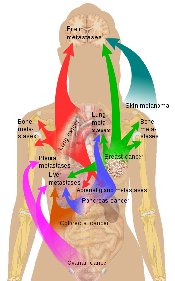
Source Mikael Häggström via Wikipedia

Paradoxically, cancer patients with a high number of macrophages have a reduced life expectancy. In a mouse model, tumor growth almost stops when the macrophages where removed.

Heiko Bruns assumes that individual tumor cells are consumed by phagocytes, but are not necessarily eliminated by them. Instead, he suspects that tumor cells are using macrophages as "Trojans horses". They could thus escape detection and travel through the body to colonize other organs.

Dr. Heiko Bruns' idea was accepted into the 'Experiment! In search of bold research ideas' based on this unusual question. This idea received funding of 120,000 euros from the Volkswagen Foundation until the end of 2021.

Les phagocytes peuvent-ils transporter des cellules tumorales?

- Posted by admin in Français

Un scientifique de l'Universitätsklinikum Erlangen, Heiko Bruns, poursuit une hypothèse innovante sur les métastases cancéreuses. Il a rédigé sa thèse de doctorat sur la survie des bactéries de la tuberculose dans les macrophages, et sa thèse postdoctorale porte sur l'importance des macrophages dans le contexte de la métastase cancéreuse.

Les macrophages sont un type de globules blancs du système immunitaire, qui engloutit et digère les débris cellulaires, les substances étrangères, les microbes, les cellules cancéreuses. Les macrophages pénètrent dans les tissus endommagés par l'endothélium d'un vaisseau sanguin, un processus connu sous le nom d'extravasation de leucocytes. Ce processus ressemble beaucoup à celui qu’on pense être celui de la métastase.

enter image description here Classical conception of the metastasis process Source: doi: 10.1038 / nri3789

Il est bien connu que les macrophages peuvent contribuer à la croissance et à la progression des cancers. Les macrophages peuvent également influencer positivement et négativement les résultats des traitements anti-cancéreux. Cependant certains agents pathogènes réussissent à vivre même à l'intérieur des macrophages. Cela permet alors à l'agent pathogène d’échapper au système immunitaire. C'est le cas de Mycobacterium tuberculosis ou du VIH.

Les métastases sont responsables de la plupart des décès causés par le cancer. Le Dr Bruns pense qu'à l'heure actuelle, on ne sait pas vraiment comment les métastases se forment. Jusqu'à présent, on a supposé qu'elles se propageaient dans tout le corps via les vaisseaux lymphatiques et la circulation sanguine.

Paradoxalement, les patients cancéreux avec un nombre élevé de macrophages ont une espérance de vie diminuée. Un modèle de souris a démontré que la croissance tumorale s'arrête pratiquement lorsque les macrophages ont été retirés.

Heiko Bruns suppose que les cellules tumorales individuelles sont consommées par les phagocytes, mais ne sont pas nécessairement éliminées par ceux-ci. Au lieu de cela, il soupçonne que les cellules tumorales utilisent les macrophages comme «chevaux de Troie». Elles pourraient ainsi échapper à la détection et voyager à travers le corps pour coloniser d'autres organes.

L’idée du docteur Heiko Bruns a été acceptée au programme de financement «Expérience! A la recherche d'idées de recherche audacieuses 'sur la base de cette question inhabituelle. Il a reçu un financement de 120 000 euros de la Fondation Volkswagen jusqu'à fin 2021. Le projet devrait démarrer au printemps.


Please, help us continue to provide valuable information: