Every month a revolutionary discovery?

Every month the specialized press informs us of decisive progress in the treatment of ALS. At the beginning of February, it was a young doctor in Scotland, who turns out to have only done in-vitro experiments showing lengthening of axons of lower motor neurons when subjected to a PGC1α inhibitor, but the press service of his university had translated this as being a great discovery allowing to hope for an ALS drug. It has been known since 2008 that PPAR-γ is a transcription factor that interacts with PGC-1α and that they are involved in ALS.

We are at the end of the month, and here is an important new discovery announcement regarding a treatment for ALS. It is a molecule that has been developed for almost ten years at Northwestern University in Illinois. Several articles have already been written about this molecule. The aim was to find a molecule which is both characterized by low toxicity, capable of crossing the blood-brain barrier and of having an action on the progression of disease. But in 10 years, extensive testing of this molecule had not been carried out on ALS model mice. It would seem that this is the case today, perhaps thanks to the collaboration between two teams, those of Professor Richard B. Silverman who is oriented "chemistry" and that of Professor Hande Ozdinler rather oriented "biology".

There is both good and bad news in this new article. The bad news is that the molecule would only be effective on upper motor neurons. This is a bit surprising and there really isn't any elaborate explanation that would be provided by the authors of the article. Usually in ALS both types of neurons are affected and restoring the functioning of the upper motor neurons will be of little help if the lower motor neurons are unable to control the muscles. In addition, the article is all about restoring the health of diseased neurons, which therefore must treated as soon as symptoms appear.

The authors use very aggressive language

The authors believe that other researchers did not seek to develop therapies for upper motor neurons. This is obviously not true, the number of studies using brain samples from deceased patients are there to testify.

The authors are also not shy about saying that other researchers are enough dumb to do their in-vitro research with different sets of cell lines, sometimes unrelated to motor neuron biology. Since the nervous system involves a lot of different types of cells, and these come from only a few lines of stem cells, on the contrary it seems a very good practice.

In addition, we could counter this focus on neurons by recalling that neurons do not perform their function in isolation, cells collaborate on the same task. Moreover, reading the article one immediately wonders why the authors did not examine the astrocytes, which are only present in the central nervous system.

They further explain that there has never been a study that examines upper motor neurons at the cellular level during the disease. The number of studies on ALS using mice models of the disease is proof of the fallacy of this claim.

A therapy that is effective regardless of the type of proteopathy.

The good news, apparently unexpected, is that the molecule is effective for both SOD1 (about 2% of patients) and TDP-43 (about 95% of patients) pathologies.

In this study, only four thin slices of motor cortex from normal control subjects without neurological disease and nine slices from ALS patients were used in addition to mice studies. Again the number is very low, in other studies several hundred tissues from deceased people are studied. There are also databases that allow data to be shared between researchers.

Histological sections of deceased patients

Upper motor neurons were only counted if their soma and apical dendrite were both visualized in the same 50 µm thick section. This assumes a severe selection of neurons, are the selected neurons representative of the upper motor neuron population?

Identification of NU-9:

The chiral 1,3-Diones cyclohexane, is a cyclohexane molecule containing two ketone groups. As said above, in fact work on NU-9 started almost ten years ago. High throughput screening of over 50,000 molecules was performed using a PC12 cell-based assay expressing SOD1G93A to identify compounds that attenuate protein aggregation and cytotoxicity. This made it possible to identify three families of compounds:

  • A first compound was selected from over 50 analogues of cyclohexan-1,3-dione, because of its ability to reduce mSOD1-mediated toxicity and inhibit protein aggregation.

  • Several rounds of optimization were performed, resulting in another compound, which also had excellent pharmaceutical properties in vitro, but did not enter neurons. Further modifications of this compound then led to the generation of NU-9, which crosses the blood-brain barrier, penetrates cortical neurons and has favorable pharmacokinetic properties.

Mouse management

The mice were derived from a cross between SOD1-type ALS model mice and mice carrying a fluorescent protein. In this study, only female mice were used for the experiments. The study involves a very small number of mice, one might wonder if it would be of interest to do a study on such a small number of mice, because no smoothing can erase the inevitable indesirable events. Mice noticeably overreact to tiny variations in their environment, we recently reported that ALS model mice were getting sick at Stanford but not at MIT!

  • Ten WT-UeGFP and six hSOD1G93A-UeGFP mice were treated with placebo,
  • Five WT-UeGFP mice and seven hSOD1G93A-UeGFP mice were treated with 20 mg / kg / day dose of NU-9.
  • Eleven WT-UeGFP mice, nine hSOD1G93A-UeGFP mice and four prpTDP-43A315T-UeGFP mice were treated with a dose of 100 mg / kg / day of NU-9.

The dose of NU-9 or placebo was administered once daily by oral gavage, starting on postnatal day P60 and continuing until P120. This NU-9 compound is able to prolong the lifespan of an ALS model mouse by 13% with a dose of 20 mg / kg. A repeated 7-day toxicological study in mice demonstrated a no-observed-adverse-effect level of 100 mg / kg.

All of the mice were sacrificed after 120 days (P120), which is considered a terminal stage for this type of ALS model mouse and approximately 60% of the upper motor neurons in the motor cortex are then lost while the motor neurons are lost. remaining upper ones have a smaller soma size and vacuolated and disintegrated apical dendrites. There does not seem to have been any test on an increase in the lifespan of the treated mice.

SOD1 qualitative analyzes

The electron microscope allowed cell type analyzes of upper motor neurons and their key organelles with great precision. At P120, the upper motor neurons of the placebo-treated hSODG93A-UeGFP mice had lost most of their cytoplasmic integrity. There were very few intact organelles left in the soma. However, the presence of disintegrated mitochondria and endoplasmic reticulum was evident. The mitochondria had mostly lost the integrity of their inner membrane, aggregated, enlarged or started to disintegrate. The endoplasmic reticulum also showed broken and scattered cisterns.

NU-9 treatment (dose of 100 mg / kg per day) showed profound improvements in both the structure and integrity of the mitochondria and endoplasmic reticulum of diseased upper motor neurons. Upon treatment, the soma of these neurons showed an intact nuclear membrane, which was devoid of any invagination or protrusion, and the detection of many organelles which were appropriate in size, location and interactions between them. The mitochondrial inner membrane was intact with proper ridges that were in close contact with the endoplasmic reticulum.

SOD1 quantitative analyzes

The authors then performed quantitative analyzes to determine whether these improvements were widely seen in diseased upper motor neurons treated with NU-9. The total number of mitochondria in upper motor neurons of hSODG93A-UeGFP mice was significantly increased after 100 mg / kg / day NU-9 treatment compared to upper motor neurons treated with placebo. The total number of mitochondria after NU-9 treatment was comparable to that of healthy mice. In addition, NU-9 treatment significantly increased the number of healthy mitochondria and endoplasmic reticulum.

present in the motor cortex of hSOD1G93A-UeGFP mice treated for 60 days with 100 mg / kg / day of NU-9 became almost comparable to that of the higher motor neuron numbers present in healthy mice

What about TDP-43?

Since NU-9 appears to improve the integrity of mitochondria and endoplasmic reticulum of upper motor neurons diseased due to mSOD1 toxicity, and these are problems that are also found in pathologies such as TDP-43, the authors speculated that NU-9 might also be effective in TDP-43 pathology. It still seems like a big conceptual step, because they are two distinct disease models. Even if NU-9 has an effect on the structure of the mitochondria and endoplasmic reticulum of higher motor neurons, it should not act on poorly formed and poorly localized protein aggregates that are already present in cytosol? But as the article assumes the drug is administered as soon as symptoms appears, they do not have to deal with that problem.

To test their new hypothesis, the scientists generated a new type of mouse, this time modeling TDP-43 type ALS.

Four mice were treated with a dose of 100 mg / kg / day of NU-9, and 3 mice of the prpTDP-43A315T-UeGFP type were used as a control. The WT-UeGFP mouse cohort was used as a healthy control for both groups.

TDP-43 qualitative analyzes

The NU-9 treatment resulted in profound improvements in both mitochondria and endoplasmic reticulums of higher motor neurons. The mitochondria, especially their internal membranes, became intact and there was no sign of mitoautophagy or mitophagy. The endoplasmic reticulum retained its structure with attached ribosomes, and there was no enlargement or disintegration of the cisterns.

TDP-43 quantitative analyzes

Quantitative analysis confirmed a significant increase in the number of total mitochondria per upper motor neuron per section after 100 mg / kg / day of NU-9 treatment. This number of mitochondria after NU-9 treatment in prpTDP-43A315T-UeGFP mice became comparable to that of healthy mice. The average percentage of healthy mitochondria also increased significantly with NU-9 treatment compared to diseased upper motor neurons. NU-9 treatment also increased the average number of intact endoplasmic reticulum cisterns in upper motor neurons.

The scientists then examined whether NU-9 treatment would also promote cell integrity and the survival of the higher motor neurons with TDP-43 pathology in vivo. The health and integrity of the apical dendrites in prpTDP-43A315T-UeGFP mice showed a profound improvement with the NU-9 treatment because the percentage of upper motor neurons with vacuolated primary apical dendrites was significantly reduced. More interestingly, the mean number of higher motor neurons in the motor cortex of prpTDP-43A315T-UeGFP mice treated with NU-9 was dramatically increased compared to that of untreated prpTDP-43A315T-UeGFP mice.

The same WT-UeGFP cohort was used as a healthy control for the hSOD1G93A-UeGFP and prpTDP-43A315T-UeGFP mice, as mentioned previously. The higher motor neuron numbers with the NU-9 treatment were comparable and almost identical to those of the healthy control mice treated with the placebo, revealing the ability of NU-9 to eliminate the continued degeneration of the higher motor neurons.

Effect of NU-9 on lower motor neurons

In order to determine whether NU-9 treatment also improves the health and survival of lower motor neurons (lower motor neurons), the authors studied the lumbar cords of hSOD1G93A-UeGFP and prpTDP-43A315T-UeGFP mice. As indicated by previous studies, there was no significant loss of lower motor neurons in the spinal cord of prpTDP-43A315T-UeGFP mice, even at P120, and therefore an investigation of NU-9 treatment on motor neuron survival inferior was not possible. This seems to indicate that TDP-43 pathology only affects the higher motor neurons, yet other studies show the opposite.

However, in the case of SOD1, there was a dramatic reduction in the number of lower motor neurons in hSOD1G93A-UeGFP mice compared to healthy mice.

Macroscopic effects

The authors quantitatively assessed the changes in the number of lower motor neurons in the lumbar spinal cord of mice that were treated with either placebo or NU-9 (20 or 100 mg / kg / day), or healthy control mice. . NU-9 treatment, regardless of dose, was not sufficient to eliminate the ongoing lower motor neuron degeneration in hSOD1G93A-UeGFP mice.

NU-9 treatment improves upper motor neuron function Even though most behavioral tests fail to properly assess upper motor neuron health and connectivity, the hanging wire test would be more specific to upper motor neuron integration.

The untreated hSOD1G93A-UeGFP mice could not stay on the hanger as the disease progressed. In contrast, hSOD1G93A-UeGFP mice treated with 100 mg / kg / day of NU-9 performed better than hSOD1G93A-UeGFP mice treated with placebo, and this performance was comparable to that of healthy mice at same age. However, treatment with NU-9 did not result in a significant improvement in performance on the rotarod test at any dose.

The prpTDP-43A315T mice performed worse than the WT litter mates on rotarod and hanging wire tests. However, when treated with 100 mg / kg / day of NU-9, they performed better on the hanging wire test, comparable to healthy WT mice at P120. Unlike the hSOD1G93A mouse model, even the Rotarod test revealed a significant improvement in the TDP-43 model only after 30 days of NU-9 treatment.

Conclusion

The possibility that NU-9 can improve the integrity of both mitochondria and endoplasmic reticulum is important, because, although the underlying causes of the disease are heterogeneous, many clues converge on the proper functioning of the mitochondria and the endoplasmic reticulum. Disruption of intracellular membrane organelles, such as the Golgi apparatus, has been suggested as a possible cause of ALS and is proposed to be upstream of endoplasmic reticulum dysfunction.

This could explain why the NU-9 treatment improves the cytoarchitecture of higher motoneurons and eliminates their progressive degeneration in hSOD1G93A and TDP-43A315T mice.

However, this does not explain why lower motor neurons would not be affected by this molecule. The explanation given that these are different lineages seems very short and has not been further investigated. At no time axons are mentioned in this article, which seems odd in an article that claims to seek a cure for ALS. It seems to me that this weakens the claim that NU-9 treatment improves upper motor neuron cytoarchitecture as this architecture is not extensively tested.

Moreover, the number of mice tested is very, very low and probably statistically meaningless. It is the same for the few histological sections of deceased patients, and the selection of motor neurons. We can also notice that it has been demonstrated for more than 15 years that other types of cells are involved in ALS, starting with astrocytes, than motor neurons. This has absolutely not been studied in this article. However, when a molecule is effective on the higher motor neurons and not on the lower motor neurons, it immediately suggests that it is not on the neurons that it acts, but on the astrocytes!.

All this considerably relativizes the very strong assertions of the press service of the university which reported that "After 60 days of treatment, diseased brain cells look like healthy cells".

For any correspondence write to me at contact [at] padiracinnovation.org

Advertisement


This book retraces the main achievements of ALS research over the last 30 years, presents the drugs under clinical trial, as well as ongoing research on future treatments likely to be able stop the disease in a few years and to provide a complete cure in a decade or two.

Tous les mois une découverte révolutionnaire ?

Tous les mois la presse spécialisée nous informe d'un progrès décisif dans le traitement de la SLA. Au début du mois de février, c'était un jeune docteur en Écosse, qui s'avère n'avoir fait que des expériences in-vitro montrant un rallongement des axones de neurones moteurs inférieurs quand ils étaient soumis à un inhibiteur de PGC1α, mais le service de presse de son université avait traduit cela comme étant une grande découverte permettant d'espérer l'arrivée prochaine d'un médicament. On sait depuis 2008 que PPAR-γ est un facteur de transcription qui interagit avec PGC-1α et qu’ils sont impliqués dans la SLA.

Nous sommes à la fin du mois, et voici une nouvelle annonce de découverte importante concernant un traitement de la SLA. Il s'agit d'une molécule qui est développée depuis presque une dizaine d'années à la Northwestern University, dans l'Illinois. Plusieurs articles ont déjà été écrits à propos de cette molécule, mais il s'agissait de trouver une molécule qui soit à la fois caractérisée par une faible toxicité, capable de passer la barrière hémato-encéphalique et d'avoir une action sur la progression de la maladie. Mais en 10 ans il n'avait pas été procédé à des essais extensifs de cette molécule sur des souris modèles de la SLA. Il semblerait que ce soit le cas aujourd'hui, peut-être grâce à la collaboration entre deux équipes, celles du professeur Richard B. Silverman qui est orientée "chimie" et celle de la professeure Hande Ozdinler plutôt orienté « biologie ».

Il y a à la fois une bonne et une mauvaise nouvelle dans ce nouvel article. La mauvaise nouvelle est que la molécule ne serait efficace que sur les neurones moteurs supérieurs. Cela est un peu surprenant et il n’y a pas vraiment d’explications élaborées qui seraient fournis par les auteurs de l’article. En général dans la SLA les deux types de neurones sont affectés et restaurer le fonctionnement des neurones moteurs supérieurs ne sera guère utile, si les neurones moteurs inférieurs sont incapable de commander les muscles. De plus dans l’article il n'est question que de restaurer la bonne santé de neurones malades, donc traités dès l'apparition des symptomes.

Les auteurs ont un langage très agressif

Les auteurs estiment que les autres chercheurs n’ont pas cherché à élaborer des thérapies pour les neurones moteurs supérieurs. C’est évidemment faux, nombres d’études utilisant des échantillons de cerveaux de malades décédés sont là pour en témoigner.

Les auteurs n’hésitent pas non plus à affirmer que les autres chercheurs effectuent leurs recherches in-vitro avec un ensemble différent de lignées cellulaires, parfois sans rapport avec la biologie des motoneurones.

Dans la mesure où le système nerveux implique un grand nombre de types différents de cellules, et que celles-ci sont originaires de seulement quelques lignées de cellules souches, au contraire cela semble très sain. Par ailleurs on pourrait contrer cette focalisation sur les neurones en rappelant que les neurones n’exercent pas leur fonction de façon isolée, un nerf c’est un ensemble de cellules qui collaborent à la même tâche. D'ailleurs à la lecture de l'article on se demande immédiatement pourquoi les auteurs n'ont pas examinés les astrocytes, qui ne sont présent que dans le système nerveux central.

Ils expliquent encore qu’il n'y a jamais eu d'étude qui examine les motoneurones supérieurs au niveau cellulaire au cours de la maladie. Le nombre d’études sur la SLA utilisant des souris modèles de la maladie est là pour témoigner de la fausseté de cette affirmation.

Une thérapie qui est efficace quel que soit le type de proteopathie.

La bonne nouvelle, apparemment inattendue, c’est que la molécule est efficace à la fois pour les pathologies de type SOD1 (environ 2 % des malades) et TDP-43 (environ 95 % des malades). Les auteurs estiment qu’il s'agit de la première étude axée sur les mécanismes et les cellules et qui jette les bases des études futures qui identifieront les composés en fonction de leur capacité à restaurer la santé des neurones. Dans cette étude, quatre fines tranches de cortex moteur issus de sujets témoins normaux sans maladie neurologique et neuf tranches issus de patients SLA ont été inclus. Là encore le nombre est très faible, dans d’autres études plusieurs centaines de tissus issus de personnes décédés sont étudiés. Il y a d’ailleurs des banques de données qui permettent de partager les données entre chercheurs.

Coupes histologiques de patients décédés

Les motoneurones supérieurs n'ont été comptés que si leur soma et leur dendrite apicale étaient tous deux visualisés dans la même section de 50 µm d'épaisseur. Cela suppose une sélection sévère des neurones, est-ce que les neurones sélectionnés sont représentatifs de la population de motoneurones supérieurs?.

Identification du NU-9 :

Le cyclohexane chiral 1,3-Diones, est une molécule de cyclohexane contenant deux groupes cétone. Comme dit plus haut, en fait les travaux sur NU-9 ont débutés il y a presque une dizaine d’années. Un criblage à haut débit de plus de 50 000 molécules avait été réalisé à l'aide d'un test à base de cellules PC12 exprimant la SOD1G93A pour identifier les composés qui atténuaient l'agrégation des protéines et la cytotoxicité. Cela a permis d’identifier trois familles de composés :

  • Un premier composé a été sélectionné parmi plus de 50 analogues de la cyclohexane-1,3-dione, à cause de sa capacité à réduire la toxicité médiée par mSOD1 et à inhiber l'agrégation des protéines.

  • Plusieurs cycles d'optimisation ont été effectués, aboutissant à un autre composé, qui a également avait d'excellentes propriétés pharmaceutiques in vitro, mais ne pénétrait pas dans les neurones. D'autres modifications de ce composé ont alors conduit à la génération de NU-9, qui pénètre les neurones corticaux, traverse la barrière hémato-encéphalique et possède des propriétés pharmacocinétiques favorables.

Gestion des souris

Les souris sont issues d’un croisement de souris modèle de la SLA de type SOD1 et de souris porteuses d’une protéine fluorescente. Dans cette étude, seules les souris femelles ont été utilisées pour les expériences. L’étude porte sur un très petit nombre de souris, on peut se demander si cela à un intérêt de faire une étude sur un nombre de souris aussi faible, car aucun lissage ne permet de gommer les inévitables mesures non significatives. Les souris surréagissent notoirement à d’infimes variations dans leur environnement, il y a peu nous avons ainsi rapporté que des souris modèles de la SLA devenaient malades à Stanford mais pas au MIT !

  • Dix souris WT-UeGFP et six hSOD1G93A-UeGFP ont été traitées avec le placebo,
  • Cinq souris WT-UeGFP et sept souris hSOD1G93A-UeGFP ont été traitées avec 20 mg / kg / jour dose de NU-9.
  • Onze souris WT-UeGFP , neuf souris hSOD1G93A-UeGFP et quatre souris prpTDP-43A315T-UeGFP ont été traitées avec une dose de 100 mg / kg / jour de NU-9.

La dose de NU-9 ou de placebo a été administrée une fois par jour par gavage oral, en commençant au jour postnatal (P) 60 et en continuant jusqu'à P120. Ce composé NU-9 est capable de prolonger la durée de vie d’une souris modèle de la SLA de 13% avec une dose de 20 mg/kg. Une étude toxicologique répétée pendant 7 jours chez la souris a mis en évidence une dose sans effet nocif observé de 100 mg/kg.

Toutes les souris ont été sacrifiées au bout de 120 jours (P120), ce qui est considéré comme un stade terminal pour ce type de souris modèle de la SLA et environ 60% des motoneurones supérieurs dans le cortex moteur sont alors perdus tandis que les motoneurones supérieurs restants ont une taille de soma plus petite et des dendrites apicales vacuolées et désintégrées. Il ne semble pas y avoir eu de test sur un accroissement de durée de vie des souris traitées.

Analyses qualitatives SOD1

Le microscope électronique a permis des analyses du type de cellule des motoneurones supérieurs et de leurs organites clés avec une grande précision. À P120, les motoneurones supérieurs des souris hSODG93A-UeGFP traitées avec le placebo avait perdu la plupart de leur intégrité cytoplasmique. Il y restait très peu d'organites intacts dans le soma. Cependant, la présence de mitochondries et de réticulum endoplasmique désintégrés étaient évidentes. Les mitochondries avaient principalement perdu l'intégrité de leur membrane interne, s’étaient agrégées, agrandies ou avaient commencé à se désintégrer. Le réticulum endoplasmique montrait également des citernes brisées et dispersées.

Le traitement NU-9 (dose de 100 mg / kg par jour) a montré de profondes améliorations à la fois de la structure et de l'intégrité des mitochondries et du réticulum endoplasmique des motoneurones supérieurs malades. Lors du traitement, le soma de ces neurones montrait une membrane nucléaire intacte, qui était dépourvue de toute invagination ou saillie, et la détection de nombreux organites qui étaient appropriés en taille, emplacement et interactions entre eux. La membrane interne mitochondriale était intacte avec des crêtes appropriées qui étaient en contact étroit avec le réticulum endoplasmique.

Analyses quantitatives SOD1

Les auteurs ont ensuite effectué des analyses quantitatives pour déterminer si ces améliorations étaient largement observées dans les motoneurones supérieurs malades traités avec NU-9. Le nombre total de mitochondries dans les motoneurones supérieurs de souris hSODG93A-UeGFP avait augmenté de manière significative après un traitement de 100 mg / kg / jour NU-9 par rapport aux motoneurones supérieurs traités avec le placebo. Le nombre total de mitochondries après traitement NU-9 était comparable à celui des souris saines. De plus, le traitement NU-9 a augmenté de manière significative le nombre de mitochondries et de réticulum endoplasmique sains dans les motoneurones supérieurs.

Et surtout, le nombre moyen d'motoneurones supérieurs présents dans le cortex moteur des souris hSOD1G93A-UeGFP traitées 60 jours avec 100 mg / kg / jour de NU-9 est devenu presque comparable à celui des nombres d'motoneurones supérieurs présents chez les souris saines

Et TDP-43 ?

Étant donné que le NU-9 semble améliorer l'intégrité des mitochondries et du réticulum endoplasmique des motoneurones supérieurs malades à cause de la toxicité de mSOD1, et qu'il s'agit de problèmes que l’on trouve aussi dans les pathologies de type TDP-43, les scientifiques ont supputé que NU-9 pourrait aussi être efficace dans la pathologie TDP-43. Cela semble quand même un grand pas conceptuel, en effet il s'agit de deux modèles de maladie distincts. Même si NU-9 a un effet sur la structure des mitochondries et du réticulum endoplasmique des motoneurones supérieurs, il ne devrait pas agir sur les agrégats de protéines mal formées et mal localisées ?

Pour tester leur nouvelle hypothèse, les scientifiques ont généré un nouveau type de souris, cette fois-ci modélisant la SLA de type TDP-43.

Quatre souris ont été traitées avec une dose de 100 mg / kg / jour de NU-9, et 3 souris de type prpTDP-43A315T-UeGFP ont été utilisées comme contrôle. La cohorte de souris WT-UeGFP a été utilisée comme témoin sain pour les deux groupes.

Analyses qualitatives TDP-43

Le traitement NU-9 a entraîné de profondes améliorations à la fois des mitochondries et des réticulums endoplasmique des motoneurones supérieurs. Les mitochondries, en particulier leurs membranes internes, sont devenues intactes et il n'y avait aucun signe de mitoautophagie ou de mitophagie. Le réticulum endoplasmique conservait sa structure avec des ribosomes attachés, et il n'y avait pas d'élargissement ou de désintégration des citernes.

Analyses quantitatives TDP-43

L'analyse quantitative a confirmé une augmentation significative du nombre de mitochondries totales par motoneurones supérieurs par section après 100 mg / kg / jour de traitement NU-9. Ce nombre de mitochondries après traitement NU-9 chez les souris prpTDP-43A315T-UeGFP est devenu comparable à celui des souris saines. Le pourcentage moyen de mitochondries saines a également augmenté de manière significative avec le traitement NU-9 par rapport aux motoneurones supérieurs malades. Le traitement NU-9 a également augmenté le nombre moyen de cisternes de réticulum endoplasmique intactes dans les motoneurones supérieurs.

Les scientifiques ont ensuite examiné si NU-9 le traitement favoriserait également l'intégrité cellulaire et la survie de l'motoneurones supérieurs avec la pathologie TDP-43 in vivo. La santé et l'intégrité des dendrites apicales chez les souris prpTDP-43A315T-UeGFP ont montré une amélioration profonde avec le traitement NU-9 car le pourcentage d'motoneurones supérieurs avec des dendrites apicales primaires vacuolées était significativement réduit. Plus intéressant encore, le nombre moyen d'motoneurones supérieurs dans le cortex moteur des souris prpTDP-43A315T-UeGFP traitées avec NU-9 a connu une augmentation profonde par rapport à celui des souris prpTDP-43A315T-UeGFP non traitées.

La même cohorte WT-UeGFP a été utilisée comme témoin sain pour les souris hSOD1G93A-UeGFP et prpTDP-43A315T-UeGFP, comme mentionné précédemment. Les nombres motoneurones supérieurs avec le traitement NU-9 étaient comparables et presque identiques à ceux des souris témoins saines traitées avec le placebo, révélant la capacité du NU-9 à éliminer la dégénérescence continue des motoneurones supérieurs.

Effet du NU-9 sur les neurones moteurs inférieurs

Dans le but de déterminer si le traitement NU-9 améliore également la santé et la survie des neurones moteurs inférieurs (motoneurones inférieurs), les auteurs ont étudié les cordons lombaires des souris hSOD1G93A-UeGFP et prpTDP-43A315T-UeGFP. Comme indiqué par les études précédentes, il n'y avait pas de perte importante de motoneurones inférieurs dans la moelle épinière des souris prpTDP-43A315T-UeGFP, même à P120, et donc une enquête sur le traitement NU-9 sur la survie des motoneurones inférieurs n'était pas possible. Cela semble indiquer que la pathologie TDP-43 n’affecterait que les motoneurones supérieurs, pourtant d’autres études montrent le contraire.

Cependant, dans le cas de SOD1, il y a eu une réduction spectaculaire du nombre de motoneurones inférieurs chez les souris hSOD1G93A-UeGFP par rapport aux souris saines.

Effets macroscopiques

Les auteurs ont évalué quantitativement les changements dans le nombre de motoneurones inférieurs dans la moelle épinière lombaire des souris qui ont été traitées soit avec le placebo, soit NU-9 (20 ou 100 mg / kg / jour), ou encore les souris saines témoins. Le traitement NU-9, quelle que soit la dose, n'était pas suffisant pour éliminer la dégénérescence motoneurones inférieurs en cours chez les souris hSOD1G93A-UeGFP.

Le traitement NU-9 améliore la fonction des neurones moteurs supérieurs Même si la plupart des tests comportementaux ne parviennent pas à évaluer correctement la santé et la connectivité motoneurones supérieurs, le test du fil suspendu serait plus spécifique à l'intégration motoneurones supérieurs.

Les souris hSOD1G93A-UeGFP non traitées n'ont pas pu rester sur le fil de suspension au fur et à mesure que la maladie progressait. Au contraire, les souris hSOD1G93A-UeGFP traitées avec une dose de 100 mg / kg / jour de NU-9 ont obtenu des résultats meilleurs que les souris hSOD1G93A-UeGFP traitées avec le placebo, et cette performance était comparable à celle de souris saines au même âge. Cependant le traitement au NU-9 n'a pas entraîné d'amélioration significative des performances au test du rotarod, quelle que soit la dose.

Les souris prpTDP-43A315T ont obtenu de moins bons résultats que les compagnons de portée WT aux tests des rotarod et des fils suspendus. Cependant, lorsqu'elles ont été traitées avec une dose de 100 mg / kg / jour de NU-9, elles ont obtenu de meilleurs résultats au test du fil suspendu, comparables à ceux des souris WT saines à P120. A la différence du modèle de souris hSOD1G93A, même le test Rotarod a révélé une amélioration significative du modèle TDP-43 seulement après 30 jours de traitement NU-9.

Conclusion

La capacité du NU-9 à améliorer l'intégrité à la fois des mitochondries et du réticulum endoplasmique est significative, car, même si les causes sous-jacentes de la maladie sont hétérogènes, de nombreuses indices convergent vers le bon fonctionnement des mitochondries et du réticulum endoplasmique. La perturbation des organites membranaires intracellulaires, telles que l'appareil de Golgi, a été suggérée comme une cause possible de l'ALS et est proposée comme étant en amont du dysfonctionnement du réticulum endoplasmique.

Cela pourrait expliquer pourquoi le traitement NU-9 améliore la cytoarchitecture motoneurones supérieurs et élimine leur dégénérescence progressive chez les souris hSOD1G93A et TDP-43A315T.

Cependant cela n’explique pas pourquoi les neurones moteurs inférieurs ne seraient pas affectés par cette molécule. L’explication donnée, qu’il s’agit de lignées différentes, semble très courte et n’a pas fait l’objet de plus d’investigation. A aucun moment on ne parle d’axones dans cet article, ce qui semble bizarre dans un article qui annonce chercher un remède à la SLA. Il me semble que cela remet en cause l’affirmation que le traitement NU-9 améliore la cytoarchitecture motoneurones supérieurs.

Par ailleurs le nombre de souris testées est très, très faible et probablement sans signification sur le plan statistique. Il en est de même pour les quelques coupes histologiques de patients décédés, et de la sélection de neurones moteurs. On peut aussi remarquer qu'il est démontré depuis plus de 15 ans que d'autres types de cellules sont impliqués dans la SLA, à commencer par les astrocytes, que les motoneurones. Cela n'a absolument pas été étudié dans cet article. Pourtant qu'une molécule soit efficace sur les motoneurones supérieurs et pas sur les motoneurones inférieurs fait immédiatement penser que ce n'est pas sur les neurones qu'elle agit mais sur les astrocytes.

Tout cela relativise considérablement les affirmations très fortes du service de presse de l’université qui rapportait que « After 60 days of treatment, diseased brain cells look like healthy cells » et les négations de la valeur du travail des autres scientifiques qui parsème l’article.

Pour toute correspondance ecrivez moi à contact [at] padiracinnovation.org

Publicité


Ce livre en Anglais retrace les principales réalisations de la recherche sur la SLA au cours des 30 dernières années. Il présente les médicaments en cours d’essai clinique ainsi que les recherches en cours sur les futurs traitements susceptibles d’ici quelques années, d’arrêter la maladie et de fournir un traitement complet en une décennie ou deux.

Parkinson’s disease (Parkinson’s disease) is a major neurodegenerative disorder. It currently lacks a clinically relevant treatment that can directly target the disease-causing processes. Current clinical approaches, like deep brain stimulation and pharmacological treatments with levodopa and dopamine agonists, only relieve symptoms. The efficacy of these treatments is largely limited by their undesirable complications and side effects. enter image description here Source: By Ajpolino via Wikipedia

Since α-synuclein is overexpressed under certain pathological conditions of PD and these upregulated proteins can interfere with many physiological processes, such as ER-to-Golgi transport, synaptic transmission, and mitochondria function and morphology, robustly knocking down the overexpressed α-synucleinmay have better neuroprotective efficacy in restoring normal cellular functions in the Parkinson’s disease brain than simply inhibiting the formation of toxic α-synuclein oligomers.

Knockdown of α-synuclein using genetic manipulations, such as antisense oligonucleotide and small interfering RNA (siRNA), has shown protection of dopaminergic neurons in various models of Parkinson’s disease.

The clinical translation of these manipulations into an efficient Parkinson’s disease therapy has however costly and uncomfortable, as it is mainly accomplished by an invasive injection or viral infection. These technologies may not be clinically practical for therapeutic use in human patients.

Here the scientists report the development of a short, BBB and plasma membrane-permeant synthetic peptide that can rapidly reduce endogenous α-synuclein via proteasomal degradation.

Using both in vitro and in vivo models of Parkinson’s disease, the scientists provide proof-of-principle evidence for using this small α-synuclein knockdown peptide as a potential Parkinson’s disease therapy.

The authors first demonstrated that the Tat-βsyn-degron peptide can specifically reduce the level of α-synuclein both in vitro and in vivo. The authors then showed that the peptide-induced α-synuclein knockdown is associated with protection of dopaminergic neurons against toxin-induced damage in a culture model of Parkinson’s disease.

Most importantly, the scientists were able to demonstrate the therapeutic potential of systemic application of the Tat-βsyn-degron peptide as an effective Parkinson’s disease treatment in two well-characterized animal models of Parkinson’s disease.

Their α-synuclein knockdown peptide (Tat-βsyn-degron) is innovative as the peptide directly targets one of the disease-causing processes, and can be expected to stop or slow down the progression of the disease.

In addition, the peptide-mediated knockdown has a clear temporal advantage over antisense or siRNA-mediated knockdown. α-synuclein is a very stable protein with a long half-life while by hijacking the endogenous proteasomal degradation system in the cell, the Tat-βsyn-degron peptide produced a rapid and robust degradation of α-synuclein protein within a few hours.

It is also interesting to note that α-synuclein is also expressed in tissues outside the central nervous system and the scientists found that a single intraperitoneal injection of the Tat-βsyn-degron peptide similarly reduced the α-synuclein expression in the kidney and the spleen of wild-type C57BL/6 mice .

A recent success in a phase 3 clinical trial has already demonstrated that a Tat-fused short peptide is not only safe, but therapeutically effective in protecting neurons against ischemic damage in humans. The authors hope this α-synuclein knockdown peptide may also have the potential to be quickly translated into the clinic as an effective disease-modifying treatment that directly targets the disease-causing process of Parkinson’s disease.

Due to the versatility of their peptide-mediated protein knockdown method, the scientists can theoretically target disease-causing cellular proteins by simply changing the protein-binding sequence of the targeting peptide. Since many human diseases, including some of the age-related neurodegenerative diseases such as ALS, Alzheimer’s disease and Huntington’s disease, are pathologically caused by gain of function of a protein due to its mutations and/or increased expression levels, the proposed study can be expected to spur the development of new therapeutics for human diseases beyond Parkinson’s disease.

Advertisement


This book retraces the main achievements of ALS research over the last 30 years, presents the drugs under clinical trial, as well as ongoing research on future treatments likely to be able stop the disease in a few years and to provide a complete cure in a decade or two.

Nucleolar stress precedes proteopathy in SLA

- Posted by admin in English

There are unfortunately many contradictory research assumptions, that wish to explain how ALS appears and progresses. One of the most serious hypothesis ensures that it is a proteopathy. The causes of these proteopathies remain unknown but many elements indicate a cellular energy deficiency. Proteopathies have been involved in several neurodegenerative diseases.

An article by Olubankole Aladeuyi Arogundade et al. shows something new and interesting. In a recent article the scientists explain that they have found events preceding proteopathy in ALS. They have studied cells from spinal cord of 11 sporadic patients, 11 patients with mutations (inducting DPR) on chromosome 9 (C9-ALS) and 11 healthy patients.

Overall, the motor neurons in the C9-ALS presented smaller nucleoli compared to controls. Surprisingly, this narrowing occurs even in neurons without pathological TDP-43. ENTER Image Description Here

Thus, the nucleolar stress that manifests itself by the nucleolar shrinking seems to be a change upstream, before many of the molecular and structural changes.

Mizielinska et al. had already shown that C9-FTLD front cortical neurons had a lower size in neurons without other pathological markers. Similar results were obtained for Alzheimer.

Thus, at least two studies have now shown coherent results of nucleolar narrowing in the absence of pathological markers such as TDP-43 or repetitions of dipeptide for chromosome 9.

Nucleolar narrowing is sometimes associated with cancer or a cellular energy deficiency. Even if it is not mentioned in the article, one may think again about the hypothesis of the cellular energy deficit in ALS.

Advertisement


This book retraces the main achievements of ALS research over the last 30 years, presents the drugs under clinical trial, as well as ongoing research on future treatments likely to be able stop the disease in a few years and to provide a complete cure in a decade or two.

Le stress nucléolaire précède la protéopathie dans la SLA

- Posted by admin in Français

Il existe malheureusement de nombreuses hypothèses contradictoires de recherche, qui souhaitent expliquer comment SLA apparaît et progresse. L'une des plus sérieuses assure qu'il s'agit d'une protéopathie. Les causes de ces protéopathies restent inconnues mais de nombreuses éléments indiquent un déficit énergétique cellulaire. Les protéopathies ont été impliqués dans plusieurs maladies neurodégénérescentes.

Un article par Olubankole Aladesuyi Arogundade et al. montre quelque chose de nouveau et d’intéressant. Dans un article récent des scientifiques expliquent qu’ils ont trouvé des événements qui précèdent la protéopathie dans la SLA. Ils ont étudié des cellules provenant de moelle épinière de 11 patients sporadiques, de 11 patients porteurs de mutations(DPR) sur le chromosome 9 (C9-ALS) et 11 patients sains.

Dans l'ensemble, les neurones moteurs dans la C9-ALS présentaient des nucléoles plus petits par rapport aux contrôles. Étonnamment, ce rétrécissement se produit dans des neurones apparemment en bonne santé tels que définis par l'absence de TDP-43 pathologique, mislocalisation ou des foyers d'ARN antisens. enter image description here

Ainsi, le stress nucléolaire qui se manifeste par le retrait nucléolaire semble être un changement en amont, avant de nombreux des principaux changements moléculaires et structurels. Mizielinska et al. avaient déjà montré que les neurones corticaux frontaux de C9-FTLD avaient une taille plus faible dans les neurones sans marqueurs pathologiques. Des résultats similaires ont été obtenus pour Alzheimer.

Ainsi, au moins deux études ont maintenant montré des résultats cohérents de rétrécissement nucléolaire en l'absence de marqueurs pathologiques tels que TDP-43 ou des répétitions de dipeptides pour le chromosome 9.

Le rétrécissement nucléolaire est parfois associé au cancer de la prostate ou à un déficit énergétique cellulaire. Même si ce n’est pas mentionné dans l’article, on pense à la piste du déficit énergétique cellulaire.

Publicité


Ce livre retrace les principales réalisations de la recherche sur la SLA au cours des 30 dernières années. Il présente les médicaments en cours d’essai clinique ainsi que les recherches en cours sur les futurs traitements susceptibles d’ici quelques années, d’arrêter la maladie et de fournir un traitement complet en une décennie ou deux.

Zn2+ modulates in vitro phase separation of TDP-43

- Posted by admin in English

Metal homeostasis plays a singular role in neurodegenerative diseases. For more than twenty years, many in vitro studies have been devoted to identifying metals' roles in protein granules, oxidative damage, and neurotoxicity.

For example Alzheimer’s plaques comprise accumulated amyloid-beta (Aβ) proteins, loaded with metal ions including copper (Cu), iron (Fe), or zinc (Zn).

A close relationship exists between increased ROS and cellular intracellular free Zn2+. Zinc promotes liquid–liquid phase separation of tau protein, forming highly dynamic liquid droplets. Zn2+ was also observed to modulate the in vitro aggregation of the TDP-43's RRM12 domains. enter image description here

TDP-43 is an essential RNA-binding protein that assembles into protein inclusions in most cases of amyotrophic lateral sclerosis (ALS). A partially helical region in the predominantly disordered C-terminal domain harbors several mutations associated with ALS and is important for TDP-43 function and liquid–liquid phase separation.

As a systematic knowledge of the TDP-43's interaction with Zn2+ is lacking, Preethi , Bharathi and Patel from Indian Institute of Technology at Hyderabad, used in silico tools to predict potential Zn2+ binding sites in TDP-43 and estimated their relative solvent accessibilities.

They predicted Zn2+ binding sites in the TDP-43's N-terminal domain, in the linker region between RRM1 and RRM2 domain, within RRM2 domain and at the junction of the RRM2 and C-terminal domain, but found none in the 311-360 region of C-terminal domain.

The observed Zn2+ promoted TDP-43 C-terminal fragments solid-like phase separation can be relevant to the Zn2+ dyshomeostasis in ALS and FTLD-TDP.

Despite the lack of strong proofs of clinical advantage so far, the conjecture that using a therapeutic metal chelator is an effective strategy for neurodegenerative diseases remains popular. However there are ongoing clinical trial for metal chelator in neurodegenerative diseases (deferiprone for Parkinson).

But not all zinc is deleterious, communities living in proximity to abandoned uranium mines have documented exposures to metals in drinking water, soil and dust. A clinical trial (NCT03908736) currently assess the effect of dietary zinc supplementation to mitigate metal toxicity in exposed populations.

Advertisement


This book retraces the main achievements of ALS research over the last 30 years, presents the drugs under clinical trial, as well as ongoing research on future treatments likely to be able stop the disease in a few years and to provide a complete cure in a decade or two.

Chronic Intermittent Hypothermia Is Therapeutic in a Mouse Model of ALS

- Posted by admin in English

Therapeutic hypothermia is now the standard of care for a variety of conditions involving cell death.

The mechanisms of action of therapeutic hypothermia are varied and poorly understood. They include mechanisms that are thought to be also involved in ALS.

However, the possibility that therapeutic hypothermia has an application in ALS had not been examined.

Lee J Martin, Mark V Niedzwiecki and Margaret Wong, the authors of the article reviewed today, therefore tested in a transgenic mouse model of ALS the hypothesis that chronic intermittent mild hypothermia has therapeutic efficacy.

Mice were observed twice and then 4 to 5 times per day in the terminal stage of the disease.

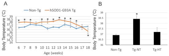
SOD1 mouse models are chronically feverish. At 6 weeks of age, the internal temperature of model SOD1 mice was significantly higher (38 ° C) compared to non-transgenic control mice (37 ° C). enter image description here

At 8 weeks of age, SOD1 mice were randomized into several groups. The SOD1 mouse model had a body temperature 1 to 2 ° C lower than the SOD1 mouse model not undergoing cooling. enter image description here

The precooling body temperatures among the SOD1 mouse model groups did not differ significantly. Warming of the mouse was slow and spontaneous at room temperature. At room temperature, motor activity was tested on a racing wheel with voluntary activity.

The onset of the disease was assessed quantitatively by a deficit of activity and descriptively by paresis of the hind limbs. To assess the effectiveness of hypothermia, a group of mice were euthanized before the final stage at 12 weeks of age.

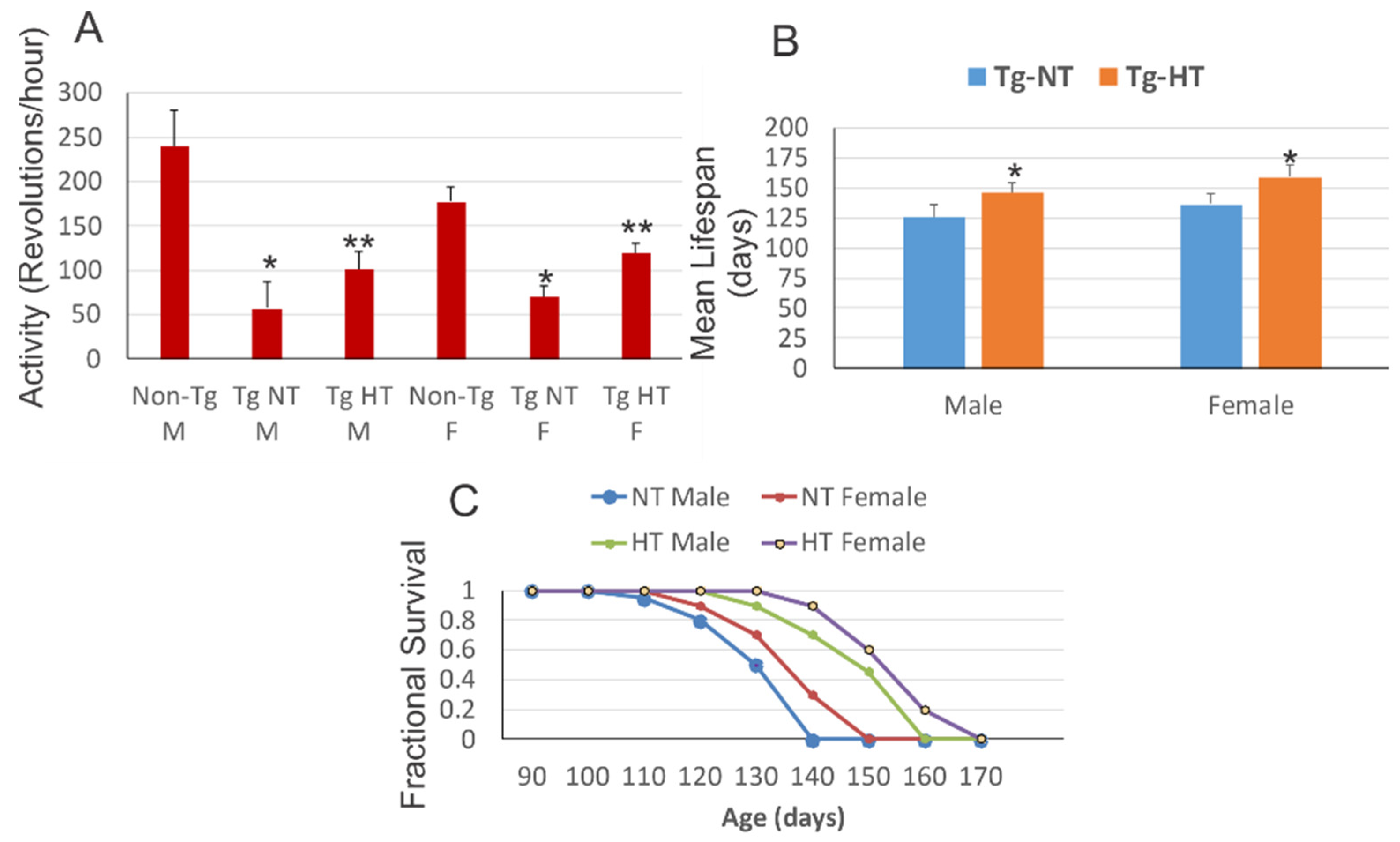
At 10 weeks of age, hypothermia improved neurological outcomes and survival in the SOD1 mouse model. Male and female SOD1 mouse models housed at room temperature exhibited a significant reduction in motor activity compared to sex-matched non-transgenic littermates. Cold acclimatized male SOD1 mice and female SOD1 mice showed significantly improved motor activity compared to the SOD1 mouse model maintained at normal temperature.

However, all groups of SOD1 mice exhibited significant motor deficits compared to non-transgenic mice of the same age. The mean lifespan of the cold acclimatized male SOD1 mouse model was significantly increased compared to the mouse model exposed to normal temperature. Cold acclimatization of the female SOD1 mouse model obtained a slightly better result on the significant increase in lifespan compared to the female SOD1 mouse model at normal temperature. 3.4.

The authors assessed hypothermic, normal-temperature SOD1 mice at 12 weeks of age for spinal cord neuropathology. Non-transgenic mice had evident spinal motor neurons with large multipolar cell bodies. In uncooled SOD1 mice, motor neurons were significantly 75% depleted and there was a secondary fulminant infiltration of small cells into the parenchyma. In cold acclimatized SOD1 mice, motor neurons were more apparent than uncooled mice.

Hypothermia rescued motor neuron counts in the lumbar spinal cord of the SOD1 mouse model (40% loss) compared to 80% loss in the uncooled SOD1 mice. Secondary small cell inflammation of the parenchyma of the spinal cord appeared to be reduced in cold acclimatized mice.

In uncooled SOD1 mice, the mitochondria were severely swollen, dysmorphic, and disrupted in motor neurons and in the neuropil. In hypothermic SOD1 mice, the mitochondria in the cell bodies of motor neurons were protected from swelling. This protection of motor neurons in SOD1 mice has been linked by an attenuation of inflammatory changes in the spinal cord.

In non-transgenic mice, diaphragm motor plate innervation was close to 100%, while in uncooled SOD1 mice, endplate innervation was significantly reduced to only about 40% at 12 weeks old.

In contrast, in cold acclimatized SOD1 mice, innervation of the neuromuscular junction was restored to about 65% of normal, but was still significantly reduced compared to diaphragm innervation from non-transgenic mice.

The cooled SOD1 mice exhibited a significant upregulation of the modified proteins HSP70, UCP3 and SUMO1 compared to the uncooled transgenic mice.

Cold acclimatization was more effective in females than in males at prolonging lifespan. The innervation of the diaphragm of the motor plates was improved by hypothermia.

Many circulating cytokines induce fever. This profile of inflammatory cytokines is consistent with their finding that ALS mice are febrile during the course of the disease. The main circulating proinflammatory pyrogenic cytokines in ALS mice are TNFα and IL6. TNFα and IL6 can cause muscle atrophy in various clinical settings. Skeletal muscle wasting is an important feature of human ALS and some mouse models of ALS.

The authors confirmed, which has been repeatedly reported, that during the course of the disease in the muscles of SOD1 mice, nitric oxide production is increased and protein nitration is elevated, y including key proteins at the neuromuscular junction.

Nitric oxide is an essential regulator of cell apoptosis. It can have an antiapoptotic effect, or, conversely, an apoptotic effect. This rocker is intimately linked to the presence or absence of cellular reducers such as glutathione.

In ALS, the thermoregulatory function could be aberrant. Hypothermia could act on the CNS, peripheral nervous system, skeletal muscle and body fat levels.

The clinical-translational application and efficacy testing of this concept in human ALS would be non-invasive and applicable, perhaps in rehabilitation spas in private and hospital settings. Cold water immersion or cryotherapy is common among athletes. Cold acclimatization protocols should be determined and empirically refined, and biomarkers of therapeutic efficacy should be identified for human ALS. Biomarkers could be based on skeletal muscle biopsy and assay for HSP70, protein sumoylation, and mPTP activation thresholds.

Advertisement


This book retraces the main achievements of ALS research over the last 30 years, presents the drugs under clinical trial, as well as ongoing research on future treatments likely to be able stop the disease in a few years and to provide a complete cure in a decade or two.

Hypothermie thérapeutique et SLA

- Posted by admin in Français

L’hypothermie thérapeutique est maintenant la norme de soins pour différentes conditions impliquant la mort cellulaire.

Les mécanismes d'action de l'hypothermie thérapeutique sont variés et mal compris. Ils comprennent des mécanismes dont on pense qu'ils sont aussi impliqués dans la SLA.

Pour autant la possibilité que l'hypothermie thérapeutique a une application à la SLA n'avait pas été examinée.

Lee J Martin, Mark V Niedzwiecki et Margaret Wong, les auteurs de l’article examiné aujourd’hui, ont donc testé dans un modèle murin transgénique de la SLA, l'hypothèse selon laquelle l'hypothermie légère chronique intermittente a une efficacité thérapeutique.

Les souris ont été observées deux fois puis 4 à 5 fois par jour au stade terminal de la maladie.

Les modèles de souris SOD1 sont chroniquement fébriles. À 6 semaines d'âge, la température interne des modèle de souris SOD1 était significativement plus élevé (38 °C) par rapport aux souris témoins non transgéniques (37 °C). enter image description here

À 8 semaines d'âge, les souris SOD1 ont été randomisés dans plusieurs groupes. Le modèle de souris SOD1 avait une température corporelle inférieure de 1 à 2 ° C par rapport au modèle de souris SOD1 ne subissant pas de refroidissement. enter image description here

Les températures corporelles de prérefroidissement parmi les groupes de modèles de souris SOD1 ne différaient pas de manière significative. Le réchauffement de la souris était lent et spontané à température ambiante. A température ambiante, l'activité motrice a été testée sur une roue de course à activité volontaire.

L'apparition de la maladie a été évaluée quantitativement par un déficit d'activité et de manière descriptive par la parésie des membres postérieurs. Pour évaluer l'efficacité de l'hypothermie, un groupe de souris a été euthanasié avant le stade final à l'âge de 12 semaines.

À 10 semaines d'âge, l'hypothermie a amélioré les résultats neurologiques et la survie du modèle de souris SOD1. Les modèles de souris SOD1 mâles et femelles logés à température ambiante présentaient une réduction significative de l'activité motrice par rapport aux compagnons de portée non transgéniques appariés au sexe. Les souris SOD1 mâles acclimatées au froid et les souris SOD1 femelles ont montré une activité motrice significativement améliorée par rapport au modèle de souris SOD1 maintenu à une température normale.

Cependant, tous les groupes de souris SOD1 présentaient des déficits moteurs significatifs par rapport aux souris non transgéniques du même âge. La durée de vie moyenne du modèle de souris SOD1 mâles acclimatées au froid a été augmentée de manière significative par rapport au modèle de souris exposé à une cempérature normale. L'acclimatation au froid du modèle de souris SOD1 femelles a obtenu un résultat légèrement meilleur sur l'augmentation significative de la durée de vie par rapport au modèle de souris SOD1 femelle à température normale. 3.4.

Les auteurs ont évalué les souris SOD1 en hypothermie et à température normale à 12 semaines d'âge pour la neuropathologie de la moelle épinière. Les souris non transgéniques avaient des neurones moteurs spinaux évidents avec de grands corps cellulaires multipolaires. Dans les souris SOD1 non refroidi, les motoneurones ont été épuisés de manière significative à 75% et il y avait une infiltration fulminante secondaire de petites cellules dans le parenchyme. Dans les souris SOD1 acclimatées au froid, les motoneurones étaient plus apparents que les souris non refroidies.

L'hypothermie a sauvé nombre de motoneurones dans la moelle épinière lombaire du modèle de souris SOD1 (perte de 40%) par rapport à la perte de 80% dans les souris SOD1 non refroidi. L'inflammation secondaire à petites cellules du parenchyme de la moelle épinière est apparue atténuée chez les souris acclimatées au froid.

Dans les souris SOD1 non refroidi, les mitochondries étaient sévèrement enflées, dysmorphiques et perturbées dans les motoneurones et dans le neuropile. Dans les souris SOD1 en hypothermie, les mitochondries dans les corps cellulaires des neurones moteurs ont été protégées du gonflement. Cette protection des motoneurones chez les souris SOD1 a été mise en parallèle par une atténuation des changements inflammatoires de la moelle épinière.

Chez les souris non transgéniques, l'innervation de la plaque motrice du diaphragme était proche de 100%, tandis que chez les souris SOD1 non refroidies, l'innervation de la plaque d'extrémité était réduite de manière significative à seulement environ 40% à l'âge de 12 semaines.

En revanche, dans les souris SOD1 acclimatées au froid, l'innervation de la jonction neuromusculaire a été restaurée à environ 65% de la normale, mais était encore significativement réduite par rapport à l'innervation du diaphragme de souris non transgénique.

Les souris SOD1 refroidies présentaient une régulation à la hausse significative des protéines HSP70, UCP3 et SUMO1 par rapport aux souris transgéniques non refroidies.

L'acclimatation au froid était plus efficace chez les femelles que chez les mâles pour prolonger la durée de vie. L'innervation du diaphragme des plateaux moteurs a été améliorée par l'hypothermie.

De nombreuses cytokines circulantes induisent une fièvre. Ce profil de cytokines inflammatoires est cohérent avec leur découverte selon laquelle les souris SLA sont fébriles au cours de l'évolution de la maladie. Les principales cytokines pyrogènes proinflammatoires circulantes chez les souris SLA sont le TNFα et l'IL6. Le TNFα et l'IL6 peuvent entraîner une atrophie musculaire dans divers contextes cliniques. La fonte musculaire squelettique est une caractéristique importante de la SLA humaine et de certains modèles murins de la SLA.

Les auteurs ont confirmé, ce qui avait été rapporté à plusieurs reprises, qu’au cours de l'évolution de la maladie dans les muscles de souris SOD1, la production de monoxyde d'azote est augmentée et la nitration des protéines est élevée, y compris les protéines clés au niveau de la jonction neuromusculaire.

Le monoxyde d'azote est un régulateur essentiel de l'apoptose cellulaire. Il peut avoir un effet antiapoptotique, ou, inversement, un effet apoptotique. Cette bascule est intimement liée à la présence ou non de réducteurs cellulaires tels de glutathion.

Lors de la SLA, la fonction thermorégulatrice pourrait être aberrante. L'hypothermie pourrait agir au niveau du SNC, du système nerveux périphérique, du muscle squelettique et des niveaux de graisse corporelle.

L'application clinique-translationnelle et les tests d'efficacité de ce concept dans la SLA humaine seraient non invasifs et applicables, peut-être dans des spas de réadaptation en milieu privé et hospitalier. L'immersion en eau froide ou la cryothérapie est courante chez les sportifs. Les protocoles d'acclimatation au froid devraient être déterminés et affinés empiriquement, et des biomarqueurs d'efficacité thérapeutique doivent être identifiés pour la SLA humaine. Les biomarqueurs pourraient être basés sur la biopsie de muscles squelettiques et le dosage de HSP70, de la sumoylation des protéines et des seuils d'activation du mPTP.

Publicité


Ce livre retrace les principales réalisations de la recherche sur la SLA au cours des 30 dernières années. Il présente les médicaments en cours d’essai clinique ainsi que les recherches en cours sur les futurs traitements susceptibles d’ici quelques années, d’arrêter la maladie et de fournir un traitement complet en une décennie ou deux.

Brain iron dyshomeostasis with iron accumulation is a known feature of brain aging. The cause of this iron accumulation is aging is unknown, but the immune system is less active in older people, and as viruses replicate more efficiently in iron-rich senescent cells, they may have developed the ability to induce this phenotype in aging host tissues.

It has been proposed several times that metal dyshomeostasis could be an underlying mechanism responsible for the initiation and progression of the pathological changes associated with neurodegenerative disorders, including the motor and extra-motor symptoms of ALS.

A new paper proposes an update of the iron hypothesis of Alzheimer's disease (Alzheimer's disease), based on large scale emerging evidence.

Iron featured historically early in Alzheimer's disease research efforts for its involvement in the amyloid and tau proteinopathies, yet iron neurochemistry remains peripheral in mainstream Alzheimer's disease research.

Much of the effort investigating iron in Alzheimer's disease has focused on the potential for iron to provoke the onset of disease, by promoting proteinopathy through increased protein expression, phosphorylation, and aggregation.

The authors provide new evidence from a large post mortem cohort that brain iron levels within the normal range are associated with accelerated ante mortem disease progression in cases with underlying proteinopathic neuropathology.

These results corroborate recent findings that argue for an additional downstream role for iron as an effector of neurodegeneration, acting independently of tau or amyloid pathologies.

The researchers hypothesize that the level of tissue iron is a trait that dictates the probability of neurodegeneration in Alzheimer's disease by ferroptosis.

Ferroptosis is a type of programmed cell death dependent on iron and characterized by the accumulation of lipid peroxides, and is genetically and biochemically distinct from other forms of regulated cell death such as apoptosis.

Ferroptosis is initiated by the failure of the glutathione-dependent antioxidant defenses, resulting in unchecked lipid peroxidation and eventual cell death. Lipophilic antioxidants and iron chelators can prevent ferroptotic cell death.

A substantial body of preclinical evidence and early clinical data has demonstrated that deferoxamine and other iron chelators have strong disease-modifying impacts in Alzheimer's disease, Parkinson's disease, ischemic stroke. Acting by the disease-nonspecific pathway of iron chelation, deferoxamine targets each of these complex diseases via multifactorial mechanisms.

Deferiprone another iron chelator is tested in clinical trials against ALS in France’s Lille CHU hospital.

Advertisement


This book retraces the main achievements of ALS research over the last 30 years, presents the drugs under clinical trial, as well as ongoing research on future treatments likely to be able stop the disease in a few years and to provide a complete cure in a decade or two.


Please, help us continue to provide valuable information: