Neuroanatomical correlates of genetic risk for obesity in children

- Posted by admin in English

Obesity has increased rapidly in recent decades to affect more than 2 billion people, making it one of the largest contributors to poor health worldwide. Despite decades of research on diet and exercise treatments, many people continue to struggle to lose weight.

Several teams think that molecular mechanisms of brain development during early life are likely a major determinant of obesity risk, in other words that obesity is a neurodevelopmental disorder.

Indeed obesity has a strong genetic component, with up to 20% of variance in body mass index (BMI) being accounted for by common polygenic variation. Most genetic polymorphisms associated with BMI are related to genes expressed in the central nervous system.

At the same time, higher BMI is associated with neurocognitive changes. However, the direct link between genetics of obesity and neurobehavioral mechanisms related to weight gain is missing. Here, authors from McGill University, Montréal, Canada, use a large sample of participants (n>4,000) from the Adolescent Brain Cognitive Development cohort to investigate how genetic risk for obesity, expressed as polygenic risk score for BMI (BMI-PRS), is related to brain and behavioral differences in adolescents.

In a series of analyses, the authors show that BMI-PRS is related to lower cortical volume and thickness in the frontal and temporal areas, relative to age-expected values.

Relatedly, using structural equation modeling, the authors find that lower overall cortical volume is associated with higher impulsivity, which in turn is related to an increase in BMI one year later.

In summay, authors' study shows that obesity might partially stem from genetic risk as expressed in brain changes in the frontal and temporal brain areas, and changes in impulsivity.

Indeed the risk for obesity among children with a high genetic susceptibility also varies with environmental and sociodemographic factors during childhood. While all children benefit from an environment that supports a healthy lifestyle, they are particularly important for children with a high genetic risk for obesity.

Read the original article on medRxiv

Results of Trial of Antisense Oligonucleotide Tofersen for SOD1 ALS

- Posted by admin in English

Tofersen is an investigational antisense oligonucleotide designed to reduce protein superoxide dismutase 1 (SOD1) synthesis through the degradation of SOD1 mRNA. This seems to me counterproductive for ALS patients, and facts seem to agree with me.

A phase 1 clinical trial (NCT01041222) tested four different doses of tofersen in 33 patients. The most common side effects were post-lumbar puncture syndrome, also known as spinal headache, injection-related back pain, and nausea. Subsequently, a second larger phase 3 trial (NCT02623699) named VALOR was initiated. Yet, in October 2021, it was announced that in this Phase 3 VALOR study, the primary endpoint measured by the Amyotrophic Lateral Sclerosis Functional Rating Scale-Revised (ALSFRS-R) did not reach statistical significance.

Surprisingly (or maybe unsurprisingly), the principal investigator stated that "The results from the VALOR study are encouraging as they show reduction of SOD1 protein, reduction of neurofilament, a potential biomarker for neurodegenerative disease, and positive signals across multiple key endpoints including measures of important aspects of the daily lives of SOD1-ALS patients”.

In particular, finding encouragement in the reduction of the SOD1 protein is bizarre at best. The SOD1 protein is what protects the central nervous system against the toxicity of metabolic end products. Indeed, in this case, these patients have SOD1 mutations, but why wasn't Torfersen engineered to modulate the mutated SOD1 gene by alternative splicing, functionally converting it to a normal SOD1 gene?

Another study was planned for 2022, which, as usual for recent ALS studies, was supported by famous ALS scientists such as Merit E Cudkowicz, Albert C. Ludolph or Pamela J Shaw.

In this phase 3 trial, the scientists randomly assigned adults with amyotrophic lateral sclerosis SOD1 in a 2:1 ratio to receive eight doses of tofersen or a placebo over a 24-week period. 
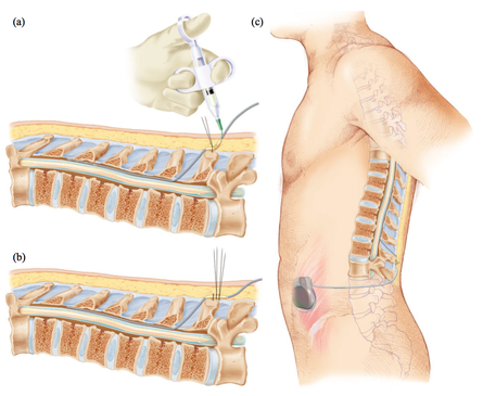
Eight doses is a lot when it's given intrathecally. Intrathecal drug delivery is the introduction of a therapeutic substance into the cerebrospinal fluid by injection into the subarachnoid space of the spinal cord to bypass the blood-brain barrier. It's an odd choice to deliver an ALS drug because it punctures the barrier that protects the central nervous system. We know that in 3% of cases, intrathecal administration of chemotherapy leads to paralysis! In this case, this barrier was perforated eight times, which means that the risk of paralysis is much higher. Serious neurological adverse events occurred in 7% of Tofersen recipients.

The primary endpoint was the change from baseline to week 28 in total score on the Revised Amyotrophic Lateral Sclerosis Functional Rating Scale among participants with more rapidly progressing disease predicted. Secondary endpoints included changes in total cerebrospinal fluid SOD1 protein concentration, plasma neurofilament light chain concentration, slow vital capacity and portable dynamometry in 16 muscles.

A combined analysis of the randomized component of the trial and its 52-week open-label extension compared outcomes in participants who started tofersen at entry into the trial with those in participants who switched from placebo to drug at week 28. A total of 72 participants received tofersen and 36 received placebo.

Morally and ethically, this means that 36 patients received eight intrathecal injections of placebo. Not only were they losing time, but they risked further health degradation by this procedure.

As in the first phase III clinical trial in the more rapidly progressing subgroup, the change at week 28 in the ALSFRS-R score (primary endpoint) was -6.98 with tofersen and -8.14 with the placebo. Administration of Tofersen also resulted in greater reductions in cerebrospinal fluid SOD1 and plasma neurofilament light chains than placebo, but overall results for secondary clinical endpoints did not differ significantly between the two groups.

At the end of the trial, 95 of the participants went on to open label extension which will last up to four and a half years. All expansion participants receive tofersen.

An analysis six months after the start of the extension found a significant difference in motor function between those who had been on tofersen from the start and those who had been on a placebo for six months before starting tofersen. After a year on the drug, the participants showed a stabilization of muscle strength and this is a remarkable finding, according to the researchers. Some scientists have even gone so far as to claim that "most of the course participants on our site regained and/or maintained a number of their activities of daily living". We see these kinds of marketing claims in all ALS clinical trials, but no one has ever encountered these lucky patients.

From a business perspective, it's hard to see Biogen's interest in Torfensen. The SOD1 gene is only mutated in about 2% of ALS patients, and there are hundreds of SOD1 mutations, so Torfensen, if effective, would be usable for less than 2% of patients.

And indeed Torfensen is not effective in ALS: If in 28 weeks the change is only 1.16 point, that means absolutely nothing in terms of improvement. Simply taking a new medication to make swallowing easier or wearing better-fitting clothing could improve the ALSFR by one or two points.

Biogen changed its strategy a few years ago in order to increase the chances of success of the clinical trials it funds. This was at a time when molecular biologists were promising wonders. Biogen could change strategy again. It would be a welcome change if human physiology were better considered in future studies.

Ce blog sur les maladies neurodégénératives assume implicitement que la médecine (classique ou non) est la seule voie qui permettra de guérir un jour de ces maladies. En particulier ce blog estime que la biologie moléculaire est au mieux un outil pour la médecine et au pire une impasse quand elle est uniquement utilisée dans l'optique de concevoir des médicaments. Les publications ayant une optique systémique ont donc un intérêt particulier pour nous.

La maladie de Parkinson est une maladie neurodégénérative courante caractérisée par une atteinte initiale du système nerveux autonome. Cela peut affecter le fonctionnement du cœur, de la vessie, des intestins, des glandes sudoripares, des pupilles et des vaisseaux sanguins. Cette atteinte initiale est suivie d'une dégénérescence nigrale, la substantia nigra est un acteur important du fonctionnement cérébral, en particulier des mouvements oculaires, de la planification motrice, de la recherche de récompenses, de l'apprentissage et de la dépendance.

Bien que l'innervation autonome affecte quasiment tous les organes, la façon dont la dysautonomie dans la maladie de Parkinson affecte le métabolisme des différents organes, n'a pas été identifiée. Dans cette étude, des scientifiques ont essayé d'estimer ce lien par des techniques de médecine nucléaire, une analyse trans-omique d'échantillons de sang et des expériences sur des cellules en culture.

Le rapport thyroïde-médiastin de la scintigraphie au MIBG a été mesuré chez 1 158 patients parkinsoniens. La scintigraphie au MIBG utilise une substance radioactive appelée iode méta-iodobenzylguanidine (MIBG) comme traceur de la circulation sanguine. La scintigraphie myocardique avec MIBG est un outil efficace pour distinguer la maladie de Parkinson des autres maladies accompagnées de parkinsonisme. Contrairement aux autres maladies parkinsoniennes, dans la maladie de Parkinson, l'accumulation de MIBG dans le cœur a tendance à diminuer.

En outre, une analyse du transcriptome des miARN de l'exosome sérique et une analyse du métabolome plasmatique suivie d'une analyse trans-omique ont été effectuées chez des patients atteints de la maladie de Parkinson de novo et des personnes témoins en bonne santé appariées selon l'âge. De plus, l'hormone thyroïdienne a été administrée à des cellules dérivées du muscle squelettique et du foie pour évaluer l'effet de l'hypothyroïdie sur ces organes.

Les chercheurs ont observé que a dénervation sympathique de la thyroïde corrélée à sa dénervation cardiaque était bien confirmée chez 1158 patients parkinsoniens par scintigraphie MIBG. Chez les patients atteints de maladie de Parkinson ne prenant pas de médicaments, une analyse complète du métabolome a révélé une diminution des niveaux de thyroxine et une β-oxydation insuffisante des acides gras, qui sont positivement corrélés les uns aux autres.

De même, les données du métabolome plasmatique et les données du transcriptome des miARN exosomaux circulants ont révélé un enrichissement spécifique de l'axe du récepteur activé par les proliférateurs de peroxysomes (PPARα). L'expression de PPAR-α est la plus élevée dans les tissus qui oxydent les acides gras à un rythme rapide. Chez les humains PPAR-α a une expression élevée dans le foie, l'intestin, le cœur, la thyroide et les reins. Enfin, l'association de l'hormone thyroïdienne avec la régulation de la β-oxydation dépendante de PPARα a été confirmée par des expériences in vitro.

Ces résultats suggèrent que les communications inter-organes entre la thyroïde et le foie sont désorganisées au stade précoce de la maladie de Parkinson. Cela pourrait suggérer de nouvelle voies médicamenteuses.

Accéder à l'article via Pubmed

The predominantly pre-synaptic intrinsically disordered protein α-synuclein is prone to misfolding and aggregation in synucleinopathies, such as Parkinson's disease and Dementia with Lewy bodies. For example molecules of the chaperone machinery are often deposited in Lewy bodies.

A new publication describes a vicious cycle in which parts of a chaperone facilitate the accumulation of toxic α-synuclein, which induces proteostatic stress that itself leads to an increase in insoluble fragments of the chaperone.

Molecular chaperones (proteins that assist the conformational folding or unfolding of large proteins) play important roles in protein misfolding diseases. Heat shock proteins are chaperones that protect cells when stressed by elevated temperatures. Heat shock protein 90 (Hsp90) is one of the most common of those chaperone proteins.

In this new publication, authors from Canada, Brazil and Israel show that STI1, the Hsp90 co-chaperone (proteins that assist chaperones ) co-immunoprecipitated α-synuclein, and co-deposited with Hsp90 and Hsp70 in phosphorylated α-synuclein in ubiquitin-positive inclusions in two mouse models of α-synuclein misfolding.

In Parkinson disease human brains, STI1 was increased, and in neurologically healthy brains, STI1 and α-synuclein location correlated. Nuclear Magnetic Resonance analyses revealed direct interaction of α-synuclein with STI1 and indicated that the STI1 domain ( a region of a protein's polypeptide chain that is self-stabilizing and that folds independently from the rest) TPR2A, but not TPR1 or TPR2B domains, interacted with the C-terminal domain of α-synuclein.

Mice over-expressing STI1 and Hsp90ß presented elevated α-synuclein S129 phosphorylation accompanied by inclusions when injected with α-synuclein pre-formed fibrils.

In contrast, reduced STI1 function decreased protein inclusion formation, S129 α-synuclein phosphorylation, while mitigating motor and cognitive deficits as well as mesoscopic brain atrophy in α-synuclein-over-expressing mice.

In conclusion the authors' findings reveal a vicious cycle in which STI1 facilitates the generation and accumulation of toxic α-synuclein conformers, while α-synuclein-induced proteostatic stress increased insoluble STI1 and Hsp90.

Read the original article on Pubmed

This new publication discusses a future (small) clinical trial (NCT05110053) of spinal cord stimulation therapy for patients with Parkinson's disease. Spinal cord stimulation is not new, there are even devices on the market for this purpose. enter image description here The imaging analyzes that this study will produce, will make it possible to define a subgroup of patients with Parkinson's disease who will have benefited from the treatment and will help to define rules about when using this therapy in order to avoid unnecessary interventions.

Parkinson's disease is a chronic neurodegenerative disease that affects nearly 8 million people worldwide. Parkinson's disease manifested by the classic triad bradykinesia (Slowness of initiation of movement (slowness of initiation of voluntary movement), rigidity and tremor. These symptoms can, at least in the early stages of the disease, be treated effectively with dopamine replacement therapy, however, as the disease progresses, more debilitating symptoms appear, including gait problems, postural instability, and falls.

Unfortunately, the onset of these symptoms represents a major step in the progression of Parkinson's disease, resulting in loss of autonomy, deterioration in quality of life and a marked increase in mortality. These disabling symptoms often respond poorly to dopamine medications and advanced therapies, including deep brain stimulation of the subthalamic nucleus (DBS). enter image description here Deep brain stimulation (DBS) is a neurosurgical procedure involving the placement of a medical device called a neurostimulator, which sends electrical impulses, via implanted electrodes, to specific targets in the brain (the cerebral nucleus) for treatment movement disorders, including Parkinson's disease. illness, essential tremor, dystonia, and other conditions such as obsessive-compulsive disorder (OCD) and epilepsy. Its underlying principles and mechanisms are not fully understood.

Other stimulation methods have been considered by other teams such as the use of infrared, ultrasound (Magnetic resonance-guided focused ultrasound) or low-frequency sounds, or magnetic fields (Transcranial Current Magnetic Stimulation) or electric current continuous or alternating (Transcranial Current Stimulation), or even radio frequencies.

Spinal cord stimulation is a surgical treatment used as a treatment for chronic neuropathic pain that is unresponsive to other conventional treatments. Several studies have shown improved walking function in patients with Parkinson's disease following spinal cord stimulation for back pain. More recently, a small number of Parkinson's disease patients with gait dysfunction (without back pain) have been treated with encouraging initial results on gait function and with few adverse events.

Spinal cord stimulation assumes that by delivering electrical current at a certain frequency, intensity, latency and specific location, the physiological functioning of targeted areas of the spinal nerve can be restored. The most common complication of spinal cord stimulation is related to lead migration, followed by infections which, sooner or later, could lead to new surgeries. CSF leak and device failure are less common complications.

The method involves introducing one or more electrodes into the epidural space through which electrical impulses are transmitted into the epidural space. The electrodes are connected to a neurostimulator placed under the skin of the abdomen. The contact between the electrodes and the neurostimulator leads to the stimulation of the posterior parts of the spinal cord and the patient then feels a "tingling sensation", where he felt intense pain. In this therapy, in which electrical impulses prevent or relieve the sensation of pain, no nerves are damaged. In addition, with a single movement of the hand, the patient can turn the device on and off, as well as regulate the force in order to obtain the desired stimulation.

This future spinal cord stimulation clinical trial, which is being planned for patients with Parkinson's disease (STEP-PD), aims to assess the safety and feasibility of burst spinal cord stimulation as a treatment gait disorders in the Parkinson's disease.

This trial will investigate possible changes after spinal cord stimulation in cholinergic activity and glucose metabolic patterns of cortex and associative cortical-subcortical loops with positron emission tomography.

A total of 14 patients will be assessed using clinical rating scales and gait assessments at baseline, and at 6 and 12 months after spinal cord stimulation implantation. They will also receive serial 18F-deoxyglucose and PET scans to assess the effects of spinal cord stimulation on cortical/subcortical activity and brain cholinergic function.

The first two patients will be included in an open-label pilot study while the others will be randomized to receive active treatment or placebo (no stimulation) for 6 months. From then on, the entire cohort will enter an open-label active treatment phase for 6 months.

Trial registration number: NCT05110053

Read the original article on Pubmed

Cette nouvelle publication traite d'un futur (petit) essai clinique (NCT05110053) sur la thérapie de stimulation de la moelle épinière pour les patients atteints de la maladie de Parkinson. La stimulation de la moelle épinière n'est pas nouvelle, il existe même sur le marché des dispositifs commercialisés à cet effet. enter image description here Les analyses d'imagerie de cette étude permettront de définir un sous-groupe de patients atteints de maladie de Parkinson qui auront bénéficié du traitement et ainsi de définir des règles quand à l'utilisation de cette thérapie, afin d'éviter des interventions inutiles.

La maladie de Parkinson est une maladie neurodégénérative chronique qui touche près de 8 millions de personnes dans le monde. La maladie de Parkinson se manifestant par la triade classique bradykinésie (Lenteur de l'initiation du mouvement (lenteur de l'initiation du mouvement volontaire), rigidité et tremblement. Ces symptômes peuvent, au moins dans les premiers stades de la maladie, être traités efficacement par une thérapie de remplacement de la dopamine. Cependant, à mesure que la maladie progresse, des symptômes plus débilitants apparaissent, notamment des problèmes de démarche, une instabilité posturale et des chutes.

Malheureusement, la survenue de ces symptômes représente une étape majeure dans la progression de la maladie de Parkinson, entraînant une perte d'autonomie, une détérioration de la qualité de vie et une augmentation marquée de la mortalité. Ces symptômes invalidants répondent souvent mal aux médicaments dopaminergiques et aux thérapies avancées, y compris la stimulation cérébrale profonde du noyau sous-thalamique (DBS). enter image description here La stimulation cérébrale profonde (DBS) est une procédure neurochirurgicale impliquant la mise en place d'un dispositif médical appelé neurostimulateur, qui envoie des impulsions électriques, via des électrodes implantées, à des cibles spécifiques dans le cerveau (le noyau cérébral) pour le traitement des troubles du mouvement, y compris la maladie de Parkinson. maladie, tremblement essentiel, dystonie, et d'autres conditions telles que le trouble obsessionnel-compulsif (TOC) et l'épilepsie. Ses principes et mécanismes sous-jacents ne sont pas entièrement compris. D'autres méthodes de stimulations comme l'utilisation d'infra-rouges, d'ultra-sons (Magnetic resonance-guided focused ultrasound ) ou de sons à basse fréquence, ou de champs magnétiques (Transcranial Current Magnetic Stimulation) ou électriques à courant continue ou alternatif (Transcranial Current Stimulation), ou encore de radio-fréquences ont été considérés.

La stimulation de la moelle épinière est un traitement chirurgical utilisé comme traitement des douleurs neuropathiques chroniques ne répondant pas aux autres traitements conventionnels. Plusieurs études ont montré une amélioration de la fonction de marche chez les patients atteints de maladie de Parkinson suite à une stimulation de la moelle épinière pour douleur dorsale. Plus récemment, un petit nombre de patients atteints de maladie de Parkinson avec un dysfonctionnement de la marche (sans douleur dorsale) ont été traités avec des résultats initiaux encourageants sur la fonction de marche et avec peu de événements indésirables.

La stimulation de la moelle épinière suppose qu'en délivrant un courant électrique à une certaine fréquence, intensité, latence et localisation spécifique, le fonctionnement physiologique des zones ciblées du nerf spinal peut être rétabli grâce à l'action neuromodulatrice. La complication la plus fréquente du stimulation de la moelle épinière est liée à la migration des dérivations, en particulier dans les dérivations quadripolaires, suivie d'infections qui, tôt ou tard, pourraient entraîner des réinterventions. La fuite de LCR et la défaillance du dispositif sont des complications moins courantes.

La méthode consiste à introduire une ou plusieurs électrodes dans l'espace épidural par lesquelles des impulsions électriques sont transmises dans l'espace épidural. Les électrodes sont reliées à un neurostimulateur ou un neuromodulateur, placé sous la peau de l'abdomen. Le contact entre les électrodes et le neurostimulateur entraîne la stimulation des parties postérieures de la moelle épinière et le patient ressent alors une "sensation de picotement", là où il ressentait une douleur intense. Dans cette thérapie, dans laquelle les impulsions électriques empêchent ou soulagent la sensation de douleur, aucun nerf n'est endommagé. En outre, d'un seul mouvement de la main, le patient peut allumer et éteindre l'appareil, ainsi que réguler la force afin d'obtenir la stimulation souhaitée.

L'essai clinique de thérapie de stimulation de la moelle épinière, qui est projeté pour les patients atteints de la maladie de Parkinson (STEP-PD), vise à évaluer l'innocuité et la faisabilité du stimulation de la moelle épinière en rafale comme traitement des troubles de la marche dans la maladie de Parkinson, tels que FoG. Cet essai étudiera les changements possibles après stimulation de la moelle épinière dans l'activité cholinergique et les schémas métaboliques du glucose du cortex et des boucles cortico-sous-corticales associatives avec tomographie par émission de positrons. Un total de 14 patients seront évalués à l'aide d'échelles d'évaluation cliniques et d'évaluations de la marche au départ, ainsi qu'à 6 et 12 mois après l'implantation de la stimulation de la moelle épinière. Ils recevront également des scans en série au 18F-désoxyglucose et au 18FEOBV PET pour évaluer les effets du stimulation de la moelle épinière sur l'activité corticale/sous-corticale et la fonction cholinergique cérébrale. Les deux premiers patients seront inclus dans une étude pilote ouverte tandis que les autres seront randomisés pour recevoir un traitement actif ou un placebo (pas de stimulation) pendant 6 mois. À partir de ce moment, l'ensemble de la cohorte entrera dans une phase de traitement actif en ouvert pendant 6 mois.

Trial registration number: NCT05110053

Read the original article on Pubmed

Resting motor threshold is the minimum intensity that evoked a visible contralateral involuntary finger twitch.

Resting motor threshold asymmetry is the absolute difference between the left and right RMT measurements.

Conflicting results have emerged from studies examining the potential of resting motor threshold as a neurophysiological marker for Alzheimer's disease diagnosis and progression.

In this study, the authors estimated the strength of the association between Resting motor threshold measurements and severity of cognitive impairment in a relatively large sample of clinical trial participants with mild to moderate Alzheimer's disease.

Resting motor threshold for each participant was determined by applying single-pulse transcranial magnetic stimulation repeated at varying intensities over left and right sides of the primary motor cortex.

Cognitive impairment was measured with the Montreal Cognitive Assessment and the Alzheimer Disease Assessment Scale - Cognitive scores.

Although the left and right resting motor threshold was lower in CDR 2 than in CDR 1 participants, neither RMT nor RMT asymmetry correlated significantly with cognitive test scores.

In conclusion, authors' study in a large sample size does not support the idea that resting motor threshold is a sensitive marker of cognitive decline/severity in Alzheimer's disease.

Read the original article on Pubmed

Alcohol Intake and dementias

- Posted by admin in English

enter image description here Alzheimer's disease (Alzheimer's disease) is progressive brain disease that affects cognition, memory and behavior.

TDP-43 limbic-predominantly age-related encephalopathy (LATE) is a recently defined common neurodegenerative disorder that mimics the clinical symptoms of Alzheimer's disease.

LATE is a very common condition, typically it affects one third of people older than 75 years of age. This is in contrast to Alzheimer's disease pathology, which tends to level off and perhaps decrease in prevalence among persons beyond age 85 years.

TDP-43 is the major disease protein found in frontotemporal dementia and in amyotrophic lateral sclerosis.

At present, the risk factors involved in LATE and those that differentiate it from Alzheimer's disease are largely unknown.

Managing cardiovascular risk factors, maintaining an active lifestyle, and eating a balanced diet are associated with a reduced risk of AD or a lower rate of cognitive decline. Alcohol consumption is widespread and socially encouraged and is rarely associated with neurodegenerative diseases.

However, alcoholism leads to brain atrophy, and long-term alcohol use can damage almost every organ and system in the body. enter image description here

Authors of a new article used an algorithmic approach to identify important factors that distinguish patients with LATE and/or Alzheimer's disease from controls with significantly imbalanced data.

They analyzed two ROSMAP and NACC datasets and found that lifetime alcohol consumption was one of the most important lifestyle and environmental factors associated with LATE and Alzheimer's disease, and that their associations varied. In particular, the authors identified a specific subpopulation consisting of APOE-e4 carriers.

The authors found that for this subpopulation, light to moderate alcohol consumption was a protective factor against Alzheimer's disease and LATE, but its protective function against Alzheimer's disease appeared to be stronger than LATE.

The codes for authors' algorithms will be (hopefully) available at https://github.com/xinxingwu-uk/PFV.


Please, help us continue to provide valuable information: