A link between a protein called IL-11 and aging in mice.

- Posted by admin in English

A new publication presents work on the link between a protein called IL-11 and aging in mice.

These studies identify IL11 as a key inflammatory factor and therapeutic target for mammalian health. What is new is that this effect is positive even for elderly subjects and for both sexes. enter image description here IL11 belongs to the IL6 family of cytokines, which bind to alpha receptors and the gp130 coreceptor to initiate intracellular signaling via JAK/STAT. This family is evolutionarily ancient, with homologs found in ascidians and fish.

The authors found that IL-11 levels increase with age and that inhibition of IL-11 provides several benefits, including improved metabolism, muscle function, and duration of life. Inhibition also appears to reduce age-related cancers.

Researchers believe that inhibiting IL-11 could be a promising approach to extending human lifespan and health, especially since drugs targeting IL-11 are already being tested for safety in the framework of clinical trials.

Initially perceived as anti-fibrotic, anti-inflammatory, and pro-regenerative, IL11 could be pro-fibrotic, pro-inflammatory, and anti-regenerative. Two misinterpretations of previous studies have shaped this incorrect understanding.

Identified in 1990 as a factor secreted by bone marrow cells, IL11 appeared to be synergistic with IL3 for the formation of megakaryocytes and the increase in platelets. This belief was validated by studies using recombinant human IL11 (rhIL11), leading to its approval by the FDA in 1998 to treat thrombocytopenia. However, as early as 1997, studies suggested that IL11 had primary functions other than hematopoiesis.

The putative role of IL11 as a hematopoietic factor has led to the development of rhIL11 as an antifibrotic, anti-inflammatory, and pro-regenerative in murine models. Clinical trials have been carried out on various diseases, but without conclusive success, suggesting a lack of effectiveness or significant toxicities.

In 2016, IL11 was shown to activate fibrogenic proteins in human fibroblasts, causing a disease: Fibrosis. This finding contradicted 20 years of data showing beneficial effects. rhIL11 appeared to inhibit the function of endogenous IL11 in mice, explaining the beneficial effects observed in previous studies. The authors of these studies did not understand that the observed effects reflected rhIL11-mediated inhibition of endogenous IL11.

From the beginning of IL11 studies, it was observed that IL11 is upregulated in tissues of aged rodents. This discovery spurred a six-year (2017-2023) study of IL11 in terms of lifespan and health. During this period, it became clear that IL11 is part of the senescence-associated secretory phenotype and can directly stimulate senescence in lung fibroblasts and epithelial cells.

Serum levels of IL11 are increased in very elderly people. Healthspan studies have identified IL11 as an inflammatory factor responsible for ERK/mTOR-mediated sarcopenia, metabolic dysfunction, and frailty in aged mice while showing that IL11 inhibition increases the mouse's health duration.

Chronic inflammation is an important feature of aging, intimately linked to senescence and implicated in the pathogenesis of age-related frailty, metabolic dysfunction, and multimorbidity. Studies in invertebrates have shown that innate immune signaling, including Jak-Stat signaling in fly adipose tissue, can impair metabolism and lifespan. The relative contributions of canonical (JAK–STAT3) and noncanonical (MEK–ERK) IL-11 signaling, alone or in combination, to aging phenotypes remain to be determined.

Inhibition of ERK or mTOR or activation of AMPK by trametinib, rapamycin, or metformin, respectively, increases lifespan in model organisms and some advocate the use of these drugs in humans. However, sometimes these agents have detrimental, effects on health and inflammation.

As mice age, IL11 gradually appears in the liver, skeletal muscle, and fat. to stimulate an ERK/AMPK/mTORC1 axis of cellular, tissue, and organism aging pathologies. In aged mice, the deletion of Il11 protects against metabolic multimorbidity, sarcopenia, and frailty. Administering anti-IL11 treatment to aged mice for six months reactivates an age-repressed white fat loss program, reverses metabolic dysfunction, restores muscle function, and reduces frailty. In all cases studied, IL11 inhibition lowers epigenetic age, reduces telomere attrition, and preserves mitochondrial function.

The metabolic effects observed with IL-11 inhibition in aged mice resemble those of young mice with white adipose-specific Raptor deletion. White adipose tissue (fat) and brown adipose tissue are the two main types of adipose tissue in mammals. The authors speculate that inhibition of IL-11 prevents mTORC1 activation in fat, thereby decreasing the amount of white adipose tissue. However, the authors did not identify the mechanism leading to weight loss with IL-11 inhibition, which in further studies could inform some of the present findings.

Beyond metabolism, IL-11 inhibition ameliorated deterministic features of aging common in vertebrates (such as frailty and sarcopenia), demonstrating generic anti-aging benefits at the organismal level. Intriguingly, some of the beneficial effects of germline Il11ra1 or Il11 deletion, notably on muscle and fat, were apparent even in young mice. The authors did not see any specificity of this effect to certain tissues.

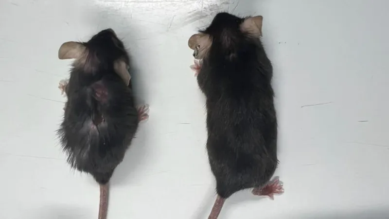
Inhibition of IL-11 increased lifespan in both male and female mice. The extent of the increase in lifespan remains unclear, but current data suggest that anti-IL-11 treatment given late in life increases median lifespan by more than 20% in both sexes. In these experiments, anti-IL-11 was injected into mice at 75 weeks (human equivalent to approximately 55 years).

Treatment with IL-11 is therefore effective in prolonging lifespan, as is the case for rapamycin. The mortality of mice in the elderly is often linked to cancer and the end-of-life autopsy data carried out by the authors support the idea that inhibition of IL-11 considerably reduces cancers linked to 'age. Of note, IL-11 is often linked to tumor onset and immune evasion. Clinical trials of anti-IL-11 in combination with immunotherapy to treat cancer are already planned.

As a first step toward creating a drug, the authors designed a high-affinity humanized neutralizing IL11 antibody.

It should be noted that some of the authors have strong links with companies that have acquired the patents resulting from this work or financed this work.

Also, the green and yellow vegetables and fruits, such as leafy greens, herbs, broccoli, peas, green bell peppers, and squash, are relatively rich sources of the oxycarotenoids lutein and zeaxanthin which may prevent the overexpression of IL-11 and ERK signaling
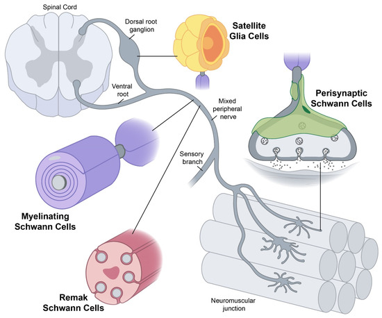
This post is about a new article about the means to rejuvenate organisms, which indeed has important implications for neurodegenerative diseases.

Telomerase restores short bits of DNA known as telomeres, which are otherwise shortened after repeated division of a cell via mitosis. In normal circumstances, where telomerase is absent, a cell can't divide in daughters indefinitely. When it reaches the Hayflick limit the cells become senescent and cell division stops. Telomerase allows each daughter cell to replace the lost bit of DNA, allowing the cell line to divide without ever reaching the limit. This same unbounded growth is a feature of cancerous growth. enter image description here Telomerase reverse transcriptase (abbreviated to TERT, or hTERT in humans) is a subunit of the enzyme telomerase. TERT inhibition has been linked directly or indirectly to all hallmarks of aging, but TERT expression is linked to the development of many cancers. Unfortunately, TERT gene is epigenetically repressed with the onset of aging markers in all tissues.

Researchers at the University of Texas MD Anderson Cancer Center have shown they can therapeutically restore "youthful" levels of TERT and this can significantly reduce the signs and symptoms of aging in preclinical models (primary human cells and naturally aged mice).

Maintenance of TERT levels in aged lab models reduced cellular senescence and tissue inflammation, spurred new neuron formation with improved memory, and enhanced neuromuscular function, which increased strength and coordination.

In the brain, scientists report that a new therapy (TAC) alleviates neuroinflammation, increases neurotrophic factors, stimulates adult neurogenesis, and preserves cognitive function without evident toxicity, including cancer risk. This is probably too beautiful to be entirely true. Enhancing TERT expression notoriously promotes cancer and some cancer drugs downregulate TERT transcription, inhibiting telomerase activity and TERT expression.

To find a suitable compound, the authors used a high-throughput screen of over 650,000 compounds. TAC restores TERT levels to promote telomere maintenance and reprogram gene expression.

The usual suspects enhance TERT levels: Physical activity, good diet, and resveratrol.

It can also be done with a genetic therapy: Scientists insert a specific DNA sequence into a safe and predictable location on the genome to insert new genes. The new DNA fragment includes, for example, a "loxP-flanked stop cassette." This cassette acts like a switch that can be turned on or off. Under the control of an enzyme, for example, the "Cre recombinase" the "loxP" sites in the inserted DNA remove the stop cassette, allowing the TERT gene to be expressed in the targeted cells.

If these findings are confirmed in clinical studies, there may be major therapeutic implications for age-related diseases such as Alzheimer's, Parkinson's, heart disease and cancer.

Are ALS or aging caused by a ribosomopathy?

- Posted by admin in English

In neurodegenerative diseases, dozens of hypotheses about etiology abound, but there are few data to support any of them; in fact, conflicting data abound. Instead of saying "We don't know," scientists use ambiguous phrases such as "It is a multistep etiology involving both genetic and environmental factors."

A little-known hypothesis is that ALS is a ribosomopathy. Ribosomes are the little machines that assemble proteins from amino acids, according to instructions provided by an mRNA frag.

In an article published in Molecular Cell, a team led by Óscar Fernández-Capetillo presents evidence indicating that the accumulation in cells of proteins linked to the assembly of ribosomes prevents the cell from functioning properly and this could have a link with aging and the onset of ALS. enter image description here Ribosomopathies encompass a wide range of syndromes. Common symptoms include a reduced number of blood cells, predisposition to cancer, skeletal abnormalities, and failure to thrive. Ribosomopathy has been associated with skeletal muscle atrophy, which brings us closer to ALS.

Neurological abnormalities are also observed, although less frequently. The link with certain genetic defects in ALS, such as C9orf72, is seemingly obvious: peptide repeats (DPR) originate from protein assembly defects. See for example this study or this one.

Yet, a complete understanding of the mechanism of toxicity of these DPRs is still lacking.

Apparently, the link with misfolded and poorly localized protein aggregates is less obvious. One study, however, showed that partially functional ribosomes might be prone to making mistakes and therefore producing misfolded proteins.

Nucleoli are large, membrane-less nuclear organelles known for their central role in ribosome biogenesis. Cells respond to growth signals, such as growth factors, by adjusting ribosome biogenesis to match the protein production required for growth. During conditions of cellular stress, such as heat shock or DNA damage, ribosome biogenesis may be temporarily reduced as the cell prioritizes other essential processes.

Ribosomes have a limited lifespan and degrade over time. Continuous ribosome biogenesis ensures a constant supply of functional ribosomes to meet the protein production needs of the cell. Although not included among the “characteristics of aging,” there is evidence that could show a role for nucleoli in aging. It has indeed been demonstrated that the size of nucleoli is inversely correlated with the lifespan of several organisms.

For the authors of the article, the accumulation of ribosomal proteins without ribosomes (R protein) in nucleoli is not limited to ribosomopathies but rather is a common outcome that occurs in response to nucleolar stress, regardless of the assault. The authors suggest the use of mTOR inhibitors as a strategy to counteract the toxicity of nucleolar stress. They are nevertheless aware of the limitations of inhibiting mTOR function in humans, as its effects are not limited to autophagy activation but also impair other important aspects such as insulin signaling or growth factors.

There is therefore a clear need to identify more selective means of stimulating the clearance of free R proteins and the identification of specific ribophagy inducers appears to be an interesting alternative.

To study the consequences of nucleolar stress in an animal model, the authors generated mice allowing generalized expression of (PR)n peptides.

The cause of death of PRKI/KI mice was the onset of an aging phenotype, as evidenced by the appearance of cataracts, hair aging, kyphosis, weight loss, skin thinning, and replacement of hematopoietic cells in the bone marrow with adipose cells. fabric. Thus for scientists, the systemic expression of (PR)n peptides leads to a generalized accumulation of nucleolar stress and accelerates aging in mice. However, contrary to what the university's PR kit suggests, this does not imply that aging is due to the accumulation of "unwanted proteins” in the nucleoli. Additionally, it's well known that any form of severe and prolonged stress in a mammal will induce the appearance of aging.

The link made with C9orf72 type ALS is even more tenuous, it is just analyses carried out on an online database, NeuroLINCS, showing an increase in the mTOR pathway and R proteins on human motor neurons derived from induced pluripotent stem cells from patients with C9orf72 type ALS.

The scientists used cancer cell lines whose biology is as foreign as possible to normal cells and generated their own mouse model, which does not help in reproducing the results. The use of commercial animal models, despite several disadvantages, at least guarantees better replicability. It is unclear whether these disease models have any connection to ALS C9orf72 or aging, so any claims in these areas should be taken with a grain of salt.

As there is little interesting news in neurodegenerative research, here is a low-quality review on one of my favorite topics and possibly a better article. You may remember that in early 2019 I made a plea for a TDP-43 genetic therapy. I wrote to roughly 250 scientists in this field, and only a handful responded. I may have absolutely no influence, but it seems to me there was more research in this area in the following years.

TDP-43 proteinopathies are pathological hallmarks in ALS/FTD. In general, aging is a risk factor for ALS/FTD adult-onset neurodegenerative diseases (NDD), as with aging comes protein misfolding and accumulation. Academics differentiate these diseases from the type of protein (or protein fragment) and the localization. In patients these distinctions do not hold: Every aging person has misfolded mislocated proteins of several kinds at the same time, so there is no pure sporadic Alzheimer's or Parkinson's diseases, or ALS. For example, scientists tell of three forms of demence that are linked to TDP-43 (FTD/FTLD, Limbic-predominant age-related TDP43 encephalopathy (LATE), and Hippocampal sclerosis of aging).

Now I think that abnormal cellular stress response, including ER stress, causes misfolded, mislocated protein aggregation so removing those deleterious protein aggregates would not really help. Just look at the 20 years of drug trials to remove amyloïds from the brains of Alzheimer patients.

Yet in this review they are still in this mindset, but they do not look after a genetic therapy, instead, the scientists explore small molecule-based approaches to enhance the clearance of pathological TDP-43.

Small molecule-based approaches are easier to implement and anyway, it fits well the academic agenda: To publish low-cost papers during one semester. enter image description here There is nothing new in the list of drugs the authors list, and they lump in the same basket many unrelated drugs.

Another article is more interesting.

The authors recognize that endoplasmic reticulum (ER) is crucial for maintaining cellular homeostasis, and the synthesis and folding of proteins and lipids. The ER is the ordered membranous network and the first compartment of the secretory pathway, which is responsible for the synthesis, modification, and delivery of biologically active proteins to their proper target sites within the cell cytoplasm and the extracellular milieu. The ER is the entry site for the majority of proteins processed in the secretory pathway. If the influx of nascent, unfolded polypeptides exceeds the folding and/or processing capacity of the ER, the normal physiological state of the ER is perturbed. ER is sensitive to stresses including viral infection, it reduces its processing capability, and results in the accumulation of unfolded/misfolded proteins.

Generation of ER stress and induction of the unfolded protein response (UPR) are generic host responses to flavivirus infection as virus replication occurs in close association with ER-derived membranes. Flaviviruses include Dengue virus, Zika, and Japanese Encephalitis virus (JEV).

Then the authors boldly link UPR activation to inflammation. Then they tell that CXCR3 in linked to inflammation. They then assume that inhibiting CXCR3 would be somewhat beneficial to alleviate UPR activity, and therefore be beneficial to patients. I have a hard time finding this as an example of clear logical reasoning, but at least these guys have done some experiments.

They investigated the molecular insights of replication and inflammation upon Japanese encephalitis(JEV) infection and how AMG487 is rescuing it. In response to this, the authors studied the UPR activation pathways leading to ER stress and the rescue effect of AMG487. AMG487 treatment decreased the phosphorylation of eIF2α, a unique ER stress marker. AMG487 treatment improved ROS-induced inflammation and cell death in JEV-infected mice brains. It's already known that Antagonists to CXCR3 including AMG487 can be useful for treatment against flaviviruses, the novelty here is that AMG487 might decrease UPR activity.

Une nouvelle publication sur la maladie d'Alzheimer se concentre sur le rôle de l'édition de l'ARN au niveau du site Q/R de GluA2 dans la régulation du nombre d'épines dendritiques des neurones et ses implications sur le déclin cognitif dans la maladie d'Alzheimer.

Une épine dendritique est une excroissance de la membrane des dendrites des neurones. Les dendrites sont des prolongements du corps cellulaire des neurones. Les épines dendritiques reçoivent les contacts synaptiques des axones des neurones présynaptiques. Bien que ces contacts puissent se faire directement sur la branche d'un dendrite, une épine dendritique constitue un espace physiquement délimité, possédant ses propres microdomaines et protéines de régulation. enter image description here Curtiss Neveu via Wikipedia

Les déséquilibres calciques ont longtemps été associée à la maladie d'Alzheimer, en particulier à « l'hypothèse du calcium ». Il y a de nombreuses hypothèses à propos de la maladie d'Alzheimer, celle-ci n'a pas la faveur d'une majorité de scientifiques.

On retrouve cette même hypothèse pour d'autres maladies neurodégénératives et même musculaires. En fait les scientifiques ne savent pas ce qui provoque chacune de ces maladies (Alzheimer, Parkinson, SLA), ni même si ces maladies sont si différentes l'une de l'autre d'où la tendance actuelle à rechercher des biomarqueurs puisqu'au niveau symptomes il n'est pas possible de les différencier clairement dans la vraie vie.

Les ions calcium en traversant la membrane présynaptique et la concentration de calcium à l'intérieur de la cellule conditionne la transmission de l'impulsion à travers le synapse. On pense que la majorité des récepteurs AMPA au niveau des synapses excitatrices contiennent du GluA2(R), qui sont des ions calcium imperméables au ca2+.

Des scientifiques Australiens s'efforcent de montrer dans un modèle de souris Alzheimer que l'édition de l'ARN précurseur de la protéine GluA2 serait dérégulée dans la maladie d'Alzheimer. L'édition de l'ARN modifie la perméabilité au Ca2+ et affecte donc le fonctionnement global du cerveau. L'article narre de multiples expériences et est très difficile à lire.

Les scientifiques Australiens ont donc étudié si rendre imperméable le Ca2+ en modifiant les mécanismes d'édition de l'ARN, aurait un effet positif sur des souris modèles de la maladie. Seules des souris males ont été utilisées par les scientifiques, les souris utilisées sont dérivées d'un modèle commercial (J20). Il n'est pas clair combien de souris ont été utilisées.

Les auteurs de l'étude ont codé génétiquement GluA2(R) pour empêcher le flux d'ions calcium ca2+ à travers les récepteurs AMPA contenant la protéine GluA2. Ils suggèrent que l'édition de l'ARN au niveau du site Q/R de GluA2 (un emplacement particulier dans la molécule) pourrait réguler le nombre d'épines dendritiques, à la fois chez les individus en bonne santé et chez ceux atteints de maladie d'Alzheimer.

Les scientifiques expliquent que ce site de GluA2 rend les récepteurs AMPA perméables aux ions calcium ca2+. Les récepteurs perméables aux ions calcium ca2+, résultant de GluA2(Q) non édité, sont principalement associés à des maladies. Ils ont noté une réduction significative de l’expression d’ADAR2, l’enzyme responsable de l’édition du site GluA2 Q/R, chez les souris J20 modèle de la maladie, ce qui suggère que l’édition réduite était la principale raison de la rectification actuelle modifiée.

Le codage génétique de GluA2(R) chez la souris a conduit à la restauration de la population neuronale et à une augmentation de la densité de la épine dendritique, ce qui suggère que GluA2(Q) non modifié pourrait être un facteur majeur d'anomalies dendritiques, de perte de épine dendritique et de dysfonctionnement neuronal dans les modèles de souris atteints de maladie d'Alzheimer. Les souris J20 modèle de la maladie présentent des déficits de mémoire et l'encodage de GluA2(R) a amélioré leur mémoire de travail spatiale et leur mémoire de référence spatiale.

Quelque chose qui intrigue dans la narration de ce texte est que les souris J20 modèle de la maladie d'Alzheimer ont un phénotype des crises, mais l'abolition de l'expression de GluA2 n'affectait pas le phénotype des crises. Ces crises chez les humains peuvent provoquer des clignements rapides des yeux ou un regard fixe dans le vide pendant quelques instants. Les malades peuvent aussi perdre connaissance et tomber. Ils peuvent ressentir des secousses musculaires ou des spasmes.

Le texte conclut en résumant les implications des résultats : l'édition de l'ARN au site Q/R de GluA2 peut être un mécanisme de perte de synapse et de neurodégénérescence dans la maladie d'Alzheimer, le ciblage de l'édition de l'ARN au site Q/R de GluA2 pourrait être une approche thérapeutique, et l’édition de l’ARN peut réguler les épines dendritiques dans les cerveaux sains et dans les maladies.

On pourrait se demander pourquoi le mécanisme décrit n'affecte pas d'autres fonctions du cerveau et aussi d'autres organes, notamment les muscles. De plus ces résultats sont obtenus à partir d'un modèle très spécifique de souris modèle de la maladie et toute étude qui prétend à des résultats extraordinairse mais qui n'est pas testée sur différents animaux modèles de la maladie n'est guère crédible. Après tout ces souris ne coutent pas très cher.

Avec le vieillissement les cellules T du système immunitaire adaptatif sont souvent épuisées et/ou deviennent sénescentes. Les personnes atteintes de lymphocytes T dysfonctionnels courent un risque élevé d’infections, de cancer, de maladies chroniques et éventuellement de mortalité.

Un texte récemment publié étudie la relation entre l'inflammation, les altérations du système immunitaire et la maladie d'Alzheimer (maladie d'Alzheimer). Alors que l'état d'esprit commun est de se demander ce qui cause la maladie d'Alzheimer (les bêta-amyloïdes), ce texte adopte une vision plus complexe : le système immunitaire vieillissant étant moins efficace, une deuxième ligne de défense entre en jeu : les bêta-amyloïdes.

Il y a de nombreuses études montrant un lien entre le système immunitaire et la maladie d'Alzheimer. Une inflammation a été observée dans des analyses cérébrales post-mortem de patients atteints de la maladie d'Alzheimer, ainsi que la présence de plaques amyloïdes et d'enchevêtrements neurofibrillaires.

L'utilisation d'anti-inflammatoires non stéroïdiens (AINS) a montré un risque plus faible de démence ou de maladie d'Alzheimer chez les adultes qui les utilisent périodiquement, bien que les résultats des essais cliniques avec les AINS aient été mitigés.

Il y a aussi un lien entre les changements cognitifs et les infections aiguës telles que la septicémie, la méningite et même le COVID-19. De même il y a un lien entre les infections chroniques et le déclin cognitif à long terme. Les herpès virus humains, en particulier le virus de l'herpès simplex-1 (HSV-1) et l'herpèsvirus humain 6 (HHV6), sont considérés comme des contributeurs potentiels à l'inflammation liée à l'infection à l'origine de la maladie d'Alzheimer.

D'autres agents pathogènes comme Porphyromonas gingivalis, Chlamydia pneumoniae et Toxoplasma gondii ont été associés au développement de la maladie d'Alzheimer en raison de leur nature chronique. Les vaccinations contre des maladies comme la grippe, le zona et le BCG ont montré des associations avec une diminution du risque de maladie d'Alzheimer dans diverses populations.

Pour comprendre comment le système immunitaire périphérique est altéré, il est intéressant d'étudier une cohorte vieillissante à différents stades de développement de la maladie d'Alzheimer.

Jason M Grayson, Suzanne Craft et leurs collègues de l'école de médecine de Winston-Salem ont donc étudié une cohorte vieillissante qui avait été évaluée pour la pathologie de la maladie d'Alzheimer.

Les scientifiques ont observé des altérations majeures du système immunitaire inné périphérique, dans le sang des membres de la cohorte vieillissante. La cytométrie en flux haute dimension, l'imagerie TEP amyloïde et les tests cognitifs ont été utilisés pour identifier les changements dans les systèmes immunitaires innés et adaptatifs à mesure que la pathologie amyloïde et les symptômes cognitifs se développaient.

Les résultats spécifiques incluent des différences dans les populations de cellules dendritiques, la différenciation des lymphocytes T et la production de cytokines chez les participants amyloïdes positifs, en particulier ceux présentant une déficience cognitive légère. enter image description here Les lymphocytes T matures sont considérés comme immunologiquement naïfs jusqu'à ce qu'ils rencontrent le peptide spécifique dans le contexte d'une molécule d'antigène leucocytaire humain (HLA) que leur récepteur reconnaît. Une fois la reconnaissance de l’antigène effectuée, les cellules reçoivent un signal prolifératif qui conduit à une expansion marquée des lymphocytes T spécifiques de l’antigène et à une réponse inflammatoire. Bien que beaucoup de ces cellules subissent l’apoptose après la réponse initiale, d’autres sont sauvées de la rétraction immunitaire et persistent sous forme de cellules T mémoire. Les lymphocytes T mémoire peuvent répondre rapidement à une nouvelle provocation spécifique d’un antigène et persister dans la circulation à long terme

Lorsque le système immunitaire adaptatif a été examiné, les participants amyloïdes positifs, quel que soit leur état cognitif, présentaient une augmentation de leurs lymphocytes T CD3. Des analyses plus approfondies des lymphocytes T CD4 et CD8 ont révélé que les membres de la cohorte vieillissante présentaient une augmentation du nombre de lymphocytes T de phénotype plus différenciés, par rapport à ceux ayant une cognition normale. C'est à dire qu'à la fois il y avait une moindre production de cellules T naïves et une forte présence de cellules T ayant été en contact avec des pathogènes.

Lorsque la fonction des lymphocytes T a été mesurée, les auteurs ont observé que les lymphocytes T des membres de la cohorte vieillissante avaient augmenté la production de cellules IFN-γ par rapport aux autres participants. L'IFN-γ, ou interféron de type II, est une cytokine essentielle à l'immunité innée et adaptative contre les infections virales, bactériennes et protozoaires. L'IFN-γ est un activateur important des macrophages et un inducteur de l'expression des molécules du complexe majeur d'histocompatibilité de classe II. L’expression aberrante de l’IFN-γ est associée à un certain nombre de maladies auto-inflammatoires et auto-immunes. plusieurs études ont observé une augmentation de l'IFNγ associée à une progression symptomatique plus lente dans la maladie d'Alzheimer.

La protéine de mort cellulaire programmée 1 (PD-1) est une protéine présente à la surface des lymphocytes T et B qui joue un rôle dans la réponse du système immunitaire aux cellules du corps humain en régulant à la baisse le système immunitaire et en favorisant l'autotolérance en supprimant l'activité inflammatoire des lymphocytes T. Cela prévient les maladies auto-immunes, mais cela peut également empêcher le système immunitaire de tuer les cellules cancéreuses. Les auteurs expliquent que les membres de la cohorte vieillissante présentaient une augmentation majeure du nombre de lymphocytes T dépourvus de production de cytokines après la restimulation et exprimaient des niveaux accrus de PD-1 et de Tox, ce qui suggère qu’il s’agit de cellules épuisées.

Compte tenu des nombreux liens entre l'infection, l'inflammation et la maladie d'Alzheimer, ces résultats suggèrent deux modèles dans lesquels les lymphocytes T pourraient être un élément déterminant dans la maladie d'Alzheimer.

  • Dans le premier modèle, la production d’amyloïde est une réponse aux infections latentes dans la périphérie et le cerveau par les multiples agents pathogènes chroniques que tous les humains sont porteurs. Les individus qui ont une forte fonction des lymphocytes T contrôlent la réplication de ces agents pathogènes et restent cognitivement normaux. Cela expliquerait pourquoi les membres de la cohorte vieillissante, qui possèdent les lymphocytes T les plus fonctionnels, ont toujours niveau cognitif élevé.

    • Mais chez les individus qui perdent la fonction des lymphocytes T, les agents pathogènes chroniques se réactivent et surstimulent les réponses innées, en particulier la production d’interféron de type I, conduisant potentiellement à des troubles cognitifs. Les auteurs suggèrent que le rajeunissement des cellules T par des inhibiteurs de points de contrôle immunitaires et d'autres traitements pourrait constituer une thérapie ex vivo plausible pour la maladie d'Alzheimer. En effet, les tests d'inhibiteurs de points de contrôle immunitaires dans le modèle murin 5X FAD de la maladie d'Alzheimer ont donné des résultats prometteurs.
  • Un modèle alternatif postule que la production de cytokines par les cellules T alors que les participants sont cognitivement normaux entraîne le développement de troubles cognitifs. Cette idée est étayée par une étude récente de Jorfi et ses collègue.

L'étude suggère que le rajeunissement de la fonction des lymphocytes T pourrait constituer un traitement potentiel pour la maladie d'Alzheimer, en particulier les thérapies contre le cancer peuvent suggérer une possibilité. La « neuro-immunothérapie adaptative personnalisée » est une nouvelle méthode permettant de rajeunir et d'améliorer les cellules T de manière sûre et puissante grâce aux neurotransmetteurs et aux neuropeptides, consistant en des protocoles diagnostiques et thérapeutiques personnalisés. Les cellules T rares et/ou dysfonctionnelles du patient sont activées ex vivo une fois par des neurotransmetteurs et/ou neuropeptides présélectionnés, testées et réinoculées au corps du patient.

How the gut microbiome influences insulin resistance.

- Posted by admin in English

Alzheimer's disease, Parkinson's disease, ALS, and FT Dementia share many biomarkers and comorbidities. One of them is insulin resistance which is found in half of patients.

Indeed insulin resistance is also a consequence of diabetes and aging. Scientists from Japan explore in a recent article how the gut microbiome influences insulin resistance. enter image description here 
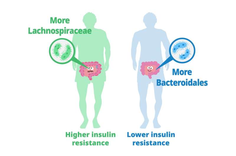
Previous studies have explored the role of gut microbiota in metabolizing nutrients in insulin resistance. This research aims to uncover the mechanisms underlying this relationship using a multi-omics approach. The study analyzes data from 306 individuals without diabetes, focusing on insulin resistance as defined by HOMA-insulin resistance scores.

The researchers used various techniques, including metabolomics, metagenomics, transcriptomics, and clinical data, to profile how the gut microbiome contributes to insulin resistance.

They found that certain carbohydrates in the feces, particularly those accessible to the host, are elevated in individuals with insulin resistance. These carbohydrates are linked to microbial carbohydrate metabolism and host inflammatory cytokines. Specific gut bacteria are associated with insulin resistance and insulin sensitivity, each displaying distinct carbohydrate metabolism patterns. In a mouse model, bacteria linked to insulin sensitivity demonstrate the potential to improve insulin resistance traits.

The study also involves analyzing metabolic syndrome (MetS) and its associations with fecal and plasma metabolites. Using human fecal cultures, the researchers discovered that Bacteroidales, a type of gut bacteria linked to insulin sensitivity, have a unique metabolic profile. These bacteria are efficient consumers of certain carbohydrates, affecting the production of fermentation products.

To explore causality, the researchers test the effects of seven candidate bacteria associated with insulin sensitivity on mice fed a high-fat diet. Several strains, notably Alistipes indistinctus, show promising results in reducing postprandial blood glucose levels and improving insulin resistance. These strains also impact body mass, lipid accumulation, and glucose intolerance.

Mechanistically, the researchers find that A. indistinctus administration reduces carbohydrate oxidation in mice, possibly due to decreased host-accessible carbohydrates in the intestine. This is supported by altered caecal metabolites, including reduced monosaccharides like fructose.

In conclusion, the study employs a comprehensive multi-omics strategy to investigate the relationship between gut microbiota and insulin resistance. It identifies specific bacteria associated with insulin resistance and sensitivity and highlights the potential of A. indistinctus in ameliorating insulin resistance in mice. However, further research is needed to understand the precise mechanisms and potential therapeutic implications of these findings.

Longevity factor klotho enhances cognition in aged Rhesus primates

- Posted by admin in English

Klotho is a protein that has been of interest in scientific research due to its potential role in aging and age-related diseases. It is believed to have several beneficial effects on various organ systems, including the brain, kidneys, and cardiovascular system. enter image description here Until now, studies on Klotho have primarily focused on its effects on animal models such as mice, rather than directly increasing its levels in primates.

This new study by scientists from the University of California, explores the potential of using the protein Klotho to enhance cognition in nonhuman primates, specifically rhesus macaques. Klotho declines with aging, and previous research has shown that increasing Klotho levels can improve cognitive function in mice. So the scientists wanted to verify whether this effect could be replicated in nonhuman primates, which have a higher genetic and functional complexity more similar to humans.

The researchers first validated the activity of the rhesus form of the Klotho protein (a variant of the Klotho protein that is found in rhesus macaques) in mice, demonstrating that it increased synaptic plasticity and cognition. They then tested different doses of Klotho in aged rhesus macaques and found that a single low-dose administration enhanced memory performance. Interestingly, higher doses did not produce the same cognitive benefits. The results suggest that systemic low-dose Klotho treatment may have therapeutic potential for aging humans.

The study emphasizes the importance of studying animal models with greater complexity, such as nonhuman primates, in the development of cognitive treatments for humans. By demonstrating the cognitive benefits of Klotho in rhesus macaques, the researchers bridge a knowledge gap and provide evidence for the potential translation of this treatment to humans.

There are several known ways to potentially increase Klotho levels in the body. Here are a few approaches that have shown promise:

  • Exercise: Regular physical exercise has been associated with increased Klotho levels.

  • Caloric restriction.

  • Pharmaceutical interventions: Certain drugs and compounds have been explored for their potential to increase Klotho levels.

  • Dietary factors: Some studies have suggested that certain dietary factors may influence Klotho levels. For example, diets rich in fruits, vegetables (rhubarb), and whole grains have been associated with higher Klotho levels.

  • Hormonal interventions: Hormonal factors, such as vitamin D and sex hormones (e.g., estrogen), have been implicated in Klotho regulation.

Yet it's not clear what is the mechanism of action of klotho on cognition. It must be stated also that there be nasty side effects to elevated levels of klotho like risks of weight gain, cancer or insulin resistance.

Improving sleep quality in neurodegeneration

- Posted by admin in English

A new publication in the Journal of Neuroscience, by Mount Sinai researchers, discusses a study conducted by researchers at Mount Sinai, which identifies a new form of treatment for rapid eye movement (REM) sleep behavior disorder. enter image description here This condition affects individuals, mostly adults over the age of 50, who physically act out their dreams during sleep, resulting in injuries to themselves or their bed partners. It's also suspected to be involved in premisses of Parkinson's disease. The study, published in the Journal of Neuroscience, presents a novel model that characterizes how REM sleep behavior disorder develops due to neurodegeneration associated with the accumulation of tau protein.

Tauopathies are a group of neurodegenerative disorders characterized by the abnormal accumulation of tau protein in the brain. DORAs have shown promise in preclinical studies as potential treatments for tauopathies, including diseases like progressive supranuclear palsy (PSP) and frontotemporal dementia with tau pathology.

DORAs have been found to modulate the activity of the orexin system, which is involved in regulating tau pathology. By targeting the orexin system, DORAs may potentially influence tau-related neurodegeneration and associated symptoms. However, further research is needed to fully understand the mechanisms and clinical implications of DORAs in tauopathies.

Using a mouse model, the researchers examined the effects of abnormal tau deposits on brain function and sleep. They analyzed various sleep-related factors, including wakefulness, REM sleep, non-REM sleep, sleep duration, and transitions between sleep stages. The study found that a significant number of older mice exhibited dream enactment behaviors similar to REM sleep behavior disorder, such as chewing and limb extension.

The researchers then administered a type of sleep medication called dual orexin receptor antagonists (DORAs) to evaluate their effects on sleep in the mice. They discovered that the medication not only improved sleep quality and duration but also significantly reduced dream enactment behaviors.

Orexin receptor antagonists (ORAs) are a class of medications that target the orexin system in the brain. Orexins, are neuropeptides that play a crucial role in regulating wakefulness and arousal. They promote wakefulness by activating orexin receptors in cells of the brain.

When orexin receptors are blocked or antagonized by medications, such as dual orexin receptor antagonists (DORAs), it leads to a decrease in wakefulness and an increase in sleepiness. DORAs are primarily used to treat insomnia, as they promote sleep initiation and maintenance.

In terms of sleep architecture, orexin receptor antagonists have been found to affect different sleep stages and sleep characteristics. Here are some of the effects observed:

  • Increased total sleep time, allowing individuals to obtain a longer and more restful sleep.

  • Increased non-rapid eye movement (NREM) sleep), which is deep and restorative sleep, and lighter stages of NREM sleep.

  • Decreased rapid eye movement (REM) sleep: DORAs have been found to suppress REM sleep, the stage associated with vivid dreaming and increased brain activity. This reduction in REM sleep may be beneficial in certain sleep disorders, such as REM sleep behavior disorder.

  • Improved sleep continuity: DORAs can enhance sleep continuity by reducing the number of awakenings during the night and decreasing the time spent awake after sleep onset. This leads to more consolidated and uninterrupted sleep.

It's important to note that the effects of orexin receptor antagonists on sleep architecture may vary depending on the specific medication, dosage, and individual factors.

These findings suggest that DORAs, which are already FDA-approved for treating insomnia, could serve as a promising new treatment for REM sleep behavior disorder. The researchers hope that their study will encourage further clinical trials of DORAs in humans with this disorder.

Overall, the study provides insights into the effects of neurodegeneration and tau protein accumulation on sleep and offers a potential therapeutic approach for managing REM sleep behavior disorder.

RAGE Inhibitors in Neurodegenerative Diseases

- Posted by admin in English

This post is about a review of the contribution of Advanced Glycation Endproducts to Neurodegenerative Diseases.

Advanced Glycation Endproducts (AGEs) are formed through non-enzymatic reactions between proteins, aminoglycosides, amino-terminal lipids, and reducing sugars like D-glucose. This process involves Amadori rearrangements and oxidative modifications. The accumulation of AGEs, especially under conditions of elevated oxidative stress, leads to various diseases. enter image description here AGEs have diverse structures, but only a limited number have been characterized. Some AGEs are small molecules formed through proteolytic degradation of protein-crosslinked or protein-modified AGEs. Imbalance between the formation and destruction of AGEs, particularly under conditions of oxidative stress, results in excessive accumulation and disease progression.

Some of the well-characterized AGEs include pentosidine, glucosepane, Argpyrimidine, and Nε-(carboxymethyl)lysine (CML). The imbalance between the formation and destruction of AGEs, triggers a cascade of signaling events, inflammation, and oxidative stress. This inflammatory signaling cascade is associated with various neurological diseases, including Alzheimer’s disease (AD), amyotrophic lateral sclerosis (ALS), diabetic neuropathy, diabetes, and atherosclerosis.

Other endogenous ligands are also involved, such as high mobility group box1 (MGB1) proteins. Additionally, exogenously ingested AGEs contribute to disease onset. The formation and accumulation of AGEs overwhelm the body's detoxification mechanisms under conditions of enhanced oxidative stress, exacerbating neurodegenerative diseases and other inflammatory-associated conditions.

RAGE (receptor for advanced glycation end-products) is a receptor that interacts with AGE (advanced glycation end-products)-derived ligands, such as CML, CEL, and MG-H1. The ligands bind to specific residues on the receptor, initiating signal transduction and pro-inflammatory signaling events.

RAGE antagonists, either endogenous or exogenous compounds, bind to RAGE and attenuate the binding interactions between AGE and RAGE, thereby preventing disease progression. These antagonists have shown potential for treating neurodegenerative diseases, diabetes, atherosclerosis, and cancers. Some small molecule-based RAGE inhibitors, like FPS-ZM1 and Azeliragon, have entered clinical trials, but none have been FDA-approved yet.

FPS-ZM1 has shown selective binding to RAGE and inhibits the formation of Aβ peptides, which are associated with brain damage in diseases like Alzheimer's. RAGE inhibitors also have potential therapeutic applications in diabetic nephropathy, cancer cell metastasis, and Parkinson's disease.

Azeliragon, currently in phase 3 clinical trials, has demonstrated decreased levels of Aβ plaques in the brain, reduces inflammation, and slower cognitive decline in Alzheimer's patients. Other compounds, such as urolithin and its analogs, have shown comparable RAGE inhibition to Azeliragon.

RAGE antagonists are also considered as therapeutics for diabetic neuropathy and retinopathy. Inhibitors of the cytoplasmic tail of RAGE (ct-RAGE) have been investigated and shown to block AGE/RAGE-mediated signaling events effectively. Some of these small molecule antagonists have structural similarities to FPS-ZM1 and have demonstrated potential for treating neurological disorders and diabetic complications.

Dietary AGEs, especially from animal-derived foods cooked at high temperatures, can also contribute to AGE accumulation and the development of diseases. RAGE antagonists, AGE inhibitors, and soluble RAGE (sRAGE) have shown promise in the treatment of age-related pathologies. Polyphenolic compounds can attenuate AGE formation and its toxic effects by reducing oxidative stress.

Therapeutic interventions targeting the AGE-RAGE axis may provide effective treatment for neurodegenerative diseases, diabetes, atherosclerosis, and cancers.

In summary, RAGE antagonists show promise as therapeutics for various diseases by attenuating the binding interactions between AGE and RAGE and preventing downstream pro-inflammatory signaling events. Clinical trials are underway for several RAGE inhibitors, and they have shown potential for treating Alzheimer's, diabetes, and other AGE-related diseases.

Overall, understanding the formation, toxicity, and interactions of AGEs with RAGE is crucial for developing therapies to combat age-related diseases.


Please, help us continue to provide valuable information: