Bats, although mammals, are not phylogenetically very close to humans, so we would not expect them to harbor many harmful viruses to our species.

enter image description here Source: Вых Пыхманн via Wikipedia

The order of bats contains more than 1,200 species. Because of this genetic diversity, information about one species may not apply to other species. In recent years, however, it has emerged that viruses have spread from bats to other mammals. For example, serological evidence and detection of viruses linked to ebolaviruses in bats suggest that bats are indeed one of the reservoirs of the virus.

However, although bats harbor many different viruses, their spread to other animals is extremely rare. One reason is that for such an event to occur, several factors should be conducive to transmission.

The ability of bats to host many viruses without showing pathology suggests that bats have developed immune mechanisms different from those of other mammals. This tolerance to viruses goes hand in hand with reduced inflammation.To explain this singular characteristic, it is necessary to compare it with another singular characteristic of bats: It is a mammal with the capacity to fly.

The metabolic rate of bats in flight is double that of running rodents of similar size. The increased metabolic rate that accompanies the flight would result in higher levels of oxygen-free radicals. To mount an immune response to the damage caused by this high metabolism, would be energetically expensive.

Bats have therefore developed mechanisms leading to reduced inflammation. It would also explain why bats of some species live longer than expected given their high metabolism and small size. Some bats can live up to 40 years, while a rodent of the same size can live only two years.

Small animals with a fast heart rate and metabolism generally have a shorter lifespan than larger animals with a slower heartbeat and a slower metabolism. But bats are unique because they have a much longer lifespan than other mammals of the same size.

But a reduced inflammatory response enables rapid replication of viruses, especially in stressful conditions that affect the immune system.

  • The awakening of hibernation is a stressful event for bats. Many large brown bats are latently infected and Gerowet and his colleagues have shown that the virus reactivates when it comes out of hibernation. This reactivation is also associated with a low level of antibodies to the virus. After hibernation, antibody levels rise, which puts the virus back to latency.

  • The virus also reactivates when bats are infected with a bacterial or fungal infection. Small brown bats are particularly susceptible to an often fatal fungal infection known as white nose syndrome. A study examining the effects of stress induced by this infection showed that bats infected with fungi had 60 times more coronavirus in their intestines than uninfected bats. The specific protection mechanisms of these bats results in a rapid response that blocks the virus outside the cells. Indeed, paradoxically a host that tolerates well the presence of viruses, because it controls inflammation, allows viruses to increase their rate of replication, by genetic selection, without damaging their host.

  • Disturbances in bat habitat also seem to stress these animals and cause them to spread even more viruses in their saliva, urine and feces which can infect other animals. "Increased environmental threats to bats can add to the threat of zoonosis," said Brook, who is also working with a Madagascar-based field project that explores the link between loss of bat habitat and spread of bat viruses to other animals and humans.

Such rapidly reproducing viruses generate extreme virulence when overflowing to hosts that do not have the same immune capabilities as bats.

When these virulent viruses travel from bats to animals without a rapid response immune system, they quickly overwhelm their new hosts.

Brook and Boots are developing a more formal model of disease progression in bats to better understand the spread of the virus to other animals and humans.

Advertisement


This book retraces the main achievements of ALS research over the last 30 years, presents the drugs under clinical trial, as well as ongoing research on future treatments likely to be able stop the disease in a few years and to provide a complete cure in a decade or two.

Le syndrome de soins post-intensifs résulte d'une inflammation continue

- Posted by admin in Français

Le syndrome de soins post-intensifs (PICS) décrit un ensemble de troubles qui sont courants chez les patients ayant subis une maladie grave et/ou des soins intensifs. Étant donné que la majorité de la littérature en médecine des soins intensifs se concentre sur les résultats à court terme (par exemple, la survie), la compréhension de l’évolution du malade sur le long terme, est relativement limitée, puisque celui-ci est alors considéré comme étant guéri.

Les troubles cognitifs comprennent des déficits de mémoire, d'attention, de vitesse de traitement mental et de résolution de problèmes. Ces déficiences touchent jusqu'à 80% des personnes ayant éprouvé une maladie grave. Les symptômes de la plupart des patients s'améliorent voire disparaissent complètement au cours de la première année qui suit le traitement en unité de soins intensifs.

La physiopathologie sous-jacente de la déficience cognitive chez les survivants des soins intensifs n'est pas bien comprise, mais une inflammation prolongée peut jouer un rôle important

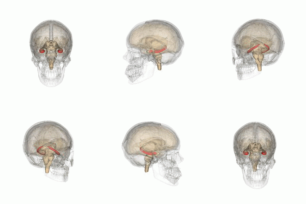
Il a été montré sur des animaux de laboratoire, que High Mobility Group Box 1 (HMGB1), une protéine libérée dans les lésions tissulaires et au cours d'une inflammation sévère, subsiste à une concentration élevée longtemps après le traumatisme et peut provoquer une inflammation hippocampique et des troubles cognitifs. enter image description here Source: Life Science Databases(LSDB) via Wikipedia.

Les humains et les autres mammifères ont deux hippocampes, un de chaque côté du cerveau. L'hippocampe fait partie du système limbique. Dans la maladie d'Alzheimer, l'hippocampe est l'une des premières régions du cerveau à subir des dommages; la perte de mémoire à court terme et la désorientation font partie des premiers symptômes. Les personnes atteintes de lésions hippocampiques bilatérales étendues peuvent souffrir d'amnésie antérograde: c'est à dire l'incapacité de former et de conserver de nouveaux souvenirs.

La forme de plasticité neurale connue sous le nom de potentialisation à long terme (LTP) a été initialement découverte dans l'hippocampe et a souvent été étudiée dans cette structure. La LTP est considérée comme l'un des principaux mécanismes neuronaux par lesquels les souvenirs sont stockés dans le cerveau.

Un traitement anti-HMGB1 administré plusieurs jours après une maladie grave peut atténuer le déclin cognitif chez la souris

Des chercheurs du Karolinska Institutet en Suède ont mené une étude de suivi prospective sur 6 mois des taux plasmatiques de HMGB1 et de la fonction cognitive chez les survivants des soins intensifs (essai clinique NCT02914756). 917 patients admis aux soins intensifs ont été dépistés, parmi ceux-ci 100 patients ont été inclus dans l’essai clinique, et ils ont été soumis à des tests de la fonction cognitive et à la mesure des taux plasmatiques de HMGB1 à 3 et 6 mois après la sortie

Les observations ont été effectuées chez ces patients montrent une élévation significative du plasma HMGB1 à 3 et 6 mois après la sortie, et est associée à un dysfonctionnement cognitif.

La source cellulaire de ce HMGB1 systémique est inconnue, mais il est à noter que le HMGB1 est habituellement sécrété par les cellules immunitaires (comme les macrophages, les monocytes et les cellules dendritiques) en tant que médiateur des cytokines de l'inflammation.

Compte tenu de ces propriétés pro-inflammatoires bien établies du HMGB1 extracellulaire, cela suggère une inflammation continue sans résolution de l’inflammation. À la lumière des résultats expérimentaux sur l'atténuation du dysfonctionnement cognitif chez des animaux de laboratoire par la thérapie anti-HMGB1, il est tentant de se demander si le blocage de l'activité pro-inflammatoire du HMGB1 chez les survivants en USI pourrait améliorer les résultats cognitifs.

Post-intensive care syndrome results from continuous inflammation

- Posted by admin in English

Post Intensive Care Syndrome (PICS) describes a set of disorders that are common in patients with severe illness and / or intensive care. Since the majority of the literature in intensive care medicine focuses on short-term outcomes (for example, survival), understanding of the patient's long-term development is relatively limited, since the latter is then considered to be healed.

Cognitive impairment includes deficits in memory, attention, speed of mental processing and problem solving. These impairments affect up to 80% of people who have experienced a serious illness. Most patients' symptoms improve or even disappear completely within the first year after treatment in the intensive care unit.

The underlying pathophysiology of cognitive impairment in critical care survivors is not well understood, but prolonged inflammation can play an important role

High Mobility Group Box 1 (HMGB1), a protein released in tissue damage and during severe inflammation, has been shown in laboratory animals to remain in high concentration long after the trauma and can cause inflammation hippocampal and cognitive impairment. enter image description here Source: Life Science Databases(LSDB) via Wikipedia.

Humans and other mammals have two seahorses, one on each side of the brain. The hippocampus is part of the limbic system. In Alzheimer's disease, the hippocampus is one of the first areas of the brain to be damaged; short-term memory loss and disorientation are among the first symptoms. People with extensive bilateral hippocampal lesions may suffer from anterograde amnesia: the inability to form and retain new memories.

The form of neural plasticity known as long-term potentiation (LTP) was originally discovered in the hippocampus and has often been studied in this structure. LTP is considered to be one of the main neural mechanisms by which memories are stored in the brain.

Anti-HMGB1 treatment given several days after a serious illness can reduce cognitive decline in mice

Researchers at the Karolinska Institutet in Sweden conducted a 6-month prospective follow-up study of HMGB1 plasma levels and cognitive function in survivors of intensive care (clinical trial NCT02914756). 917 patients admitted to intensive care were screened, of which 100 patients were included in the clinical trial, and they were subjected to cognitive function tests and to the measurement of plasma levels of HMGB1 at 3 and 6 months after discharge

The observations were made in these patients show a significant elevation of HMGB1 plasma at 3 and 6 months after discharge, and is associated with cognitive dysfunction.

The cellular source of this systemic HMGB1 is unknown, but it should be noted that HMGB1 is usually secreted by immune cells (such as macrophages, monocytes and dendritic cells) as a mediator of the cytokines of inflammation.

Given these well-established pro-inflammatory properties of extracellular HMGB1, this suggests continued inflammation without resolution of the inflammation. In light of the experimental results on the attenuation of cognitive dysfunction in laboratory animals by anti-HMGB1 therapy, it is tempting to ask whether blocking the pro-inflammatory activity of HMGB1 in ICU survivors could improve cognitive outcomes.


Please, help us continue to provide valuable information: