Motor Imagery and rehabilitation

- Posted by admin in English

Recently I learned something new in anatomy (I am a noob in anatomy). It's known for some times there are mirror neurons, but there is an interesting subset: The mirror motor neurons. It was mentioned on a forum by a pALS (patient with ALS) who told he rehabilitated his bladder thanks to mirror motor neurons. Near my home in France, there is a center that uses similar techniques for people who unfortunately experienced strokes. enter image description here Bladder issues are unfortunately common in ALS patients. These issues arise because ALS affects the nerve cells that control the muscles in bladder and sphincter.

This forum post picked my curiosity and I looked in Pubmed. Indeed there are many sudies that discuss about this kind of rehabilitation technique in ALS and Parkinson's disease. It's called Motor Imagery and there is even a Wikipedia about it.

If you pardon me for grave oversimplification, it's a matter of showing some action to the subject, for example someone is walking, and then asking to the subject to imagine doing the same thing.

The ALS patient told it needed years to regain control of his sphincter, so it's not possible to find some scientific litterature that would enlight us on the results wa can expect with this long duration of rehabilitation, without mentioning that unfortunately many ALS patients do not live that long. Scientists are usually busy people, their studies last between days and a few months as it must coincide with academic time. But there are many studies that mention that even after a few tests, a positive effect can be detected. Here is an example.

A review on Parkinson's disease is less optimistic, it tells of ~5% motor improvement.

I wonder to which extend this Motor Imagery could help to regain some important functionality. Maybe some reinforcement could be added when the imaging process is detected. Apparently even a simple EEG is able to detect this mental state. The article cited above has some additional details on this. It calls also in question why those mirror motor neurons are not striked by the disease as they are probably colocated with upper motor neurons.

Maladie de Parkinson, glucides et fer

- Posted by admin in Français

Introduction

L'article (en pre-print) discuté aujourd'ui traite de nutrition et de la maladie de Parkinson. La maladie de Parkinson est le deuxième trouble neurodégénératif le plus répandu dans le monde et entraîne une réduction significative de la qualité de vie. Les tendances actuelles en matière d'incidence, de prévalence et de charge de morbidité montrent que le fardeau mondial de la maladie de Parkinson a augmenté.

Les connaissances actuelles suggèrent de façon très générales que la maladie de Parkinson est probablement causée par une interaction entre une prédisposition génétique et la présence de facteurs environnementaux qui peuvent s'accumuler tout au long de la vie, c'est à dire que les scientifiques ont de grandes difficultés à identifier les facteurs à risques. Même le diagnostic semble ne pas faire l'unanimité des chercheurs, pour certains le problème est le manque de production de dopamine dans une certaine zone du cerveau, pour d'autres il s'agit d'une maladie liée à l'accumulation d'une protéine mal-formée: L'alpha-synucléine.

Le fer a peut-être un rôle particulier dans la maladie de Parkinson car il est nécessaire à l'enzyme limitant la production de dopamine. En effet celle-ci, la tyrosine hydroxylase, catalyze la conversion de l'amino acide L-tyrosine vers le L-3,4-dihydroxyphenylalanine (L-DOPA). Pour cela elle a besoin d'oxygène et de fer et aussi de tetrahydrobiopterine comme cofactors. L-DOPA est un precurseur de la dopamine, qui à son tour est un precurseur des neurotransmitters norepinephrine (noradrenaline) and epinephrine (adrenaline). enter image description here De même il existe une relation entre le métabolisme des glucides et celui du fer. Ce qui suggère qu'un métabolisme anormal des glucides puisse avoir une relation avec l'apparition de la maladie de Parkinson.

Les facteurs environnementaux qui se sont avérés liés au risque de maladie de Parkinson comprennent : * Les facteurs associés à un risque réduit de maladie de Parkinson sont le tabagisme, la consommation d'alcool, et la consommation de caféine. De façon moins claire on trouve aussi notamment la vitamine E, les flavinoïdes et le β-carotène ou encore la viande rouge. * Tandis que l'exposition aux pesticides ou aux herbicides et aux blessures à la tête1 sont associés à un risque accru de maladie de Parkinson. Il a également été suggéré que les produits laitiers pourraient augmenter le risque de maladie de Parkinson.

Il existe une relation bien établie entre le microbiome intestinal et la pathogenèse de la maladie de Parkinson. Certaines théories développées sur l'étiologie de la maladie de Parkinson, telles que l'hypothèse de Braak, affirment que la maladie de Parkinson peut commencer dans le système nerveux entérique de l'intestin avant de remonter jusqu'au cerveau. Ces théories sont étayées par des associations connues entre les problèmes gastro-intestinaux prodromiques et le biomarqueur caractéristique de la maladie de Parkinson, l'α-synucléine, dans le système nerveux entérique avant le diagnostic de la maladie de Parkinson.

Fer et maladie de Parkinson

Une accumulation accrue de fer dans des régions comme la substance noire et les noyaux gris centraux et une dyshoméostasie du métabolisme du fer sont des constatations courantes chez les patients parkinsoniens. Malgré cela, les recherches sur le fer alimentaire ont donné des résultats mitigés. Dans l’ensemble, l’apport alimentaire en fer ne semble pas être associé au risque de maladie de Parkinson, mais des analyses de sous-groupes dans les sous-populations occidentales et masculines ont révélé que, dans ces sous-groupes, le fer alimentaire était associé à une augmentation significative du risque de maladie de Parkinson.

Comme souvent, certaines études signalent que l'anémie est associée à un risque accru de maladie de Parkinson, tandis que d'autres rapportant le contraire. Ces résultats apparemment contradictoires peuvent suggérer une relation non linéaire dans lequel à la fois, des niveaux faibles et élevés de fer cérébral dans les circuits moteurs sont associés à un risque accru de maladie de Parkinson

Motivation

Bien que des travaux antérieurs aient étudié les habitudes alimentaires des patients atteints de maladie de Parkinson, cela ne fournit pas de compréhension mécaniste de la façon dont les différences alimentaires peuvent entraîner des profils de risque différentiels de maladie de Parkinson. Dans ce nouvel article, des chercheurs étudient les différences en matière de fer dans le cerveau liées aux facteurs alimentaires et liés au mode de vie liés au risque de maladie de Parkinson en utilisant un biomarqueur cérébral spécifique du fer de la maladie de Parkinson, qu'ils appellent PVS cérébral d'hémochromatose. Ce biomarqueur regroupe les signaux d'accumulation de fer provenant des IRM cérébrales T2-w des régions motrices, notamment le cervelet, le thalamus, le caudé et le putamen.

Les auteurs ont cherché à comprendre comment les facteurs alimentaires et ceux liés au mode de vie influence la présence de ce biomarqueur spécifique du fer et comment cela est lié au risque de maladie de Parkinson.

Leurs investigations montrent que les préférences alimentaires en faveur (bizarrement) de l'alcool et la consommation de produits frais sont associées à un risque réduit de maladie de Parkinson, et que l'apport alimentaire et les préférences en faveur des aliments sucrés sont associés à un risque accru de maladie de Parkinson.

Il semblerait qu'une relation existe dans laquelle les nutriments et les préférences alimentaires liés à des niveaux de fer cérébraux plus faibles seraient associés à un risque accru de maladie de Parkinson.

La dérégulation du fer est une caractéristique courante du maladie de Parkinson, et des essais cliniques de la chélation sont en cours comme voie de traitement potentielle pour le maladie de Parkinson et d'autres maladies neurodégénératives.

Par exemple les essais cliniques concernant le défériprone comme traitement de la neurodégénérescence associée à la pantothénate kinase, une maladie génétique liée à une accumulation accrue de fer dans le cerveau et à des symptômes liés au mouvement. Mais ces essais cliniques ont montré que le défériprone n'entraînait qu'une réduction très faible de la progression de la maladie.

Les essais sur la défériprone, un chélateur du fer, pour le traitement de la maladie de Parkinson ont donné des résultats mitigés. La aussi les essais ont montré une réduction de l'accumulation de fer dans certaines zones du cerveau, mais aucune amélioration significative des symptômes.

Sucres et glucides

Dans toutes leurs analyses, les scientifiques ont constaté que les facteurs liés aux glucides dans les préférences et l'apport nutritionnel estimé étaient associés à une diminution du PVS cérébral de l'hémochromatose, à une augmentation du risque de maladie de Parkinson.

La littérature suggère un effet bidirectionnel avec : * a) un taux élevé de fer influençant la régulation glycémique et augmentant le risque de diabète de type II, une maladie principalement causée par une altération aiguë de la régulation glycémique, * b) l'ingestion orale de glucose entraînant des modifications des facteurs du métabolisme du fer résultant en fer périphérique.

Le fer peut jouer un rôle dans le développement de la résistance à l’insuline. La littérature scientifique à ce jour, montre que la dérégulation glycémique et les maladies associées sont liées à un risque accru de maladie de Parkinson et à de pires résultats de la maladie de Parkinson.

Un métabolisme dérégulé du fer pourrait conduire à des envies inadaptées de glucides, ce qui pourrait déréguler conjointement le métabolisme du glucose et du fer, conduisant à une boucle de rétroaction. La dérégulation fragmentée du métabolisme du fer et du glucose peut expliquer pourquoi une manifestation occasionnelle de carence en fer comme le pika, incite à manger des aliments riches en glucides comme l'amidon, le riz non cuit et les pâtes non cuites.

Les sucres et les glucides peuvent également avoir un impact sur le risque de maladie de Parkinson et l’accumulation de fer au niveau du microbiome. Une forte préférence alimentaire pour les sucreries et les glucides peut augmenter les niveaux de bactéries pathogènes opportunistes pro-inflammatoires dans l’intestin, ce qui est fortement lié à un risque accru de maladie de Parkinson. Ce modèle alimentaire est également en corrélation avec la pathologie de la ɑ-synucléine, qui peut émerger d’un état intestinal dysbiotique et pro-inflammatoire.

Alcool

Il existe également une littérature importante selon laquelle la consommation d’alcool a un impact sur l’absorption et l’accumulation du fer. L’alcool peut entraîner une accumulation accrue de fer dans le cerveau82. L’alcool est connu pour réguler négativement la synthèse de l’hepcidine, une hormone régulatrice du fer, et, en cas de consommation excessive, peut provoquer une surcharge en fer chez des individus par ailleurs hémodynamiquement typiques. En particulier chez les personnes atteintes d’hémochromatose héréditaire, la consommation d’alcool est largement associée à de pires résultats en matière de santé. Des effets similaires sont observés dans d’autres troubles de surcharge en fer comme la bêta-thalasémie.

Exercice physique

Les auteurs ont constaté que les préférences liées à l'exercice sont associées de manière significative à une réduction du risque de maladie de Parkinson et à une réduction du fer cérébral, telles que mesurées avec le PVS cérébral de l'hémochromatose. Evidemment les principaux déficits moteurs de la maladie de Parkinson rendent l'activité physique moins attractive. Mais des niveaux d’activité modérés à élevés se sont avérés associés à un risque plus faible de développer une maladie de Parkinson plus tard dans la vie et les personnes atteintes de maladie de Parkinson qui déclarent une activité physique plus élevée ont une progression plus lente des symptômes et une meilleure qualité de vie.

Céréales et fruits

Les préférences pour les légumes et les fruits étaient associées à une diminution du risque de maladie de Parkinson et à aucune association significative avec le PVS. Ces résultats concordent avec les conclusions d’études antérieures selon lesquelles une consommation élevée de fruits et légumes est liée à un risque plus faible de maladie de Parkinson. Une consommation élevée de fruits et légumes pourrait expliquer une partie des effets protecteurs de la maladie de Parkinson observés dans le régime méditerranéen.

Les préférences liées aux céréales étaient associées à une réduction du fer cérébral. Ce résultat est surprenant étant donné que les céréales sont un véhicule courant pour l’enrichissement en fer. Similairement les grains de céréales peuvent contenir entre 50 et 80 % de glucides en poids, on pourrait donc penser que ce type de nourriture a un effet défavorable sur la maladie de Parkinson. Mais, les céréales et les produits laitiers, généralement consommés avec les céréales, sont riches en inhibiteurs de l'absorption du fer comme l'acide phytique et le calcium, qui réduisent la biodisponibilité du fer en chélatant et en cloîtrant le fer dans le tube digestif. L'interprétation actuelle des auteurs est que les niveaux plus faibles de fer dans le cerveau observés chez les individus préférant les céréales sont dus aux inhibiteurs de l'absorption du fer présents dans ces repas et que l'association qui est observée se produit malgré l'enrichissement en fer des céréales et non à cause de celui-ci.

Conclusion

En conclusion, c'est une étude intéressante, qui montre bien la complexité de la biologie humaine et que les maladies ne se réduisent pas à la carence ou l'excès de quelque molécule, contrairement à ce que le biologie moléculaire laisse entendre.

For a patient, participating in a clinical trial is complicated to organize, in addition, the drugs tested are very rarely effective in the field of neurodegenerative diseases. However, it seems that there are unexpected benefits to volunteering for a clinical trial.

Clinical trials in neurodegenerative diseases are often disappointing, there are probably many reasons for that situation. A core aspect of clinical trials is that the population who received the treatment should be representative of the general population. For logistic reasons, biotechs that have few employees have to subcontract clinical trials. Principal investigators and subcontractors have every reason to select patients who present a textbook-like disease.

Suspecting that the clinical trial population is not representative of real-life patients, an international team wanted to characterize the progression of Parkinson's disease using real-world data to guide the design of clinical trials and identify subpopulations.

The increasing availability of real-world data, and recent advances in natural language processing, particularly large language models, allow for an easier and more granular comparison of populations than before.

This study includes two research populations and two populations derived from real-world data.

The research populations are the Harvard Biomarkers Study (935 patients), which is a longitudinal biomarker cohort study with structured in-person study visits, and finally Fox Insights (36,660 patients), a research study based on the Michael J. Fox Foundation online self-survey.

The real-world cohorts are Optum Integrated Claims electronic health records (157,475 patients), representing large-scale linked medical and claims data and de-identified data from Mass General Brigham (Mass General Brigham, 22,949 patients), a University Hospital.

Structured, anonymized data from Mass General Brigham's electronic health records is augmented using natural language processing with a large language model to extract measures of Parkinson's disease progression. This extraction process is manually validated to verify accuracy.

Motor and cognitive progression scores change more rapidly in the Mass General Brigham than in the Harvard Biomarkers Study (median survival to H&Y scale: 5.6 years versus more than 10 years); median decline to mini-exam of mental status 0.28 versus 0.11. In real-world populations, patients are diagnosed more than eleven years later! After diagnosis, in real-world cohorts, treatment with Parkinson's drugs is initiated 2.3 years later on average than for patients in clinical trials.

This study provides a detailed characterization of Parkinson's disease progression in various populations. It delineates systemic discrepancies between patient populations enrolled in research settings and real-world patients. enter image description here The study shows systematic differences and potential directional biases between research and real-world datasets. Patients in research populations are diagnosed much earlier, start levodopa and other Parkinson's medications earlier, and show slower changes in clinical scales of motor and cognitive progression. Real-world-based populations are diagnosed at older ages, start medications later than research cohorts, and experience more rapid changes in clinical scales.

These discrepancies are likely due to a combination of selection bias, but exact attribution of causes is difficult using existing data. This study emphasizes the need to diligently consider potential biases when planning a clinical trial.

This post is about an interesting hypothesis. Hypotheses abound, yet few a convincing.

Half of patients with Alzheimer's disease, Parkinson's disease, or ALS have insulin resistance. Obesity and diabetes have been linked to neurodegenerative diseases like multiple sclerosis (MS), Alzheimer's (AD), and Parkinson's (PD). This means the cells of their body cannot let the glucose enter them. Glucose is the main energy source as it is converted into ATP. Glucose is for short-term (day) energy needs. Another source of energy is lipids (fat). Lipids are even more dense than glucose energy-wise.

The body needs an enormous amount of energy. With all the lipids in the body of a healthy person, you could charge two Tesla cars! The brain (a part of the CNS) needs 20% of all energy intake.

A new paper argues that cells shift their metabolism from glucose to lipids under stressors. It tells that one notable distinction between glucose and lipid metabolism is in the quantity of oxygen required to generate each ATP molecule. Lipid metabolism needs two times more oxygen than glucose metabolism. The result is two times more damaging ROS (a by-product of metabolism). enter image description here Studies have shown that oxidative stress and endoplasmic reticulum stress are correlated and can lead to protein misfolding (Abramov et al., 2020). Accumulation of misfolded proteins causes cellular damage and mitochondrial dysfunction and is associated with a range of neurodegenerative diseases, including ALS (misfolded SOD1, TDP-43, C9orf72) (McAlary et al., 2020), Parkinson's disease (misfolded α-synuclein) and Alzheimer disease (misfolded Aβ and Tau) (Abramov et al., 2020).

It explains also the accumulation of iron in patients' brains: To transport oxygen the blood cells need iron, and as the glucose in the blood is not absorbed in cells, it induces a change in microbiota.

It's also well known that SCFAs (including butyrate) have a positive effect on neurodegenerative diseases by their action on microbiota. SCFAs help to restore glucose as the preferred energy substrate. Authors say there are other means to restore glucose as the main source of energy.

What to think about this paper? First, some authors belong to a biotech so we can expect they want to promote their drug: Mitometin. Second, this is a review, this is not even a pre-clinical study, yet some of the authors were involved in pre-clinical studies on this topic. Other groups have written on this topic. What to make of this? Acetyl-CoA carboxylase might be of interest as they produce malonyl-CoA which inhibits the CPT1 gene that regulates lipid metabolism. B7 vitamin is known to convert acetyl-CoA to malonyl-CoA for fatty acid synthesis.

Open innovation challenge

- Posted by admin in English

Now you have an opportunity to fast-track the discovery of drugs in some neuro-degenerative diseases.

AstraZeneca, the pharmaceutical company, proposes a challenge in the area of nucleotide repeat expansion disorders, which includes ALS (Charcot/Gehrig) disease, but also Huntington's disease, etc...

Typically you have to be a startup or an academic with a plan to launch a startup because your solution must be translated into practice within a 12–18-month timeframe.

I am willing to provide reasonable help if you hesitate. This will be mostly in terms of shaping your proposal. I believe that AstraZeneca, like other large companies, is not interested in unproven, hypothetical ideas. On the contrary, IMO they search for people with energy and time (and intellectual right) to give blood to ideas where there is a consensus but where nobody cared to develop it in the pre-clinical stage.

I guess also AstraZeneca would prefer a simple implementation, instead of a complex one. So for example, a small molecule proposal would be preferred to a genetic therapy. A compound of two or three existing drugs to an untested drug. You get the idea.

Good luck!

Jean-Pierre Le Rouzic You will find the link to email me at the bottom of this page.

The changing landscape of disease diagnostic.

- Posted by admin in English

Unfortunately, after decades of research and hundreds of unsuccessful phase III clinical trials, it's clear that the pharmacological industry and a cohort of academic laboratories are unable to create drugs that slow significantly the progression of neurodegenerative diseases.

Some courageous scientists interrogate basic hypotheses or design longer, more complex better clinical trials. For example, Alzheimer's disease can't seriously be attributed to any molecular dysfunction, as it would mean that a lot of cerebral functions would be affected, not only memory issues, and anyway, memory issues in Alzheimer's are much more complex than described in textbooks: They did not simply vanish: The patient looks to be living today in the context of the past. Sometimes the patient can discuss simultaneously at two levels: In the context of the past (when they were infants) and in the context of today.

Others are currently busy breaking the thermometer. If the clinical diagnosis makes it impossible to validate current clinical trials, then change the way success is defined: Abandon clinical criteria and use molecular criteria. They did it recently for Alzheimer's disease and now they propose it for Parkinson's disease..

The immediate consequence will be a flurry of successful clinical trials, even if patients get no improvements, as they did for Aducanumab.

There will also be false positives, people will be diagnosed sick because of the presence of a molecule but without any clinical signs.

Alpha-synuclein, the protein implicated in Parkinson's disease, is primarily localized in presynaptic terminals of neurons. In particular, it is involved in the regulation of neurotransmitter release, maintenance of synaptic integrity, and plasticity of synapses and, consequently, cognitive processes. But it's not simply the presence of a protein that is important, much more important is the place and time a protein changes, either in location, composition, or in shape. Those changes are caused by a chemical signal for example: phosphorylation. Phosphorylation events are omnipresent in our bodies. For example, phosphorylation of muscles' myosin, enables the contraction of muscle filaments. Phosphorylation is catalyzed by enzymes known as kinases which regulate various cellular functions by adding phosphate groups to specific target molecules. The phosphorylation-dephosphorylation cycle is dynamic and reversible, providing cells with a rapid and precise means of responding to external cues. Importantly, the balance between kinase and phosphatase activity (enzymes that remove phosphate groups) determines the overall phosphorylation status of a cell and influences its physiological state.

The phosphorylation of alpha-synuclein is associated with the formation of aggregates like Lewy bodies, contributing to the neurodegenerative process characteristic of Parkinson's disease and Lewy body dementia. It is not known if the phosphorylation of alpha-synuclein is causative of Parkinson's disease and Lewy body dementia.

This addition of a phosphate group is done at specific sites in the molecules, in the case of alpha-synuclein, it is done at a site called Ser129. Previous research suggested that abnormal accumulation of alpha-synuclein, when phosphorylated at Ser129, contributes to the formation of Lewy bodies—protein aggregates commonly found in the brains of individuals with Parkinson's. These Lewy bodies are associated with the degeneration of dopaminergic neurons, a hallmark of Parkinson's disease.

A new publication by Leonardo Parra-Rivas, Subhojit Roy, and colleagues at the University of California, San Diego, investigates the phosphorylation of alpha-synuclein (α-syn) at the Ser129 site and its implications in Parkinson's disease (PD) and related synucleinopathies.

Their study challenges the view that Ser129 phosphorylation directly causes toxicity and proposes a physiologic role in synaptic function. Indeed this has consequences Their new research somehow has consequences for common statements that abnormal accumulation of phosphorylated alpha-synuclein, contributes to the formation of Lewy bodies—protein.

Almost all pathologically aggregated α-syn in Lewy bodies is phosphorylated at Ser129. Antibodies to Ser129 α-syn are sensitive markers for neuropathologic diagnosis in synucleinopathies. Studies often use Ser129 phosphorylation as a surrogate marker for pathology. Intrigually, inhibitors against Polo-like kinase 2 (PLK2), responsible for α-syn phosphorylation, were explored as a PD drug target. However, PLK2 over-expression surprisingly suppressed neurodegeneration in vivo, challenging expectations. This hints that the physiological role of phosphorylated α-syn at Ser129 is misunderstood.

For the authors of this new publication, under normal physiological conditions, only a small fraction (∼4%) of α-syn is phosphorylated at Ser129. Despite its low frequency, Ser129P is produced during normal metabolism, raising questions about its exclusive pathological role. A recent study suggests that increasing neuronal activity augments Ser129 phosphorylation, implying that it may be linked to an increase in cognitive works and that the role of α-synuclein protein may be to regulate excessive neuronal firing. This suggests that α-syn might have a role in healthy brains, which had not previously been investigated. The researchers give an interesting example: "In hindsight, we hadn't been looking at synuclein phosphorylation the right way, take, for instance, the circuits in the olfactory bulb, which according to our data has high levels of phosphorylated α-synuclein. The nose never stops smelling, so it needs to be active all the time. One hypothesis is that synuclein phosphorylation may have evolved as a safety mechanism to protect neuronal circuits that need to be hyperactive."

The authors systematically examined α-syn Ser129 phosphorylation using various assays, in vivo studies, cell-free assays, mass spectrometry, electron microscopy, and dynamic simulations. when α-synuclein is phosphorylated, its structure changes in a way that promotes interactions with other proteins in healthy brains. The scientists propose a model where Ser129 phosphorylation induces conformational changes at the α-syn C terminus, facilitating its association with functional interacting partners and eliciting α-syn function.

Conclusion: The study challenges the view that Ser129 phosphorylation directly causes toxicity and proposes a physiologic role in synaptic function. Yet it is done on cultured cells and in mouse brain tissue, so those findings are far from being confirmed in humans. As we all know you can find contradictory studies in scientific literature and only clinical trials are credible (when authorities do not bypass their findings).

Early nonmotor symptoms in Lewy body disease

Parkinson's disease is a progressive neurodegenerative movement disorder with symptoms of rest tremor, muscular rigidity, slowness of movement, postural impairment, and later on often dementia which is sometimes attributed to drugs. Many people not diagnosed with Parkinson's have parkinsonism and others have only Lewy body dementia.

This medical characterization parallels ALS which has many mimics and shares a strong genetic and molecular background with fronto-temporal dementia. There is also a distinction between “body-first” and “brain-first” disease in ALS and Parkinson's disease. Both diseases mostly affect older people and the prevalence of Parkinson's disease is expected to continue to increase with the aging of the population in most developed countries.

Pathologically, Parkinson's disease is characterized by intraneuronal Lewy body inclusions prominently containing misfolded α-synuclein, which normally functions as an intracellular trafficking protein, as well as prominent neuronal death of dopaminergic neurons in the midbrain substantia nigra pars compacta. This disrupts dopamine neurotransmitter production and signaling in the basal ganglia circuit, a deep, central part of the brain.

While clinical findings of Parkinson's disease and Lewy body dementia are prominently associated with aberrant α-synuclein deposits in the central nervous system, pathologic α-synuclein can also be found in the peripheral nervous system. It has been postulated that central α-synucleinopathies may begin in the peripheral nervous system before spreading to the central nervous system.

Common early nonmotor symptoms in these disorders, such as orthostatic hypotension, constipation, and erectile dysfunction in men prefigure impairment of the peripheral and autonomic nervous systems rather than impairment of the CNS (the brain and spinal cord). Anosmia or hyposmia is also an early sign of Parkinson's disease. Despite these insights about the peripheral neurons, the main focus on central nervous system pathology and related symptoms drove the field.

Yet it is difficult to assess the presence of misfolded α-synuclein in the central nervous system (CNS). An ability to identify patients at risk with low-cost and easy-to-interpret biomarkers would facilitate the testing and implementation of disease-modifying therapies.

Cardiac noradrenergic dysfunction as an early marker There is an ongoing search for biomarkers in many chronic diseases. The well-publicized rationale is that it would help to diagnose early diseases. Indeed many neurodegenerative diseases are not diagnosed quickly, often it need several years of examinations which causes distress to patients and families. A less publicized rationale is that it would help companies quickly get market authorizations from authorities, a good example is the ongoing proposal to revise the criteria for Alzheimer's diagnosis.

Goldstein et al. report the results of the prospective, longitudinal PDRisk study (ClinicalTrials.gov NCT00775853).

The team investigated an NIH-developed positron emission tomography (PET) tracer, 18F-dopamine, to assess dysfunction of the cardiac noradrenergic system as a prelude to the development of Parkinson's disease and Lewy body dementia. Though relatively small, this study demonstrated a remarkably accurate predictive value of low cardiac uptake of 18F-dopamine for central Lewy body disease. This finding was further corroborated by cerebrospinal fluid 3,4-dihydroxyphenylacetic acid (DOPAC).

Goldstein et al. demonstrate that a combination of cardiac noradrenergic cell loss and inefficient sequestration of catecholamines in residual cardiac sympathetic nerves precedes the onset of central Lewy body diseases, at least in a population with several identified nonmotor Parkinson's disease risk factors. The precise timing of cardiac noradrenergic cell loss to central nervous system dopaminergic cell loss remains to be elucidated. As the authors acknowledge, this paradigm may be more effective in identifying preclinical disease in patients with “body-first” rather than “brain-first” central Lewy body disease (10), since the choice of nonmotor risk factors for entry into the PDRisk study probably biases toward the “body-first” paradigm.

The catecholaldehyde hypothesis

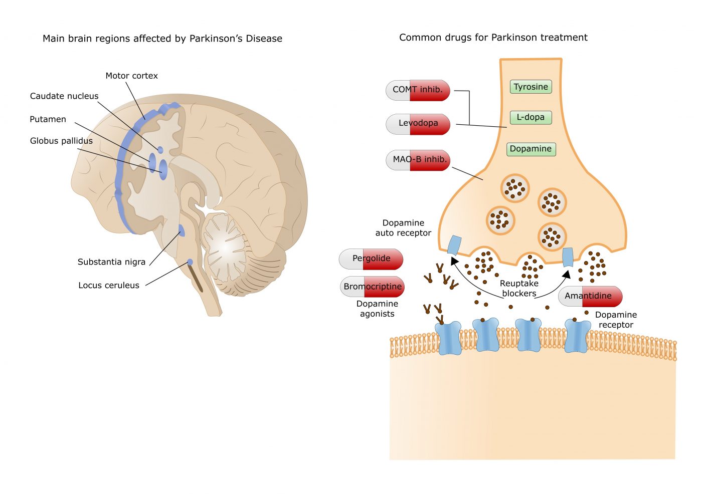
There is a hypothesis for the pathogenesis of Parkinson’s disease that centers on an accumulation of 3,4-dihydroxyphenylacetaldehyde (DOPAL) in dopaminergic neurons, this hypothesis is sometimes called the catecholaldehyde hypothesis. DOPAC is a metabolite (a by-product) of the neurotransmitter dopamine which the authors had previously demonstrated is low in individuals with preclinical Parkinson's disease. DOPAC can be oxidized by hydrogen peroxide, leading to the formation of toxic metabolites which destroy dopamine storage vesicles in the substantia nigra. This may contribute to the failure of levodopa treatment of Parkinson's disease. A MAO-B inhibitor can prevent this from happening. enter image description here It is also possible that there is an association of Parkinson's disease with a higher risk of important cardiovascular events like stroke and myocardial infarction (MI). 18F-dopamine is not the first tracer to demonstrate in identifying cardiac sympathetic dysfunction, however, Goldstein et al. are the first to demonstrate this relationship in a longitudinal, long-term prospective study of cardiac noradrenergic imaging in individuals with specific, self-reported nonmotor risk factors for Parkinson's disease.

Timing of therapeutic interventions

At this time, there are no interventions that can convincingly prevent the development or progression of central Lewy body diseases. Instead, treatment for both Parkinson's disease and Lewy body dementia relies mostly on symptomatic therapies, typically replacing or augmenting the progressively declining levels of the neurotransmitter dopamine. The amount of dopamine replacement needed increases as the disease progresses, and at a certain point, it creates dreadful adverse effects.

It is estimated that 50% to 80% of dopaminergic neurons in the substantia nigra are lost by the time noticeable clinical symptoms appear in Parkinson's disease. The peripheral nervous system presents an attractive target for developing methods of detecting α-synuclein pathology early, before the onset of widespread central nervous system damage.

Future implications

The application of this longitudinal, prospective protocol, will stimulate further studies. For example, 123I-MIBG SPECT, skin and gut α-synuclein immunofluorescence, or seed amplification assays from cerebrospinal fluid or nasal secretions also have the potential to identify prodromal or early disease in at-risk individuals.

It is commonly accepted that Parkinson's disease results from the loss of dopamine neurons in the substantia nigra in the midbrain, but the underlying cause of this loss is largely unknown. Parkinson's disease is most often treated with the precursor dopamine. levodopa (L-Dopa) or a dopamine receptor agonist.

However, these drugs lack specificity due to the wide distribution of dopamine receptors in the brain and peripheral organs which contribute to the disruption of other dopaminergic systems leading to many serious side effects. Thus, new precision therapies for Parkinson's disease allowing selective modulation of circuits affected by Parkinson's disease are highly sought after.

In this study, scientists developed a gene therapy to treat the main symptoms of Parkinson's disease in non-human primates. As humans are primates, there is a significant probability that this approach could be applied to humans, whereas research using animals more distant from humans, such as mice, is very often not transferable to humans.

The approach they developed takes advantage of a highly innovative strategy that does not require significant genetic modification and therefore has high potential for clinical applications in humans enter image description here (Source: university of Toronto via Wikipedia)

Cellular receptors are a kind of molecular valves on the surface of cells that are controlled by certain signals (for example insulin to bring glucose into cells). A way of modifying the behavior of a cell, more natural than modifying its genome, therefore consists of acting on the signal controlling the receptor.

The researchers are using a very particular technique in this study: They are using cell receptors activated only by a synthetic ligand (RASSL) or a synthetic receptor activated exclusively by synthetic drugs (DREADD). This currently state-of-the-art technology is used in biomedical research, particularly in neuroscience, to manipulate the activity of neurons.

RASSL and DREADD are families of G protein-coupled receptors (GPCRs). These gene therapy-modified receptors do not respond to endogenous ligands, but can be activated by nanomolar concentrations of pharmacologically inert small molecules. There are several types of these receptors, derived from muscarinic or κ-opioid receptors. One of the first DREADDs was based on the human muscarinic M3 (hM3) receptor.

The innovative approach described in this article can precisely modulate the direct blood glucose pathway without affecting other dopamine pathways and can likely prevent the occurrence of many L-Dopa-induced side effects.

Additionally, for L-Dopa administration to be effective, it requires the survival of at least some nigral dopamine neurons to convert it to dopamine, which contributes to the decline in its effectiveness after long-term use in patients. parkinsonians. The method described here, in contrast, does not require the survival of nigral dopamine neurons and may provide a treatment option for advanced non-human primate patients who have lost most or all of their nigral dopamine neurons. To give an image, it's as if they had grafted new receptors on other neurons than the ones that are dying.

The article is very comprehensive and probably the key point for Parkinson's patients is that this approach was able to reverse Parkinson's symptoms in Parkinson's primates after 8 months of treatment.

Another key feature of this approach is its significantly extended window of effectiveness compared to a standard 6-hour window for L-Dopa. This new method is effective 24 hours after drug administration in Parkinson's monkeys and shows no signs of downtime during the extended therapeutic window.

In addition, benefits are to be expected in terms of improvement in depression which often affects Parkinson's patients. Not only, the precision gene therapy approach that scientists have developed has the potential to transform the treatment landscape for Parkinson's disease, but it can be adapted and may be adapted to other brain disorders.

Il est couramment admis que la maladie de Parkinson, résulte de la perte de neurones dopaminergiques dans le mésencéphale, mais la cause sous-jacente de cette perte est largement inconnue. La maladie de Parkinson est le plus souvent traités avec le précurseur de la dopamine. la lévodopa (L-Dopa) ou encore un agoniste des récepteurs de la dopamine.

Cependant, ces médicaments manquent de spécificité en raison de la large distribution des récepteurs de la dopamine dans le cerveau et les organes périphériques ce qui contribuent à la perturbation d'autres systèmes dopaminergiques. Ainsi, de nouvelles thérapies de précision pour la maladie de Parkinson permettant une modulation sélective des circuits affectés par la maladie de Parkinson sont très recherchées.

Dans cette étude, des scientifiques ont développé une thérapie génique pour traiter les principaux symptômes de la maladie de Parkinson chez des primates non-humain. Comme les humains sont des primates, il existe une probabilité importante que cette approche puisse être appliquée aux humains alors que les recherches utilisant des animaux plus éloignés de l'humain comme les souris, sont très souvent non-transposables chez l'humain.

L'approche qu'ils ont développée tire parti des propriétés distinctes de ciblage axonal de différents sous-types de neurones de projection, qui sont couramment observés dans de nombreuses régions et sous-régions du cerveau.

Les scientifiques ont utilisé une stratégie très novatrice qui ne nécessite pas de modification génétique importante et présente donc un potentiel d'applications cliniques chez l'homme. enter image description here (Source: university of Toronto via Wikipedia)

Les récepteurs cellulaires sont des sortes de vannes moléculaires à la surface des cellules qui sont commandés par certains signaux (par exemple l'insuline pour faire pénétrer du glucose dans les cellules). Une façon de modifier le comportement d'une cellule, plus naturelle que modifier son génome, consiste donc à agir sur le signal commandant le récepteur.

Les chercheurs en effet utilisent dans cette étude une technique très particulière: Ils utilisent des récepteurs de cellules activé uniquement par un ligand synthétique (RASSL) ou un récepteur de synthèse activé exclusivement par des drogues de synthèse (DREADD). Cette technologie à la pointe de l'art actuellement, est utilisés dans la recherche biomédicale, notamment en neurosciences pour manipuler l'activité des neurones.

Les RASSL et DREADD sont des familles de récepteurs couplés aux protéines G (GPCR). Ces récepteurs modifiés par une thérapie génétique ne répondent pas aux ligands endogènes, mais peuvent être activés par des concentrations nanomolaires de petites molécules pharmacologiquement inertes. Il existe plusieurs types de ces récepteurs, dérivés des récepteurs muscariniques ou κ-opioïdes. L'un des premiers DREADD était basé sur le récepteur muscarinique M3 humain (hM3).

L'administration systémique de L-Dopa est la principale méthode de traitement pour les patients parkinsoniens. L'action de la L-Dopa sur les systèmes dopaminergiques centraux non glycémiques et périphériques contribue à l'apparition de nombreux effets secondaires. l'approche innovante décrite dans cet article peut moduler avec précision la voie directe de la glycémie sans affecter les autres voies de la dopamine et peut probablement prévenir l'apparition de nombreux effets secondaires induits par la L-Dopa.

De plus, pour que l'administration de L-Dopa soit efficace, elle nécessite la survie d'au moins certains neurones dopaminergiques nigraux pour la convertir en dopamine, ce qui contribue au déclin de son efficacité après une utilisation à long terme chez les patients parkinsoniens. La méthode décrite ici, en revanche, ne nécessite pas la survie des neurones dopaminergiques nigraux et peut constituer une option de traitement pour les patients atteints de primates non-humain à un stade avancé qui ont perdu la plupart ou la totalité de leurs neurones dopaminergiques nigraux.

L'article est très complet et le point essentiel pour les patients de la maladie de Parkinson est probablement que cette approche a permis d'inverser les symptômes parkinsoniens chez les primates de la maladie de Parkinson après un traitement de 8 mois.

Une autre caractéristique essentielle de cette approche est sa fenêtre d'efficacité considérablement étendue par rapport à une fenêtre standard de 6 heures pour la L-Dopa. Cette nouvelle méthode est efficace 24 heures après l'administration du médicament chez les singes parkinsoniens et ne montre aucun signe de temps d'arrêt pendant la fenêtre thérapeutique étendue.

De plus des bénéfices sont à attendre en terme d'amélioration de la dépression qui atteint souvent les malades. Dans l’ensemble, l’approche de thérapie génique de précision que les scientifiques ont développé a le potentiel de transformer le paysage du traitement de la maladie de Parkinson et peut être adaptée à d’autres troubles cérébraux.


Please, help us continue to provide valuable information: