Acoustofluidic multimodal diagnostic system for Alzheimer's disease.

- Posted by admin in English

Alzheimer's disease (AD) is a progressive and irreversible neurodegenerative brain disorder. Despite its prevalence and severity, early diagnosis of AD remains a considerable challenge. enter image description here
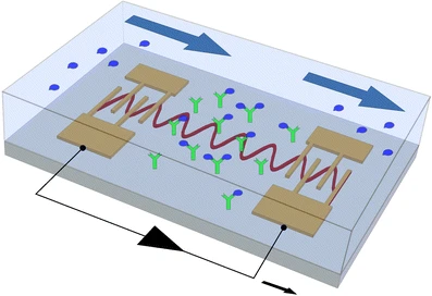
Scientists from Duke University, report an integrated acoustofluidics-based diagnostic system (ADx), which combines triple functions of acoustics, microfluidics, and orthogonal biosensors for clinically accurate, sensitive, and rapid detection of AD biomarkers from human plasma.

They also designed and built a surface acoustic wave-based acoustofluidic separation device to isolate and purify Alzheimer's disease biomarkers to increase the signal-to-noise ratio.

Semiconductor metal oxide nanostructures have various compositions and morphologies such as single crystals, one dimensional, and thin or thick film form. Recently, 1D nanoarchitectors have much attraction for sensing due to their aspect ratio beside their thermal and chemical stabilities.

Nanostructures grown by these techniques could adopt the shape of nanowires, nanobelts, nanotubes, nanoneedles, nanorods, etc...

The modifications in surface morphology result in sensing of various kinds of oxidizing and reducing gases like CO, NH3, NO2, H2, O2, H2S, LPG, xylene, propane, toluene, triethylamine, methanol, and acetone.

Multimodal biosensors within the integrated acoustofluidics-based Alzheimer disease diagnostic system were fabricated by in-situ patterning of the ZnO nanorod array and deposition of Ag nanoparticles onto the ZnO nanorods for surface-enhanced Raman scattering (SERS) and electrochemical immunosensors.

This diagnostic system enabled the label-free detections of SERS and electrochemical immunoassay of clinical plasma samples from AD patients and healthy controls with high sensitivity and specificity.

The researchers believe that this efficient integration provides promising solutions for the early diagnosis of AD.

Read the original article on Pubmed

Scientists aimed to investigate the effects and potential mechanisms underlying the action of Shenqi Fuzheng Injection (a Chinese traditional medicine) on a well-established transgenic mouse model of Amyotrophic Lateral Sclerosis.

Shenqi Fuzheng Injection (SFI) is concocted from two kinds of Chinese medicinal herbs, Radix Codonopsis (the root of Codonopsis pilosula; Chinese name: Dangshen) and Radix Astragali (the root of Astragalus; Chinese name: Huangqi).

It is a drug approved by the State Food and Drug Administration of the People’s Republic of China primarily as an antitumor auxiliary injection and marketed in China by Livzon Pharmaceutical Group Inc.

Transgenic SOD1-G93A mice were intraperitoneally injected with SFI three times a week from 87 days of age.

Motor function, survival, pathological manifestations in the brain, and Nrf2 pathway-related assessments of the mice were performed.

SFI marginally reduced motor neuron loss and astrocytic activation in the motor cortex of the brain of SOD1-G93A mice at 130 days of age.

There was a decrease of the level of malondialdehyde and an increase of the levels of superoxide dismutase, Nrf2, heme oxygenase-1, and glutathione S-transferase in the SOD1-G93A mice.

The SFI treatment did not significantly extended the overall survival but improved the pathological manifestations of the brain alleviating the oxydative stress injury and activating the Nrf2 pathway in the animal model of Amyotrophic Lateral Sclerosis.

Read the original article on Pubmed

CRISPR/Cas9-mediated gene activation is a potential therapeutic strategy that does not induce double-strand break (DSB) DNA damage. However, in vivo gene activation via a Cas9 activator remains a challenge, currently limiting its therapeutic applications.

Scientists developed a Cas9 activator nanocomplex that efficiently activates an endogenous gene in the brain in vivo, suggesting its possible application in novel therapeutics. They demonstrated a potential treatment application of the Cas9 activator nanocomplex by activating Adam10 in the mouse brain without introducing insertions and deletions (inDels).

Remarkably, in vivo activation of Adam10 with the Cas9 activator nanocomplex improved cognitive deficits in an Alzheimer's disease (AD) mouse model. These results demonstrate the therapeutic potential of Cas9 activator nanocomplexes for a wide range of neurological diseases.

Read the original article on Pubmed

Cardiac output is dependent on stroke volume and heart rate. A significant portion patients with chronotropic incompetence are unable to increase heart rate to compensate for increased output demand.

Parkinson's disease (PD) as well as Parkinsonism (PS)are of the most common age-related neurodegenerative disorders. Epidemiological studies have shown that Parkinson's disease is accompanied by high rates of premature death compared with the general population.

In general, death in PD/PS is usually caused by determinant factors such as pneumonia, cerebrovascular, and cardiovascular disease. There is a significant body of literature demonstrating involvement of the heart in PD/PS.

In this study, the authors tried to clarify the link between CI and UPDRS part II (off-on), which was calculated by subtracting part II (on) from part II (off), in patients with Parkinson's disease (PD).

Thirty-six hospitalized patients were examined by using cardiopulmonary exercise testing (CPET) for exercise tolerance (ΔVO2/ΔWR and peak VO2/W) and the presence of CI (ΔHR/ΔWR), and using electrocardiogram for heart rate variability.

The patients with Parkinson's disease (PD) who had Chronotropic incompetence, indeed experienced impaired exercise tolerance.

Those patients with Chronotropic incompetence had a difference in the UPDRS part II score when they were on and off. They felt the difference between ‘on’ and ‘off’ in activities of daily living.

The authors conclusions is that in PD patients, the difference between 'on' and 'off' in activities of daily living might be predicted by using ΔHR/ΔWR x100 obtained from CPET as an index.

Read the original article on Pubmed

Alzheimer's disease (AD), a prevalent neurodegenerative disease with progressive dementia in older adults. The dominant hypothesis tells it is caused by abnormal Amyloid-β (Aβ) peptide in extracellular plaques in the brain. A competing hypothesis ponts to the culprit is intracellular Tau aggregates, which would make Alzheimer's disease more similar to other neurodegenerative diseases.

Drugs to cure AD are not in sight, there were more than 2500 unsuccessful clinical trials in Alzheimer's disease.

The authors of a recent article wanted to determine the effect of various neurotransmission-altering compounds including fenobam, quisqualic acid, and dimethyl sulfoxide in the protection against Amyloid-β toxicity.

The well-known C. elegans Alzheimer's disease model, CL4176, in which human Amyloid-β expression is turned on upon a temperature shift to 25 °C that leads to paralysis, was screened for protection/delay in paralysis because of Amyloid-β toxicity.

Still it looks that scientists choose their animal model more because of availability and costs than credibility. Nematode last ancestors in common with Chordate (Protostomia) lived 650 millions years ago, before the Cambian explosion. In contrast last common ancestors between humans and rodents are 60 million years. Nematode's neurons do not fire action potentials, and do not express any voltage-gated sodium channels. A weird choice of animal model for a neurodegenerative disease!

While screening the compounds, dimethyl sulfoxide (DMSO), a universal solvent used to solubilize compounds, was identified to provide protection. DMSO has been examined for the treatment of numerous conditions including dubious alternative medicine "cures".

The scientists conclusion is that DMSO and Fenobam protect against Amyloid-β toxicity through modulation of neurotransmission. Yet this seems far fetched because of the choice of animal model.

Read the original article on Pubmed

Lifelong bilingualism is associated with delayed dementia onset, suggesting a protective effect on the brain. Here, the authors aim to study the effects of lifelong bilingualism as a dichotomous and continuous phenomenon, on brain metabolism and connectivity in individuals with Alzheimer's dementia.

In the present study, the scientists from Italy and Switzerland modeled bilingualism with two complementary approaches, as either a dichotomous or a continuous phenomenon. They employed FDG-PET to evaluate brain glucose metabolism and brain dysfunction in large series of bilingual and monolingual individuals with AD to investigate the neural effects of lifelong bilingualism.

According to the brain reserve hypothesis, they expected more severe cerebral hypometabolism in the group of bilinguals with AD in comparison to the monolingual AD patients, at comparable levels of dementia severity.

A language background questionnaire measured the level of language use for conversation and reading. Severity of brain hypometabolism and strength of connectivity of the major neurocognitive networks was compared across monolingual and bilingual individuals, and tested against the frequency of second language life-long usage.

Cerebral hypometabolism was more severe in bilingual compared to monolingual patients; severity of hypometabolism positively correlated with the degree of second language use.

The metabolic connectivity analyses showed increased connectivity in the executive, language, and anterior default mode networks in bilingual compared to monolingual patients. The change in neuronal connectivity was stronger in subjects with higher second language use.

The neuroprotective effects of lifelong bilingualism act both against neurodegenerative processes and through the modulation of brain networks connectivity.

These findings highlight the relevance of lifelong bilingualism in brain reserve and compensation, supporting bilingual education and social interventions aimed at usage, and maintenance of two or more languages, including dialects, especially crucial in the elderly people.

Read the original article on Pubmed

The electronic health record is designed principally to support the provision and documentation of clinical care, as well as billing and insurance claims.

Broad implementation of the EHR, however, also yields an opportunity to use EHR data for other purposes, including research and quality improvement. Indeed, effective use of clinical data for research purposes has been a longstanding goal of physicians who provide care for patients with Amyotrophic Lateral Sclerosis, but the quality and completeness of clinical data, as well as the burden of double data entry into the EHR and into a research database, have been persistent barriers.

These factors provided motivation for the development of the Amyotrophic Lateral Sclerosis Toolkit, a set of interactive digital forms within the EHR that enable easy, consistent, and structured capture of information relevant to Amyotrophic Lateral Sclerosis patient care during clinical encounters.

Routine use of the Amyotrophic Lateral Sclerosis Toolkit within the context of the CReATe Consortium's IRB-approved Clinical Procedures to Support Research in Amyotrophic Lateral Sclerosis study protocol, permits aggregation of structured Amyotrophic Lateral Sclerosis patient data, with the goals of empowering research and driving quality improvement.

Widespread use of the Amyotrophic Lateral Sclerosis Toolkit through the CAPTURE-Amyotrophic Lateral Sclerosis protocol will help to ensure that Amyotrophic Lateral Sclerosis clinics become a driving force for collecting and aggregating clinical data in a way that reflects the true diversity of the populations affected by this disease, rather than the restricted subset of patients that currently participate in dedicated research studies.

Read the original article on Pubmed

MRi is a new way to diagnose ALS. It replaces the unreliable practice of old school neurologists with instantaneous, factual information.

Yet there is a need of specialists to analyze MRi pictures. In the long term it will disrupt the way neurologists work, and they may take a back seat in their relation with neurodegenerescent patients.

MRi also reintroduces anatomy in the global picture and scientists may notice what is obvious to patients: That muscle wasting is a central feature of ALS, and not a epiphenomenon.

MRi brings also a new vocabulary, T1, T2, relaxivity, FLAIR, STIR, STRATE. enter image description here Source: KieranMaher at English Wikibooks
T relaxivity contrast imaging may serve as a potential imaging biomarker for amyotrophic lateral sclerosis by noninvasively quantifying the tissue microstructure.

In this preliminary longitudinal study, the authors investigated the Transverse Relaxivity at Tracer Equilibrium (TRATE, an MRI technique) in three muscle groups between SOD1-G93A rat and a control population at two different timepoints. The control group was time matched to the Amyotrophic Lateral Sclerosis group such that the second timepoint was the onset of disease. Other groups have as well experimented this new way to diagnose ALS with MRi on muscles.

They observed a statistically significant decrease in TRATE over time in the gastrocnemius, tibialis, and digital flexor muscles in the SOD1-G93A model, whereas TRATE did not change over time in the control group.

Immunofluorescent staining revealed a decrease in minimum fiber area and cell density in the SOD1-G93A model when compared to the control group. These microstructural changes observed from histology align with the theorized biophysical properties of TRATE.

The scientists here demonstrate that TRATE can longitudinally differentiate disease associated atrophy from healthy muscle and has potential to serve as a biomarker for disease progression and ultimately therapy response in patients with Amyotrophic Lateral Sclerosis.

Let's hope the usage of MRi whill shorten the diagnosis "black hole" that characterize old school neurologists practice.

Let's hope MRi will help to shift the paradigm of "ALS as a Motor Neuron Disease". Many signs hint a a disease striking the skeletal muscles as well, including the presence of TDP-43 aggregates.

Read the original article here

Contact the author

Advertisement


This book retraces the main achievements of ALS research over the last 30 years, presents the drugs under clinical trial, as well as ongoing research on future treatments likely to be able stop the disease in a few years and to provide a complete cure in a decade or two.

Neurodegenerative diseases are characterized by the selective degeneration of neuronal populations in different brain regions and frequently the formation of distinct protein aggregates that often overlap between diseases.

Contrary to most other cells, neurons cannot divide, and therefore cannot dilute misfolded/aggregated proteins that accumulate in their cytosol. Their removal is a complex task involving recognition proteins, chaperones, and eventually activation of degradation pathways such as the ubiquitin-proteasome system and the autophagy-lysosomal pathway.

Mechanisms to recycle proteins are complex, one of them uses lysosomes. A lysosome is a membrane-bound organelle found in many animal cells. They are spherical vesicles that contain hydrolytic enzymes that can break down many kinds of biomolecules.

Lysosomal health is crucial for the degradation of dysfunctional proteins and in particular for the clearance of autophagic vacuoles. An impaired lysosomal system contributes to autophagy stress, accumulation of damaged mitochondria, and restricts clearance of proteins aggregates.

Vacuolar protein sorting ortholog 35 (VPS35) is a protein involved in autophagy and is implicated in neurodegenerative diseases, such as Parkinson's disease (PD) and Alzheimer's disease (AD). Yet it was not commonly associated with ALS.

VPS35 is part of a complex called the retromer, which is responsible for transporting select cargo proteins between vesicular structures (e.g., endosomes, lysosomes, vacuoles) and the Golgi apparatus.

So any defects in VPS35 is associated with neurodegenerative diseases. Indeed it is certainly not the unique cause of those diseases. Mutations in the VPS35 gene have been identified to cause late-onset, autosomal dominant familial Parkinson's disease, whereas reduced VPS35 protein levels are reported in vulnerable brain regions of subjects with Alzheimer's disease, neurodegenerative tauopathies such as progressive supranuclear palsy and Pick's disease, and amyotrophic lateral sclerosis.

Here, Dorian Sargent and colleagues develop conditional knockout mice with the selective deletion of in neurons to better elucidate its role in neuronal viability and its connection to neurodegenerative diseases.

Surprisingly, the pan-neuronal deletion of induces a progressive and rapid disease with motor deficits and early post-natal lethality. Neuronal loss is accompanied and preceded by the formation of p62-positive protein inclusions and robust reactive astrogliosis.

The authors' study reveals a critical yet unappreciated role for VPS35 function in the normal maintenance and survival of motor neurons during post-natal development that has important implications for neurodegenerative diseases, particularly amyotrophic lateral sclerosis.

In 2020 another team achieved a substantial reduction of CRC proteins in motor neurons of SOD1 G93A mice. They designed, synthetize and characterize a small array of bis-guanylhydrazones. Such retromer stabilizers possess good in vivo bioavailability, potency, and stability.

Read the original article on Pubmed

Evolution of pure autonomic failure towards Parkinson or MSA

- Posted by admin in English

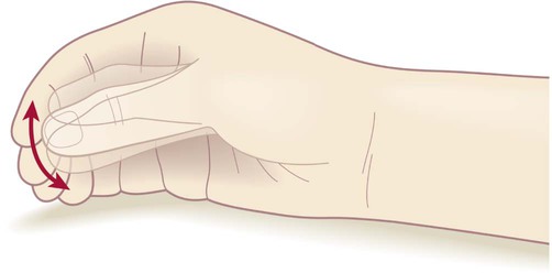
Pure autonomic failure is a rare degenerative disease of the autonomic nervous system. Symptoms include dizziness and fainting (caused by orthostatic hypotension), visual disturbances and neck pain. Chest pain, fatigue and sexual dysfunction are less common symptoms that may also occur. enter image description here ‘Pill-rolling’ rest tremor as found in Parkinson’s disease.

The autonomic nervous system is a control system that acts largely unconsciously and regulates bodily functions. It has a sequential organization: The preganglionic neuron synapse onto a postganglionic neuron before innervating the target organ. It's a bit similar to the upper/lower motor neuron/muscle assemblage.

Longitudinal studies have reported that Pure autonomic failure can phenoconvert to a central synucleinopathy with motor or cognitive involvement-i.e., to Parkinson disease, dementia with Lewy bodies, or multiple system atrophy.

Synucleinopathies are neurodegenerative diseases characterised by the abnormal accumulation of aggregates of alpha-synuclein protein in neurons, nerve fibres or glial cells.

These longitudinal studies have classified patients clinically as having Pure autonomic failure based on neurogenic orthostatic hypotension without an identified secondary cause or clinical evidence of motor or cognitive impairment due to central neurodegeneration.

This approach lumps together two neurogenic orthostatic hypotension syndromes that are pathologically and neurochemically distinct.

  • One is characterized by intraneuronal cytoplasmic alpha-synuclein aggregates and degeneration of postganglionic sympathetic neurons, as in Parkinson disease and Dementia with Lewy Bodies.
  • The other is not, as in multiple system atrophy.

Clinical and postmortem data show that the form of Pure autonomic failure that involves sympathetic intraneuronal synucleinopathy and noradrenergic deficiency can phenoconvert to Parkinson disease or Dementia with Lewy Bodies-but not to multiple system atrophy.

Conversely, Pure autonomic failure without these features leaves open the possibility of premotor multiple system atrophy.

Multiple system atrophy is a rare neurodegenerative disorder characterized by autonomic dysfunction, tremors, slow movement, muscle rigidity, and postural instability (collectively known as parkinsonism) and ataxia. This is caused by progressive degeneration of neurons in several parts of the brain including the basal ganglia, inferior olivary nucleus, and cerebellum.

MSA generally show little response to the dopamine medications used to treat Parkinson's disease and only about 9% of MSA patients with tremor exhibit a true parkinsonian pill-rolling tremor.

Read the original article on Pubmed


Please, help us continue to provide valuable information: