A new publication indicates that amyotrophic lateral sclerosis, a chronic and progressive neurodegenerative disease of the upper and lower motor neurons, is associated with an inflammatory attack of the brain and spinal cord.

This is certainly not a common view among scientists, although some related views have long been held for example by Dr. Stanley Appel. On contrary this view, that in some patients the disease begins as a result of trauma or severe infection, has long been advocated by patients on Internet forums.

For dr Appel, initially, motor neurons sustain injuries causing disruptions of critical intracellular pathways. But as injuries add up, motor neurons release distress signals, which induce inflammatory processes produced by surrounding glial cells in the CNS as well as peripheral innate and adaptive immune cells. In addition there are infiltrating peripheral monocytes and lymphocytes

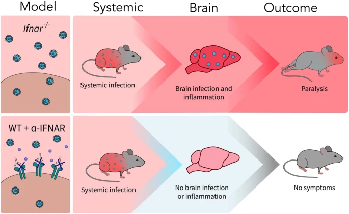
In a rare perspective, the authors, led by Milan Fiala of UCLA's David Geffen School of Medicine, report that while the cause of ALS is unknown, epidemiological data show an association of ALS with trauma antecedents and microbial infections.

For example, the role of microbial infection was noted by the lead author in a study of a genetically identical pair of twins with both twins possessing common ALS mutations, but only the twin suffering from a cat bite and from a severe systemic Pasteurella infection later developed ALS, while the other twin remains healthy 11 years later. The ALS twin exhibited spontaneous production of the cytokines IL-6 and TNFα and epigenetic modification through differential methylation of immune-related regions near the EGFR and TNFRSF11A genes.

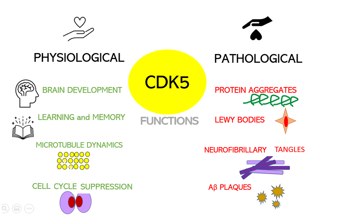
For the authors, TDP-43, a nuclear DNA/RNA binding protein present in 95% of ALS patients in an aggregated, poorly localized and unfolded form, induces inflammation. It is not clear from the publication why TDP-43 should have this deleterious behavior. TDP-43 is an essential protein for correcting DNA errors, such as those induced by HIV infections. Interestingly, leading scientists believe it's the opposite: inflammation promotes TDP-43 aggregation.

Scientists studied the peripheral blood mononuclear cell (PBMC) transcriptome of eight patients with sporadic ALS (sALS) over the course of the disease. They also analyzed the relationship between inflammation and autoimmunity in PBMC and NK cells from a pair of ALS-discordant identical twins. But they only followed 2 patients for a year and the results they claim came mostly from one patient (#10). So take all of this with a pinch of salt. enter image description here The authors initially hypothesized that activation of the inhibitory co-receptor PD-1 by recombinant PD-L1 would be anti-inflammatory. However, recombinant PD-L1 ligand and recombinant PD-1 receptor were strongly pro-inflammatory. Then they abandoned the PD-L1 strategy and chose a strategy with dimethyl fumarate (DMF), a drug approved against two autoimmune diseases, multiple sclerosis and psoriasis, and the cGAS- STING H-151 involved in autoimmunity.

While DMF and H-151 have been shown to inhibit inflammatory and autoimmune signaling in the immune cells of some ALS patients, the results are really inconclusive. Scientists suggest that due to the heterogeneity of inflammatory mechanisms in ALS, various immunotherapeutic strategies may be needed.

Des acides gras polyinsaturés ralentiraient la progression de la SLA

- Posted by admin in Français

Même si les mécanismes moléculaires responsables des maladies neurodégénératives ne sont pas totalement élucidés, la dérégulation de l'autophagie et de la voie endosomal-lysosomal, se sont avérés associés à ces troubles. Au sein de ces voies, la cible mammalienne de la rapamycine (mTOR) entre autres inhibe les mécanismes autophagiques, qui sont nécessaires pour dégrader et éliminer les protéines anormales ou agrégées. enter image description here Les inhibiteurs classiques de mTOR tels que la rapamycine ou la metformine peuvent améliorer les dommages aux neurones. Une autre approche importante pour lutter contre les ND est l'utilisation de produits/composés naturels comprenant des antioxydants, des flavonoïdes, des polyphénols et des acides gras polyinsaturés (PUFA). Diverses études ces dernières années ont montré les effets bénéfiques des acides gras poly insaturés (AGPI) oméga-3 à travers divers mécanismes dont des effets anti-inflammatoires. Les acides gras oméga-3 les mieux étudiés sont l'acide docosahexaénoïque (DHA), l'acide eicosapentaénoïque (EPA) et l'acide α-linolénique (ALA).

Une nouvelle étude dirigée par la Harvard T.H. Chan School of Public Health indique que la consommation d'acides gras oméga-3, en particulier l'acide alpha-linolénique (ALA), un nutriment que l'on retrouve dans des aliments tels que les graines de lin, les noix, l'huile de colza, l'huile de soja et les graines de chia, pourrait aider à ralentir la progression de la maladie chez les patients atteints de sclérose latérale amyotrophique (SLA). Cette étude a été publiée le 21 juin 2023 dans la revue Neurology.

"Les résultats antérieurs de notre groupe de recherche ont montré qu'un régime alimentaire riche en ALA et des taux sanguins élevés de cet acide gras pouvaient réduire le risque de développer la SLA. Dans cette étude, nous avons constaté que chez les personnes atteintes de SLA, des taux sanguins plus élevés d'ALA étaient également associés à une progression plus lente de la maladie et à un risque de décès plus faible pendant la période de l'étude", a déclaré Kjetil Bjornevik, auteur principal de l'étude et professeur adjoint d'épidémiologie et de nutrition. "Ces résultats, ainsi que nos recherches précédentes, suggèrent que cet acide gras pourrait avoir des effets neuroprotecteurs bénéfiques pour les personnes atteintes de SLA."

Les chercheurs ont mené une étude auprès de 449 personnes atteintes de SLA qui ont participé à un essai clinique. Dans le cadre de cet essai, la gravité de leurs symptômes et la progression de leur maladie ont été évaluées et notées de 0 à 40, les scores les plus élevés indiquant des symptômes moins graves de la maladie. Les chercheurs ont mesuré les niveaux d'acides gras oméga-3 dans le sang des participants et les ont répartis en quatre groupes, des niveaux les plus élevés aux niveaux les plus faibles. Ils ont ensuite effectué un suivi 18 mois plus tard pour évaluer la fonctionnalité physique des groupes et la survie en fonction de l'essai clinique.

Ils ont constaté que l'ALA présentait le plus d'avantages parmi tous les acides gras oméga-3, car il était le plus étroitement lié à un déclin plus lent et à un risque de décès réduit. Sur les 126 participants décédés dans les 18 mois suivant le début de l'étude, 33 % appartenaient au groupe ayant les niveaux les plus faibles d'ALA, tandis que 19 % appartenaient au groupe ayant les niveaux les plus élevés d'ALA. En ajustant les facteurs tels que l'âge, le sexe, l'origine ethnique, l'IMC, la durée des symptômes et les antécédents familiaux de SLA, les chercheurs ont calculé que les participants ayant les niveaux les plus élevés d'ALA présentaient un risque de décès réduit de 50 % pendant la période de l'étude par rapport aux participants ayant les niveaux les plus faibles d'ALA.

Deux autres acides gras étaient également associés à une réduction du risque de décès pendant la période de l'étude : l'acide eicosapentaénoïque, un autre acide gras oméga-3 que l'on trouve dans les poissons gras et l'huile de poisson, et l'acide linoléique, un acide gras oméga-6 présent dans les huiles végétales, les noix et les graines.

"Le lien que notre étude a trouvé entre l'alimentation et la SLA est intrigant", a déclaré Alberto Ascherio, auteur principal de l'étude et professeur d'épidémiologie et de nutrition. "Nous contactons maintenant les chercheurs cliniques pour promouvoir un essai randomisé visant à déterminer si l'ALA est bénéfique pour les personnes atteintes de SLA. Obtenir un financement sera un défi, car l'ALA n'est pas un médicament brevetable, mais nous espérons y parvenir."

Malheureusement il est habituel pour l'industrie pharmaceutique de délibérément négliger les médicaments potentiels qui ne sont pas brevetables.

CuATSM, also known as copper (II) diacetylbis(N4-methylthiosemicarbazone), is a chemical compound that has been studied for its potential therapeutic effects in patients with amyotrophic lateral sclerosis (ALS). enter image description here SOD1, a protein involved in 2% of ALS cases contains a binuclear Cu/Zn site in each subunit. So it seems there is some relation between copper and ALS. It is believed that CuATSM's mechanism of action involves restoring the balance of copper ions and preventing the buildup of toxic forms of copper. But this is at best highly speculative.

CuATSM is one of those drugs highly promoted on social networks as an effective treatment for SOD1-ALS, yet the evidence was unclear. There were several pre-clinical studies that were very encouraging ... by the same team.

There were clinical studies. On January 7th, 2019, television spots and a media release stated that CuATSM slowed disease progression by 70%! in the phase 1 clinical trial participants. This resulted in an international outcry for people with ALS/MND to access CuATSM. The Phase 2/3 trial was completed in late 2021 although the outcome of the clinical trial has yet to be announced.

Up to 80 patients were planned to be treated with CuATSM or placebo for 24 weeks. The studied drug is administered orally, once a day before breakfast.

The present study set out to perform a comparative analysis of Amyotrophic Lateral Sclerosis pathology.

  • In 6 ALS patients that had been administered CuATSM and riluzole (4 X ALS-TDP and 2 X ALS-SOD1)
  • In 6 ALS patients that had been administered riluzole only ( (4 X ALS-TDP and 2 X ALS-SOD1) ).

The authors' results revealed no significant difference in neuron density or TDP-43 burden in the motor cortex and spinal cord of patients that had received CuATSM compared to patients that had not.

In patients that had received CuATSM, immunoreactive astrocytes were observed in the motor cortex and reduced Iba1 density (Iba1/ AIF-1 is usually found in macrophages and microglia) in the spinal cord.

However, no significant difference in measures of astrocytic activity and SOD1 immunoreactivity was found with CuATSM treatment.

These findings, in this first postmortem investigation of patients with Amyotrophic Lateral Sclerosis in CuATSM trials, demonstrate that in contrast to that seen in preclinical models of disease, CuATSM does not significantly alleviate neuronal pathology or astrogliosis in patients with Amyotrophic Lateral Sclerosis.

Un article de scientifiques de Lisbonne et de Londres décrit une étude rétrospective monocentrique menée sur des patients atteints d'une des maladies du motoneurone sur une période de 27 ans, la SLA était le phénotype le plus répandu, qui était présent chez 82 % des patients. enter image description here L'étude visait à analyser les caractéristiques démographiques et cliniques des patients, y compris les différents sous-types de SLA, la progression de la maladie, les retards de diagnostic, l'intervention de ventilation non invasive (VNI) et les taux de survie. L'étude a inclus un total de 1 550 patients qui ont visité la clinique entre 1994 et 2020.

Les résultats ont montré que l'âge moyen au début de la maladie était de 62 ans, avec une prévalence légèrement plus élevée chez les hommes. La distribution des sous-types SLA variait, certains sous-types montrant une prédominance masculine et d'autres montrant une prédominance féminine. L'âge médian d'apparition différait selon les sous-types. La fréquence globale des cas familiaux de SLA était de 8,0 %, certains sous-types ayant une proportion plus élevée de cas familiaux.

Le délai de diagnostic variait selon les sous-types, la SLA cognitive (c-ALS) ayant le délai le plus long et la SLA bulbaire (b-ALS) ayant le plus court. Des différences dans les taux de déclin fonctionnel ont également été observées entre les sous-types, certains sous-types progressant plus lentement que d'autres. Après un an de suivi, les patients atteints de SLA ont perdu en moyenne 11 points sur l'échelle d'évaluation fonctionnelle de la SLA (ALSFRS-R), la c-ALS affichant la plus forte baisse.

Le taux et le moment de l'intervention de la VNI variaient selon les sous-types, certains sous-types étant prescrits plus tôt et plus fréquemment que d'autres. Les taux de survie différaient également entre les sous-types, certains sous-types ayant des durées de survie plus longues que d'autres. L'analyse de survie comparant différentes périodes de temps a montré une augmentation de la survie médiane au fil du temps, en particulier dans les périodes les plus récentes.

Les caractéristiques démographiques et cliniques de différents phénotypes de la maladie de la motoneurone (MND) tels que la sclérose latérale amyotrophique (SLA), l'amyotrophie spinale proximale (PMA), le syndrome de la motoneurone progressive (PLS) et les formes monoméliques (qui n'affectent qu'un seul membre) sont en résumé les suivantes :

  • La SLA était le phénotype le plus répandu, représentant 82 % des cas étudiés.
  • La prédominance masculine était observée dans la SLA, la PMA et la SLA monomélique, mais pas dans le PLS.
  • La SLA et la PMA avaient tendance à se manifester plus tard dans la vie par rapport au PLS et aux formes monoméliques.
  • La progression de la SLA était décrite comme une courbe sigmoïde (en S), avec une diminution progressive des scores ALSFRS-R suivie d'une phase de progression uniforme.
  • La zone d'apparition de la maladie était une caractéristique prédictive du pronostic, avec des différences significatives entre les phénotypes.
  • Les patients présentant des modifications cognitives isolées (c-ALS) étaient diagnostiqués plus tardivement que les autres groupes mais avaient une survie plus longue.
  • La survie dans la SLA a augmenté au fil du temps, en particulier dans la sclérose latérale amyotrophique sporadique (s-ALS), probablement en raison d'un diagnostic plus rapide, d'une utilisation plus répandue de la ventilation non invasive et de l'utilisation du riluzole.

Dans l'ensemble, l'étude a révélé des changements dans la distribution des sous-types SLA au fil des ans, avec une augmentation de la proportion de patients atteints d'amyotrophie progressive (PMA) et une diminution de ceux atteints de SLA sporadique (s-ALS). Il y avait aussi une tendance vers un diagnostic plus précoce au fil du temps. La déficience fonctionnelle, l'intervention VNI et les taux de survie variaient entre les différentes périodes.

The retina is considered an anatomical projection of the central nervous system with the same embryological origins. Unmyelinated axons of retinal ganglion cells form the retinal nerve fiber layer (RNFL) which extends like the optic nerve and connects to the lateral geniculate nucleus (LGN) in the thalamus, which serves as the first relay center of the visual pathway.

The presence of retinal pathology is closely linked to cognitive deficits in patients with Alzheimer's disease (AD), the deposition of amyloid-β plaques leading to retinal thickening. Specifically, peripapillary thinning of the retinal nerve fiber layer is the most common finding in many neurological conditions such as multiple sclerosis, stroke, neuromyelitis optica, dementia with Lewy bodies, Parkinson's, and Alzheimer's disease. Contradictory results are described in amyotrophic lateral sclerosis.

Even though some biomarkers have been validated and incorporated into new clinical diagnostic criteria, most have suboptimal test accuracy and are either very expensive, such as detecting Aβ and Tau deposits in the brain by positron emission tomography (PET), or quite invasive such as as a measure of tau protein and Aβ peptide levels in cerebrospinal fluid analysis. These retinal changes are clinically measurable using existing non-invasive techniques and can be used for the early diagnosis of neurodegenerative disorders.

Optical coherence tomography (OCT) is a relatively inexpensive, harmless, and rapid technique that allows objective retinal measurements and quantification in vivo. OCT is commonly used in ophthalmology to assess retinal integrity through high-resolution cross-sectional scans of retinal layers such as RNFL and CGL at different locations such as the macula or papilla. It is also an interesting tool for neurological research.

A new study sheds light on the pathological significance and clinical value of retinal atrophy in patients with amyotrophic lateral sclerosis and Kennedy disease (spinal and bulbar muscular atrophy).

Authors from Padua, Italy recruited thirty-five patients with amyotrophic lateral sclerosis, thirty-seven with Kennedy disease, and forty-nine age-matched healthy controls were included in a longitudinal study of a year. Spectrum-domain optical coherence tomography was performed at the study entry and after 12 months.

Peripapillary retinal nerve fiber layer thickness was significantly thinner in amyotrophic lateral sclerosis and Kennedy disease compared to healthy controls. The peripapillary retinal nerve fiber layer was thinner in Kennedy disease compared to amyotrophic lateral sclerosis, but the difference was not statistically significant. In Kennedy's disease, pRNFL atrophy was significantly correlated with both disease severity and duration, whereas no significant correlation was found in amyotrophic lateral sclerosis.

During follow-up, the thickness of the peripapillary retinal nerve fiber layer remained stable in Kennedy disease whereas it decreased significantly in amyotrophic lateral sclerosis. : The authors conclude that retinal atrophy in amyotrophic lateral sclerosis and Kennedy disease. However, the study has no statistical value because the sample is much too small, and we know that the Amyotrophic Lateral Sclerosis Functional Rating Scale Revised (ALSFRS-R) and the functional assessment of bulbar spinal muscular atrophy (SBMA-FRS) are not reliable markers of disease progression. In fact, the use of tools to turn over, to clear the bronchi, or respiratory assistance leads to an improvement in this type of measurement, without the progression of the disease being changed.

So as usual, while the authors obviously did a lot of work, we do not know much more after this study than before reading it.

Essai clinique de phase 1/2a dans la SLA avec le ropinirol

- Posted by admin in Français

Le 1er juin dans la revue Cell Stem Cell, des chercheurs japonais montrent dans un essai clinique de phase I/IIa que le ropinirole, un médicament contre la maladie de Parkinson, peut être utilisé en toute sécurité chez les patients SLA et retarde la progression de la maladie de 27, semaines en moyenne. enter image description here Le développement de médicaments pour les maladies du système nerveux a un faible taux de réussite et les modèles animaux précliniques ont un potentiel translationnel limité.

L'essai de faisabilité de phase 1/2a monocentrique, randomisé, contrôlé par placebo, évaluait l'innocuité, la tolérabilité et l'efficacité du ropinirole.

L'équipe a recruté 20 patients recevant des soins à l'hôpital universitaire de Keio au Japon. Aucun des patients n'était porteur de gènes prédisposant à la maladie et, en moyenne, ils vivaient avec la SLA depuis 20 mois.

L'essai était en double aveugle pendant les 24 premières semaines, puis pendant les 24 semaines suivantes, tous les patients qui souhaitaient continuer ont reçu du ropinirole. Cependant de nombreux patients ont abandonné en cours de route, en partie à cause de la pandémie de COVID-19, de sorte que seuls sept des 13 patients traités au ropinirole et un des sept patients traités par placebo suivi par le ropinirole ont été suivis pendant toute l'année. Cependant, aucun patient n'a abandonné pour des raisons de sécurité.

Pour déterminer si le médicament était efficace pour ralentir la progression de la SLA, l'équipe a effectué un vaste ensemble de mesures tout au long de l'essai et pendant quatre semaines après la fin du traitement. Celles-ci comprenaient des changements dans l'activité physique autodéclarée des patients et leur capacité à manger et à boire de manière autonome, des données d'activité provenant d'appareils portables et des changements mesurés par le médecin dans la mobilité, la force musculaire et la fonction pulmonaire.

Certains patients étaient plus sensibles au traitement au ropinirole que d'autres. Cependant, les patients du groupe placebo qui ont commencé à prendre du ropinirole à mi-parcours de l'essai n'ont pas connu un ralentissement de la progression de la maladie, ce qui suggère que le traitement au ropinirole ne peut être utile que si le traitement est commencé plus tôt et administré sur une durée plus longue.

Pour pallier à la très faible taille de l'échantillon et sa valeur nulle sur le plan statistique, les chercheurs ont essayé de corroborer ces résultats in vitro à l'aide de motoneurones dérivés de cellules souches de patients.

Pour ce faire, ils ont généré des cellules souches pluripotentes induites à partir du sang des patients et ont transformé ces cellules en motoneurones en laboratoire. Par rapport aux motoneurones sains, ils ont découvert que les motoneurones des patients SLA présentaient des différences distinctes dans la structure, l'expression des gènes et les concentrations de métabolites, mais le traitement au ropinirole réduisait ces différences.

Plus précisément, les motoneurones issus de patients SLA avaient des neurites plus courts que les motoneurones sains, mais ces axones ont atteint une longueur plus normale lorsque les cellules ont été traitées avec du ropinirole. On ne sait pas pourquoi certains patients sont plus sensibles au ropinirole que d'autres, mais les chercheurs pensent que cela est probablement dû à des différences génétiques qu'ils espèrent identifier dans de futures études.

L'ampleur de l'effet estimé du ropinirole sur le score ALSFRS-R sur 48 semaines était de 1 à 9, points, entraînant un déclin fonctionnel de 21 % à 60 % plus lent, ce qui est considéré comme cliniquement significatif. Le mécanisme d'action précis du ropinirole dans la SLA mérite une enquête plus approfondie ; par conséquent, les scientifiques ont également mené une recherche translationnelle inverse en parallèle avec l'essai clinique.

Les interprétations des analyses d'efficacité dans cette étude étaient limitées par la petite taille de l'échantillon de 20 participants. Cela a été encore aggravé par le taux d'abandon étonnamment plus élevé dans cette étude par rapport au taux historique dans les essais cliniques sur la SLA (∼20 %)19 ; 46, % (6/13) des participants du groupe ropinirole et 85, % (6/7) des participants du groupe placebo ont arrêté l'essai. Le ratio de participants dans le groupe ropinirole versus placebo a changé, en particulier dans la période d'extension en ouvert.

Ces taux d'abandon, en particulier pendant la phase d'extension en ouvert, étaient attribuables, au moins en partie, à l'épidémie de maladie à coronavirus 2019 (COVID-19), qui représentait 23 % et 29 % des participants des groupes ropinirole et placebo , respectivement.

De façon questionnable, les scientifiques ont utilisé le modèle mixte pour mesures répétées (MMRM) pour déduire les données manquantes. Les enquêteurs ont utilisé des données historiques issues d'autres études. Les scientifiques ont effectué une analyse post hoc en utilisant les données du registre PRO-ACT. Après toutes ces manipulations statistiques il semblait que le traitement des patients SLA avec du chlorhydrate de ropinirole pendant 48 semaines réduisait significativement la baisse de l'ALSFRS-R après au moins 6 mois d'administration.

Les résultats ont montré que le NfL du liquide céphalo-rachidien, un marqueur des dommages neuronaux, était diminué dans le groupe dans lequel le peroxyde lipidique du liquide céphalo-rachidien était diminué par le traitement au ropinirole.

En résumé, les scientifiques suggérent que la régulation négative de la voie de synthèse du cholestérol au niveau neuronal, plutôt que la réduction du cholestérol circulant systémique, peut être un traitement efficace pour les patients SLA. Cependant, ses mécanismes sous-jacents restent obscurs. Notamment, les taux systémiques de cholestérol circulant n'ont pas été modifiés par le ropinirole dans cet essai clinique. Cela peut être un avantage médicamenteux car le ropinirole abaisse le taux de cholestérol au niveau des motoneurones sans abaisser le taux de cholestérol circulant systémique, qui est un facteur important dans la survie à la SLA.

En effet, in vitro, les scientifiques ont constaté que l'inhibition directe du système voie de synthèse du cholestérol a un effet protecteur sur les motoneurones dérivés d'iPSC d'un patient atteint de SLA familiale (en dehors de l'essai).

Les scientifiques ont proposé que la suppression de la production de cholestérol au niveau du motoneurone plutôt que la voie de synthèse du cholestérol systémique par le ropinirole puisse avoir des effets anti-SLA.

L'essai ROPALS de phase I était donc de très petite taille, les méthodes statistiques d'analyse des résultats peu conventionneles (mais ce genre de pratique semble de plus en plus répandu dans le milieu scientifique et pharmaceutique où des sommes importantes sont en cause) et donc peu signicatif, malgré tout il est encourageant. De plus, il y avait un taux d'attrition ou d'abandon très élevé pendant la période d'extension.

Ces résultats nécessitent donc une étude approfondie avant d'être intégrés à la pratique clinique. Seule une étude clinique de phase III sur un nombre de participants supérieur à 500 pourra démontrer un effet réel de ce médicament sur la maladie.

What could be learned from Wave's ALS drug failure?

- Posted by admin in English

enter image description here When Arimoclomol debacle was announced I was stunned. After all, it was supposed to have a precise mechanism of action in ALS, and this was not so frequent in ALS clinical trial. Most ALS cases are characterized by misfolded, mislocated TDP-43 protein accumulation in granules. This is similar to what appears in Alzheimer's and other neurodegenerative diseases. Arimoclomol was supposed to activate the cellular heat shock response by activating molecular chaperones. As molecular chaperones' role is to fold proteins correctly, it seemed evident that they would correctly fold the misfolded proteins. Yet this ignores that those corrected proteins are still mislocated in the cytosol and as they do not enter the ER and Golgi apparatus, they are not sent to the nucleus. The phase III ALS clinical trial was a failure, but the drug works in other diseases.

Wave's WVE-004 targets C9orf72 mutations in ALS. C9orf72 hexanucleotide GGGGCC repeat expansion is the most common genetic cause of amyotrophic lateral sclerosis (ALS) and frontotemporal dementia (FTD) and WvE-004 targets them in a very smart manner.

C9 mutations are much more complicated than SOD1 mutations which are already numerous and present very different phenotypes, with some patients dying one year after diagnosis, and others living two decades. An ASO therapy, Tofersen, was conditionally approved for medical use in the United States in April 2023. Yet it is useful for only one mutation in the SOD1 gene, found in less than 2% of all ALS cases, and therefore it will be useless for most ALS patients. But for those patients that are targeted, it works, kind of.

WVE-004 uses an approach known as ‘antisense’, where the drug directly interferes with the faulty instructions for making a protein.

So an ASO for C9orf72 which targets mutations in the C9 chromosome, should in principle result in good results.

The C9orf72 gene contains three different sets of instructions (RNA) to make the C9orf72 protein and these three RNA are called V1, V2, and V3. In some forms of MND, mutations in the C9orf72 gene cause the V1 and V3 instructions to be faulty and this leads to the production of toxic proteins which build up in the neurons. WVE-004 targets the faulty V1 and V3 RNA and signals to the cell that they need to be destroyed, leaving the V2 instructions functional so that a healthy version of the C9orf72 protein can still be made.

So what went wrong? We will never know as Wave will discontinue developing the therapy, and its open-label extension study will be stopped. Yet strong reductions in poly(GP) were observed, with patients on a single 20 mg dose and those in the multiple dose groups all having a roughly 50% decrease in poly(GP) levels over the start of the study.

This is hard to understand, there were more than 500 clinical trials on ALS drugs, and most of them did not have a theoretical principle of action. Here is a drug that has a sound principle of action, that apparently works at biomarkers levels. Yet it is stopped in phase I!

What if, as common sense tells us, there is a need for much more than 24 weeks to heal an ALS patient? Even at a purely statistical level, 42 (patients) is a too small number to rise any meaningful conclusion.

It's a financial reasoning, for investors in biotechs it's a game, biotechs often lose but if they win then it's the jackpot for investors. In this paradigm, you have to invest small amounts and know how to move on to another target if there are no quick results.

Here most likely the biotech Wave was unable to convince its investors of the value of their drug and the investors decided it was time to stop funding.

Assessing the effectiveness of GluA2 mRNA editing in CSF can provide valuable insight into the diagnosis and prognosis of ALS, as well as guide the development of potential therapeutic approaches, a new study from Tokyo Medical University suggests. enter image description here The disease known as ALS is multifaceted, in fact not only are there believed to be dozens of ALS subtypes, but there are also dozens of diseases that have similar symptoms to ALS. This greatly complicates not only the diagnosis, which is always long and uncertain, but also the clinical trials which are based on the implicit assumption that all the patients treated have the same disease, which is probably not the case for the trials of drugs for degenerative diseases.

The publication by researchers at the University of Tsukuba that interests us today discusses the need for reliable diagnostic biomarkers and treatment predictors for amyotrophic lateral sclerosis. The authors highlight the role of dysregulated glutamatergic signaling and RNA editing in the pathogenesis of ALS. They focus specifically on the glutamine/arginine (Q/R) site of the mRNA of GluA2, a subunit of AMPA receptors, which is normally edited by RNA-acting adenosine deaminase 2 (ADAR2) in healthy neurons. In sporadic ALS, downregulation of ADAR2 leads to expression of unedited GluA2 mRNA at the Q/R site, resulting in motor neuron death.

Researchers at the University of Tsukuba studied the mRNA editing efficiency of GluA2 in the cerebrospinal fluid (CSF) of patients with sporadic ALS and compared it to non-ALS controls. They found a significant reduction in the editing efficiency at the Q/R site of GluA2 mRNA in the CSF of ALS patients, suggesting that it could serve as a diagnostic biomarker for ALS. They also observed a correlation between reduced editing efficiency and clinical parameters such as longer disease duration and more advanced symptoms, particularly affecting the lower extremities.

GluA2 mRNA is an RNA molecule that contains instructions for the production of the protein GluA2, which is a subunit of AMPA receptors, glutamate receptors.

Editing efficiency refers to the extent to which the Q/R site of GluA2 mRNA is modified by the enzyme ADAR2. In healthy neurons, ADAR2 edits GluA2 mRNA at the Q/R site, converting adenosine to inosine. However, in sporadic ALS, ADAR2 is downregulated, resulting in unedited GluA2 mRNA expression at the Q/R site. This decrease in editing efficiency suggests impaired GluA2 mRNA regulation in sporadic ALS.

Specifically, ALS patients with reduced editing efficiency experienced longer disease duration and more advanced symptoms, including impaired lower limb functions. These results suggest that GluA2 mRNA editing efficiency in CSF may be associated with the progression and severity of sporadic ALS.

The study implies that the efficiency of editing at the Q/R site of GluA2 mRNA could not only help diagnose ALS, but also help identify treatable ALS cases. Therapies targeting dysregulation of RNA editing are currently being developed, and this biomarker could potentially help predict treatment efficacy.

Misdiagnosis in Amyotrophic Lateral Sclerosis

- Posted by admin in English

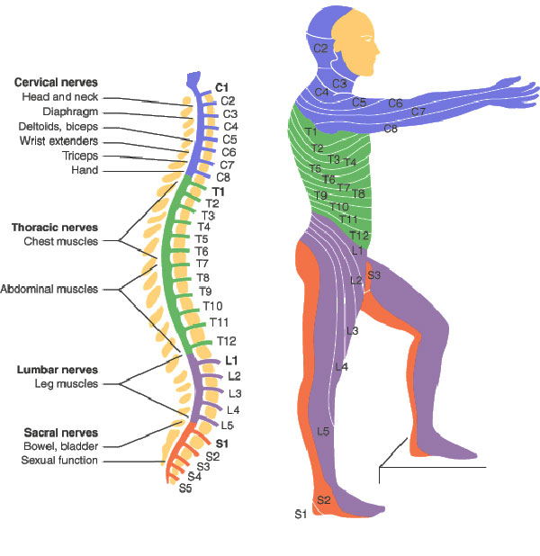
This post is about a review of misdiagnosis in Amyotrophic Lateral Sclerosis (ALS). This refers to cases where individuals are incorrectly diagnosed with ALS when they actually have a different condition or disease. ALS is a complex and challenging disease to diagnose accurately, as it shares some similarities with other neurological disorders. Misdiagnosis can lead to delays in appropriate treatment and management of the actual condition, resulting in potential harm to patients. enter image description here Hand surgeons are commonly the first providers to evaluate patients with undiagnosed ALS (the famous split-hand syndrome). So in early phases, the symptoms of amyotrophic lateral sclerosis (ALS) can mimic those of compressive neuropathies, such as carpal and cubital tunnel syndromes, especially early in a patient's clinical course. Scientists surveyed members of the American Society for Surgery of the Hand and found that 11% of active and retired members have performed nerve decompression surgeries on patients later diagnosed with ALS. Other scientists found that in 95% of cases, the diagnostic errors are, related to physician cognitive error that results from a lack of knowledge about the disease and inadequate decisions

Several factors contribute to the potential for misdiagnosis in ALS:

  • Overlapping symptoms: ALS symptoms, such as muscle weakness, muscle atrophy, fasciculations (muscle twitches), and difficulty speaking or swallowing, can resemble symptoms of other neuromuscular or neurodegenerative disorders. Conditions such as spinal muscular atrophy, primary lateral sclerosis, Kennedy's disease, multifocal motor neuropathy, and certain forms of muscular dystrophy may exhibit similar symptoms, leading to confusion in diagnosis.

  • Variability in symptom progression: ALS symptoms can vary in their presentation and progression among individuals. Some patients may experience rapid progression, while others have a slower disease course. This variability can make it challenging to differentiate ALS from other conditions that may share similar symptom patterns.

  • The diagnosis of ALS is primarily based on clinical examination, medical history, and the exclusion of other potential causes. There is no single definitive test for ALS, making the diagnosis subjective and reliant on the judgment and experience of healthcare professionals. This subjectivity can increase the risk of misdiagnosis.

  • Limited accessibility to specialized testing: Certain diagnostic tests, such as electromyography (EMG) and nerve conduction studies, can help in confirming ALS by evaluating the electrical activity of muscles and nerves. However, these tests may not be readily available in all healthcare settings, leading to potential delays or errors in diagnosis.

  • Disease heterogeneity: ALS is a heterogeneous disease, meaning it can manifest differently in different individuals. Some cases may present with atypical features or unusual disease progression, making it more challenging to reach an accurate diagnosis.

To minimize the risk of misdiagnosis in ALS, it is crucial to follow a comprehensive diagnostic approach that includes thorough clinical evaluations, specialized testing (such as EMG), and consultation with neurologists or neuromuscular specialists experienced in diagnosing and managing ALS. Additionally, regular follow-up assessments and monitoring of symptoms are important to ensure accurate diagnosis and appropriate management throughout the disease course.

It is important to be aware of the history, signs, and symptoms of ALS to provide an accurate diagnosis and prevent unnecessary morbidities, such as nerve decompression surgery, which invariably results in poor outcomes. The major "red flag" symptoms warranting further work-up include weakness without sensory symptoms, profound weakness and atrophy in multiple nerve distributions, progressively bilateral and global symptoms, presence of bulbar symptoms (such as tongue fasciculations and speech/swallowing difficulties), and if surgery is performed, failure to improve. If any of these red flags are present, we recommend neurodiagnostic testing and prompt referral to a neurologist for further work-up and treatment.

It is worth noting that misdiagnosis can also occur in the opposite direction, where individuals with other conditions are mistakenly diagnosed with ALS. Therefore, seeking multiple medical opinions and consulting with specialists can help reduce the likelihood of misdiagnosis and ensure the most accurate diagnosis and appropriate care.

This review discusses the potential therapeutic approach of targeting mitochondrial dysdynamism in amyotrophic lateral sclerosis (ALS) and other neurodegenerative diseases.

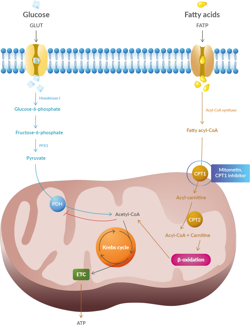
Mitochondria, which are responsible for producing energy in cells, play a crucial role in neuronal function. Neurons have high energy requirements, and mitochondrial dysfunction can contribute to the selective degeneration of motor neurons in ALS. In ALS, there is evidence of altered mitochondrial metabolism and impaired mitochondrial transport within neurons. enter image description here The primary function of mitochondria in normal cells is to employ oxidative phosphorylation of ADP via the electron transport chain to produce ATP. Compared to non-excitable cells such as fibroblasts (a standard experimental platform for studying mitochondrial dynamics), neurons are especially dependent upon mitochondrial ATP because of the high energy requirements for electrochemical neurotransmission.

In ALS, the requirement for constant ATP production may be compounded by altered or impaired mitochondrial metabolism, as reported in pre-clinical models. The unique elongated anatomy of upper and lower motor neurons poses special challenges for ATP delivery, which may contribute to the motor neuron selectivity of ALS.

Because ATP undergoes spontaneous hydrolysis in an aqueous solution at physiological pH, mitochondria need to be positioned at subcellular locations proximate to ATP utilization.

While the generation of “new” mitochondria to replace those lost to damage or senescence necessarily occurs in neuronal soma, mitochondrial residence within the distal synaptic branches of normal neurons (where ATP production fuels neuronal signaling) and in the terminal growth buds of damaged/regenerating axons (where ATP production fuels neuronal repair and regrowth) is essential for neuronal homeostasis.. Thus, the mitochondrial supply chain is long and fragile.

Mitochondrial dysdynamism refers to the imbalance between mitochondrial fusion (the joining of mitochondria) and fission (the division of mitochondria) processes. In ALS, there is a decrease in fusion-promoting proteins and an increase in fission-related proteins, suggesting an imbalance in mitochondrial dynamics. Additionally, impaired mitochondrial motility is observed in ALS, which further affects their proper distribution within neurons.

Like everything, mitochondria wear out. Because >99% of their protein components are transcribed in the nucleus and translated in neuronal soma, the functional lifespan of a mitochondrion located at a distal synaptic terminus can be abbreviated. Moreover, as neurons are terminally differentiated cells, mitochondrial renewal during cell proliferation does not occur.

Healthy mitochondria face a formidable physical challenges in the anterograde transport of young mitochondria, from the soma to distal motor neuron synapses which must be balanced by removing damaged, potentially cytotoxic mitochondria from distal neuronal termini through retrograde transport. It is not surprising that mitochondrial dysmotility and clumping within neuronal soma (like traffic congestion, a sign of impaired transport) are observed in many neurodegenerative diseases, including ALS

In conclusion, one approach is to inhibit mitochondrial fission using small molecules or peptides, which has shown promise in reversing mitochondrial fragmentation and improving motor function in animal models of ALS. Another approach is to enhance mitochondrial fusion by activating mitofusin proteins, which has also demonstrated positive effects in improving mitochondrial morphology and delaying disease progression in animal models.


Please, help us continue to provide valuable information: