There's a new post on one of my favorite topics: ER (endoplasmic reticulum) stress and ALS. If ER stress becomes chronic, as it does in many neurological diseases, it can cause proteins to misfold and accumulate in the cytosol, exactly what is found in ALS.

There are already drugs that target ER stress in ALS, for example Sephin1 also known as IFB-088 or icerguastat. https://inflectisbioscience.com/our-pipeline/

This new publication comes from an independent group in Finland.

One of the interesting aspects of this work is that they tested their drug candidate on 3 types of animal models (a fast TDP-43 model and a slow model, 1 SOD1). However, TDP-43 animal models are not commercial and it is therefore impossible to quickly reproduce their results. As usual with ALS animal models, the mice die quickly which does not reflect the human disease.

The authors used transgenic technology to produce brain dopamine neurotrophic factor (CDNF) in vivo. Activation of the transgene was as usual conditioned on the withdrawal of doxycycline in the diet. The method of administration was quite intrusive and would be difficult to replicate in human patients. A continuous infusion of 6 µg/day of CDNF or phosphate-buffered saline (PBS) as vehicle into the lateral ventricle of the brain (where the motor neurons are located).

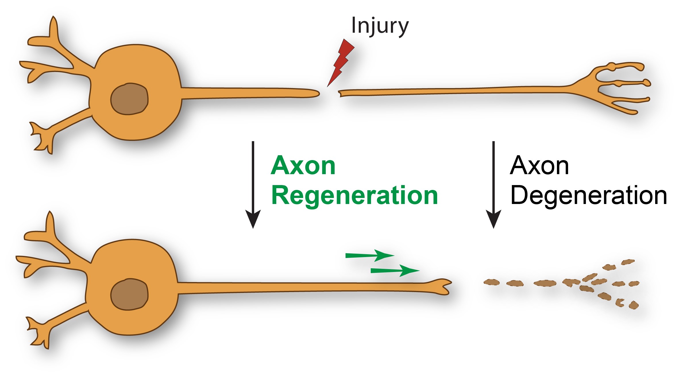
Neurotrophic factors support the survival of dopamine neurons. Brain dopamine neurotrophic factor (CDNF) is a novel neurotrophic factor with strong trophic activity on dopamine neurons comparable to that of glial cell line-derived neurotrophic factor (GDNF). It is often cited in articles on Parkinson's disease. The CDNF protein is found primarily in the endoplasmic reticulum (ER) of cells. ER is an important cellular organelle primarily involved in the folding of approximately one third of all proteins in the cell. enter image description here The authors' claims are impressive: "We found that administering CDNF to ALS mice and rats significantly improved their motor behavior and stopped the progression of paralysis symptoms. The improvement in symptoms was reflected in an increased number of surviving motor neurons in the spinal cord. spinal cord of animals compared to rodents that did not receive “CDNF. Our experiments suggest that CDNF could rescue motor neurons by reducing the ER stress response and, consequently, cell death. Importantly, ER stress was present in all of our animal models, regardless of specific genetic mutations,” explains study lead author Dr. Francesca De Lorenzo.

However, if we read the text carefully, only one animal model (SOD1) showed benefits, and the progression was slowed by 8 days or about a year for a human, which is an impressive result.

  • In SOD1-G93A mice, the median survival time for females was 148 days for CDNF-treated mice and 140 days for PBS-treated mice, with an increase of 8 days. In males, median survival was 140.5 days for CDNF-treated mice and 132 days for PBS-treated mice, with an increase of 8.5 days. This likely corresponds to SOD1-G93A mice treated daily with riluzole in drinking water.

  • Based on the text and supplementary materials, it appears that there was no benefit in survival time for either TDP-43 mouse models.

Yet, and this is a bit worrying, the abstract states "We show that intracerebroventricular administration of brain dopamine neurotrophic factor significantly arrests disease progression and improves motor behavior in the TDP43-M337V and SOD1 rodent models -G93A amyotrophic lateral sclerosis."

Since most people only read the summary or popular science articles, they are misled.

Is there a connection between stroke, TDP-43 and ALS?

- Posted by admin in English

Is there a connection between stroke, TDP-42 and ALS?

Amyotrophic lateral sclerosis (ALS or Lou Gehrig disease) is a representative neurodegenerative disease that affects upper and lower motor neurons. The mechanism of ALS is not fully understood, but mislocalization and aggregation of the TDP-43 protein in the cytoplasm play an important role. A stroke in the motor area could cause symptoms similar to those of ALS, with one big difference: a stroke is a sudden accident, and ALS is a disease that develops slowly.

A stroke in the motor area could cause symptoms similar to those of ALS

TDP-43 proteinopathy is thus associated with several chronic neurodegenerative diseases and is common in the elderly. We have known for several years that there is an association between TDP-43 and ischemic stroke.

The TDP-43 protein is normally expressed in the nucleus of cells, but under pathological conditions, it forms inclusions in the cytoplasm.

Historically, the prevalence of stroke in patients with ALS ranged from 1.6 to 8.0% in case-control studies, with inconsistent results.

Thus, in a study of 500 patients with ALS in Portugal, the prevalence of strokes (hemorrhagic and ischemic) did not differ from that of controls, regardless of the region of onset.

However, another study of 200 patients in Germany suggested that the prevalence of ischemic stroke was higher among patients in control groups.

On the other hand, previous ischemic stroke has been reported to increase the risk of ALS.

In a cohort study carried out in England, the relative risk of ALS was found to be 1.31 times higher than the expected number.

A new study in Korea

In a recent study published on MedRXiv, the authors studied the risk of developing ischemic stroke in Korean ALS patients compared to a control population using the Korean National Health Insurance Service (NHIS) database and to what extent. the degree of disability may influence TDP-43 proteinopathy.

The type and severity of disability are legally defined in Korea by the degree of disability recorded in the National Disability Registration System (NDRS) of the Ministry of Health and Welfare. Korean scientists defined and studied three groups, the control group, the group of ALS patients without disability, and that of ALS patients with disability.

Risk of ischemic stroke in the ALS group compared to the control group

During the follow-up period, 13 ischemic strokes were recorded in the ALS group and 204 in the control group. Incidence rates were 7.8/1000 person-years in the ALS group and 3.2/1000 PY in the control group. Incidence rates of ischemic stroke were similar in the disabled and non-disabled ALS groups.

There are several possible explanations for the increased risk of ischemic stroke not explained by vascular risk factors. First, ischemic stroke can be caused by paradoxical embolism.

Venous thromboembolism is common in ALS patients due to reduced mobility, and the risk of deep vein thrombosis is 3.2 times higher than in people without ALS.

This increased risk of thromboembolism in ALS may explain the higher risk of ischemic stroke. Second, increased systemic inflammation in ALS can lead to ischemic stroke.

The lack of expected effect of disability on the risk of ischemic stroke in ALS patients may be due to the small number of events.

Detailed clinical characteristics regarding the region of onset, disease duration, and medications for the treatment of ALS such as riluzole and edaravone, which may be neuroprotective in cerebral ischemia were not included in Analyses.

Our conclusion

This study, like most, leaves us wanting more. Making a causal link between strokes which would lead to TDP-43 inclusions which would ultimately cause ALS is attractive. The existence of stroke or TIA (micro/mini-stroke) is common as we age over fifty. For the cells concerned this is an enormous stress, that of no longer being supplied with oxygen and nutrients. We know that stressed cells sometimes develop proteinopathies.

However, this article does not further explore the causal link between stroke and proteinopathy.

Another article published in 2018 can serve as a complement to this recent article. Scientists studied the age-related expression of TDP-43 in neurons and glial cells and its role as a modulator of inflammation following ischemic injury. To do this, they created artificial strokes in wild-type and TDP-43 transgenic mice of different age groups.

These authors reported an age-related increase and formation of cytoplasmic inclusions of TDP-43 after artificial strokes. The dysregulation observed in TDP-43 expression patterns was associated with increased microglial activation and innate immune signaling.

The presence of aggregates of ubiquitinated TDP-43 and its cleaved fragments of TDP-35 and TDP-25a was markedly increased in mice aged 12 months, leading to larger infarcts and a significant increase in neuronal death.

Overexpression of cytoplasmic TDP-43 also drove the pathogenic NF-κB response and further increased the levels of pro-inflammatory markers and ischemic injury.

Regardless, the causes of ALS are probably multiple and, once neurons or muscles are lost, unfortunately, nothing currently known will be able to replenish them.

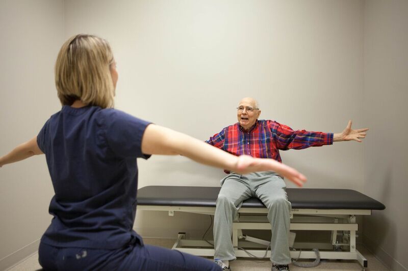
A recently published text discusses the application of electrical stimulation of the neuro-muscular system in combination with rehabilitation strategies based on the mirror neuron system (Mirror Neuron System) to improve the rehabilitation of function motor of the upper limbs and of the hand. enter image description here Motor dysfunctions of the upper limbs and hands have a significant impact on the daily lives of people with neurological diseases. Neuroplasticity is the ability of the nervous system to find other nerve circuits in response to external stimuli to activate specific muscles.

Electrical stimulation of the neuro-muscular system uses low-frequency electrical currents through surface electrodes to induce involuntary movements and facilitate motor rehabilitation. It is a common physiotherapy method, based on neuroplasticity and supposed to work by coupling between the sensory system and the motor system. We electrically activate a muscle (remember Volta's frog?), via a device controlled by the patient, the sensation of this activation reaches the brain through the sensory system, if this is not disturbed and the brain learns after numerous tests, there is a parallel path which activates this motor system. In a way it's similar to learning to drive a car, we activate different devices (brakes, accelerator, steering wheel, shifters), we have sensory feedback (the car accelerates, brakes, turns), and little by little these are maneuvers that we do instinctively.

Electrical stimulation of the neuromuscular system involves the use of electrodes placed on the skin over target muscles. These electrodes deliver controlled electrical impulses to the muscles, causing them to contract. Electrical stimulation of the neuromuscular system can be used to:

  • Muscle Activation: Electrical stimulation of the neuro-muscular system can activate muscles that are weak or paralyzed due to neurological problems or injuries. This is particularly useful when voluntary muscle activation is limited or impossible.

  • Build Strength and Endurance: Electrical stimulation of the neuro-muscular system can help strengthen muscles and improve endurance, which is important for regaining functional motor skills.

  • Prevent Atrophy: In cases of muscle disuse or atrophy, such as after surgery or during prolonged immobility, electrical stimulation of the neuro-muscular system can prevent muscle loss by maintaining muscle contractions.

*Improve blood flow: Electrical stimulation of the neuro-muscular system can promote blood circulation in the stimulated area, which facilitates tissue healing and recovery.

Although electrical stimulation of the neuro-muscular system has benefits, it is passive and can lead to limited patient engagement. The combination of electrical stimulation of the neuro-muscular system with active rehabilitation strategies or methods based on the mirror neuron system can improve the results.

The mirror neuron system plays an essential role in neuronal plasticity linked to motor learning. It is activated when a person performs an action but also when they observe a similar action. The mirror neuron system plays a crucial role in learning.

Various rehabilitation techniques, such as Action Observation Therapy (AOT), Mirror Therapy (MT), Motor Imagery (MI), and Virtual Reality (VR), are based on the system theory of mirror neurons and are widely used in neurological rehabilitation. The study described in this post uses functional near-infrared spectroscopy (fNIRS) to measure cortical activation patterns related to electrical stimulation of the neuromuscular system combined with strategies based on the mirror neuron system.

The study involved 66 healthy adults in various experimental tasks combining electrical stimulation of the neuro-muscular system with different mirror neuron system strategies, such as action observation, action execution, and action imitation.

The scientists used an fNIRS device with multiple channels to measure changes in blood oxygen levels in the brain during different tasks. This method is slower to acquire data than EEG, but it is also more reliable.

Results showed that combining electrical stimulation of the neuromuscular system with strategies based on the mirror neuron system activated brain areas, with active exercises showing the most significant activation. This suggests a potential for enhanced rehabilitation effects.

As our editorial policy concerns neurodegenerative diseases, among them ALS (Lou Gehrig's disease), we immediately consider which benefits a patient could derive from this technology.

Spinal cord injuries cause symptoms quite similar to those of ALS. Recently patients who had severed spinal cords have been able to walk again thanks to similar technologies.

The study suggests that brain-computer interface (BCI) systems based on fNIRS could be developed to aid rehabilitation, especially in cases where patients have lost the ability to perform active exercises.

Neuromuscular electrical stimulation (Electrical stimulation of the neuro-muscular system) may have some potential benefits for people with amyotrophic lateral sclerosis (ALS), but it is important to understand its limitations and consider it as part of an approach. comprehensive disease management. One would think that she might be of particular interest for the following points:

  • Muscle Preservation: Electrical stimulation of the neuro-muscular system may help slow muscle atrophy and maintain muscle function in people with ALS. This is particularly relevant when voluntary muscle activation becomes difficult or impossible.

  • Management of pain and spasticity: Some people with ALS may experience muscle pain and spasticity. Electrical stimulation of the neuro-muscular system can help alleviate these symptoms by promoting muscle relaxation and blood circulation.

In the future, one could even imagine that devices for electrical stimulation of the neuro-muscular system can be used as assistive devices to facilitate daily activities, such as grasping objects or walking, by stimulating specific muscle groups.

However, it is unlikely that this technology will be able to slow the progression of the disease: ALS is a progressive disease and, although electrical stimulation of the neuromuscular system can provide temporary relief and muscle preservation, it is unlikely to stop not the underlying neurodegenerative process.

There is also the problem of individual variability which is very broad in the context of ALS. ALS is more of a syndrome than a disease, each patient is unique which makes clinical trials terribly complicated, and in most patients, their health and motor skills evolve terribly quickly. As the response to electrical stimulation of the neuromuscular system can vary greatly among individuals with ALS, some may find it beneficial, while others may not benefit or may even experience accelerated deterioration.

Electrical stimulation of the neuromuscular system could be considered part of a comprehensive approach to managing ALS, which may include physiotherapy, occupational therapy, speech therapy, and medical treatments.

In summary, electrical stimulation of the neuromuscular system may be helpful in managing specific symptoms and preserving muscle function in people with ALS. However, this should be part of a broader, multidisciplinary approach to ALS care. The effectiveness of electrical stimulation of the neuromuscular system can vary from person to person, so it is crucial to work closely with healthcare professionals to determine the most appropriate interventions and therapies for individual needs and the stage of the disease.

*Affiliate link*

Un texte récemment publié traite de l'application de la stimulation électrique du système neuro-musculaire en combinaison avec des stratégies de rééducation basées sur le système de neurones miroirs (Système de neurones miroirs) pour améliorer la rééducation de la fonction motrice des membres supérieurs et de la main. enter image description here Les dysfonctionnements moteurs des membres supérieurs et des mains ont un impact significatif sur la vie quotidienne des personnes atteintes de maladies neurologiques. La neuroplasticité est la capacité du système nerveux à trouver d'autres circuits nerveux en réponse à des stimuli externes pour activer certains muscles.

La stimulation électrique du système neuro-musculaire utilise des courants électriques basse fréquence à travers des électrodes de surface pour induire des mouvements involontaires et faciliter la rééducation motrice. La stimulation électrique du système neuro-musculaire est une méthode de physiothérapie courante, reposant sur la neuroplasticité et supposée fonctionner par couplage entre le système sensoriel et le système moteur. On active électriquement un muscle (vous vous rappelez la grenouille de Volta?), via un dispositif commandé par le patient, la sensation de cette activation arrive au cerveau par le système sensoriel, si celui-ci n'est pas perturbé et le cerveau apprend au bout de nombreux essais qu'il y a un chemin parrallèle qui active ce système moteur.

D'une certaine façon c'est similaire à l'apprentissage de la conduite d'une voiture, on active différents dispositifs (freins, accélérateur, volant, manettes), on a un retour sensoriel (la voiture accélère, freine, tourne), et petit à petit ce sont des maneuvres que l'on fait instinctivement.

La stimulation électrique du système neuro-musculaire implique l’utilisation d’électrodes placées sur la peau sur les muscles cibles. Ces électrodes délivrent des impulsions électriques contrôlées aux muscles, provoquant leur contraction. La stimulation électrique du système neuro-musculaire peut être utilisé pour :

  • L'activation musculaire : La stimulation électrique du système neuro-musculaire peut activer les muscles faibles ou paralysés en raison de problèmes neurologiques ou de blessures. C'est particulièrement utile lorsque l'activation musculaire volontaire est limitée ou impossible.

  • Renforcer la force et l'endurance : La stimulation électrique du système neuro-musculaire peut aider à renforcer les muscles et à améliorer l’endurance, ce qui est important pour retrouver les capacités motrices fonctionnelles.

  • Prévenir l'atrophie : En cas de désuétude ou d'atrophie musculaire, comme après une intervention chirurgicale ou lors d'une immobilité prolongée, la stimulation électrique du système neuro-musculaire peut prévenir la perte musculaire en maintenant les contractions musculaires.

  • Améliorer le flux sanguin : La stimulation électrique du système neuro-musculaire peut favoriser la circulation sanguine dans la zone stimulée, ce qui facilite la guérison et la récupération des tissus.

Bien que la stimulation électrique du système neuro-musculaire présente des avantages, elle est passive et peut conduire à un engagement limité des patients. La combinaison de la stimulation électrique du système neuro-musculaire avec des stratégies de rééducation active ou des méthodes basées sur le système de neurones miroirs peut améliorer les résultats.

Le système de neurones miroirs joue un rôle essentiel dans la plasticité neuronale liée à l’apprentissage moteur. Il est activé lorsqu’une personne effectue une action mais également lorsqu’elle observe une action similaire. Le système de neurones miroirs joue un rôle crucial dans l'apprentissage.

Diverses techniques de réadaptation, telles que la thérapie par observation d'action (AOT), la thérapie par le miroir (MT), l'imagerie motrice (IM) et la réalité virtuelle (VR), sont basées sur la théorie du système de neurones miroirs et sont largement utilisées en réadaptation neurologique. L'étude que décrit ce post utilise la spectroscopie fonctionnelle proche infrarouge (fNIRS) pour mesurer les modèles d'activation corticale liés au La stimulation électrique du système neuro-musculaire combinés à des stratégies basées sur le système de neurones miroirs.

L'étude a impliqué 66 adultes en bonne santé dans diverses tâches expérimentales combinant la stimulation électrique du système neuro-musculaire avec différentes stratégies de système de neurones miroirs, telles que l'observation d'action (AO), l'exécution d'action (AE) et l'imitation d'action (IA).

Les scientifiques ont utilisé un appareil fNIRS doté de plusieurs canaux pour mesurer les changements dans les niveaux d’oxygène dans le sang dans le cerveau au cours de différentes tâches. Cette méthode est plus lente à acquérir des données que l'EEG, mais elle est aussi plus fiable.

Les résultats ont montré que la combinaison de la stimulation électrique du système neuro-musculaire avec des stratégies basées sur le système de neurones miroirs activait des zones cérébrales, les exercices actifs montrant l'activation la plus significative. Cela suggère un potentiel d’effets de réhabilitation accrus.

Notre politique éditoriale concernant les maladies neurodégénératives et entre autre la SLA (maladie de Charcot), on pense immédiatement quels bénéfices un patient pourrait retirer de ce type de technologie. Les lésions de la moëlle épinière occasionnent des symptomes assez semblables à ceux de la SLA. Récemment des patients qui avaient une moëlle épinière coupée ont pu remarcher grâce à une technologie similaire.

L'étude suggère que des systèmes d'interface cerveau-ordinateur (BCI) basés sur fNIRS pourraient être développés pour faciliter la rééducation, en particulier dans les cas où les patients ont perdu la capacité d'effectuer des exercices actifs.

La stimulation électrique neuromusculaire (La stimulation électrique du système neuro-musculaire ) peut présenter certains avantages potentiels pour les personnes atteintes de sclérose latérale amyotrophique (SLA), mais il est important de comprendre ses limites et de la considérer comme faisant partie d'une approche globale de gestion de la maladie. On pourrait penser qu'elle pourrait avoir un présenter des un intérêt particulier pour les points suivants :

  • Préservation musculaire : La stimulation électrique du système neuro-musculaire peut aider à ralentir l’atrophie musculaire et à maintenir la fonction musculaire chez les personnes atteintes de SLA. Ceci est particulièrement pertinent lorsque l’activation musculaire volontaire devient difficile, voire impossible.

  • Gestion de la douleur et de la spasticité : Certaines personnes atteintes de SLA peuvent ressentir des douleurs musculaires et de la spasticité. La stimulation électrique du système neuro-musculaire peut aider à atténuer ces symptômes en favorisant la relaxation musculaire et la circulation sanguine.

Dans le futur on pourrait même imaginer que les appareils de stimulation électrique du système neuro-musculaire peuvent être utilisés comme appareils d'assistance pour faciliter les activités quotidiennes, telles que saisir des objets ou marcher, en stimulant des groupes musculaires spécifiques.

Cependant il est peu probable que cette technologie puisse ralentir le progression de la maladie : La SLA est une maladie évolutive et, même si la stimulation électrique du système neuro-musculaire peut apporter un soulagement temporaire et une préservation musculaire, elle n'arrêtera sans doute pas le processus neurodégénératif sous-jacent.

Il y a aussi le problème de la variabilité individuelle qui est très large dans le cadre de la SLA. La SLA est plus un syndrome qu'une maladie, chaque patient est unique ce qui complique teriblement les essais cliniques et chez la plupart des patients leur santé et leurs capacitées motrices évoluent terriblement rapidement. Comme la réponse à la stimulation électrique du système neuro-musculaire peut varier considérablement selon les individus atteints de SLA, certains pourraient trouver cela bénéfique, tandis que d’autres pourraient ne pas en bénéficier, voire assister à une dégradation accélérée.

La stimulation électrique du système neuro-musculaire pourrait être considéré comme faisant partie d'une approche globale de gestion de la SLA, qui peut inclure la physiothérapie, l'ergothérapie, l'orthophonie et les traitements médicaux.

En résumé, la stimulation électrique du système neuro-musculaire peut être utile dans la gestion de symptômes spécifiques et dans la préservation de la fonction musculaire chez les personnes atteintes de SLA. Cependant, cela devrait s’inscrire dans une approche plus large et multidisciplinaire des soins de la SLA. L'efficacité de la stimulation électrique du système neuro-musculaire peut varier d'une personne à l'autre, il est donc crucial de travailler en étroite collaboration avec les professionnels de la santé pour déterminer les interventions et les thérapies les plus adaptées aux besoins uniques de chaque individu et au stade de la maladie.

Brain implant to vocalize thoughts

- Posted by admin in English

For ALS patients, failing to access a cure, it is important to overcome certain important problems. Alas a chair that can be controlled by gaze, telemanipulation arms, intelligent ventilation, and a device for vocalizing by thought, will not be marketed for another decade or two.

The press echoed two recent articles about devices for vocalizing through thought. One acquires the data via a deep cranial implant, the other via a surface cranial implant. enter image description here I analyze below the first article because the data and programs to implement this experiment are publicly available. Which is apparently not the case for the second.

I asked one of the authors if it was possible to use his program with an EEG helmet. We'll see what his answer will be, but I anticipate that the results of this course of action will be very disappointing.

The study focused on understanding how facial movement and speech production are organized in the motor cortex at the level of individual neurons. Neural activity was recorded from microelectrode arrays implanted in the brain of a participant with amyotrophic lateral sclerosis (ALS) who had limited facial movements and an ability to vocalize but not produce intelligible speech.

The results indicated neural solid agreement on various facial movements in a region of the brain called area 6v, and this activity was very distinct for different movements. In contrast, area 44, traditionally associated with speech production, appears (in this experiment) to contain little information about facial actions or speech.

The ventral premotor cortex, which is located in the middle of the upper part of the brain has been involved in motor vocabularies in both speech and manual gestures. A recent prospective fMRI study demonstrated adaptation effects in the ventral premotor cortex to repeating syllables.

Broca's area, is a region in the frontal lobe of the dominant hemisphere, usually the left, of the brain with functions linked to speech production.

The researchers developed a decoder using a recurrent neural network (RNN) to translate neural activity into speech. The participant attempted to speak sentences and the RNN decoded the predicted words in real-time, achieving a word error rate of 9.1% for a vocabulary of 50 words and 23.8% for a vocabulary of 125 000 words. This demonstrated the feasibility of decoding speech attempts using neural signals.

The neural representation of speech sounds in the brain was analyzed, showing that the activity patterns reflected the articulatory features of the phonemes. This suggests that even after years of paralysis, the detailed articulatory code of phonemes remains preserved in the brain.

The study also discussed design considerations for improving the accuracy of speech brain-computer interfaces (BCIs), including vocabulary size, number of electrodes used, and size of the training data set. The researchers noted that while their results were promising, there was still room for further optimization and improvements in the technology.

Overall, the study presented a proof of concept for a speech BCI that could potentially enable people with severe motor impairments to communicate more effectively by translating their intended speech into text from neural signals.

Grafting muscles to heal from ALS?

- Posted by admin in English

Scientists tell that ALS is a disease striking upper motor neurons, which in a cascade effect, affects the lower motor neurons and muscles. Yet this does not match the patient's experience. enter image description here For them, symptoms often start with weakness, unreliability, and thinning of the thumb or calf, and with time the disease progresses to other (skeletal) muscles.

Scientific approaches are focussing on healing upper motor neurons (with a complete lack of success, which hints that we understand nearly nothing in this area) or replacing them (there are still no clinical studies).

For most scientists, it would be meaningless to try to replace muscles in ALS patients, as the upper motor neurons are dead (despite evidence they are not) new muscles would never be usable and therefore waste and die quickly.

And anyway grafting muscles on neuromuscular junctions is an extraordinarily difficult problem.

Yet there are contrarians: Scientists have recently unveiled an approach to address the devastating effects of amyotrophic lateral sclerosis (ALS), on muscle weakness. Their research introduces a novel technique involving a combination of grafted replacement motor neurons and optical nerve stimulation.

By employing a two-pronged strategy—grafting modified motor neurons and using light-based nerve stimulation—the researchers managed to rejuvenate muscle function, marking a tiny step toward devising effective therapies for ALS.

Led by Dr. Barney Bryson, the team of scientists had shown potential in restoring muscle denervation in a mouse model by utilizing optical nerve stimulation. This method hinged on grafting replacement motor neurons that were engineered to be light-sensitive, enabling their activation through an external light source.

In this latest study, the initial objective was to ensure the survival of donor motor neurons during the grafting process, overcoming potential immune system rejection. After determining that conventional immunosuppressive drugs were not viable for ALS mice, the researchers experimented with a specific antibody known as H57-597, which successfully prevented graft rejection and began reconnecting nerves to target muscles. Despite these positive outcomes, the force generated by muscle contractions was relatively weak, prompting the team to further refine their strategy.

Recognizing that neuromuscular junctions—the connection points between nerves and muscles—are influenced by regular stimulation, the scientists introduced a wireless optical stimulation system to enforce consistent muscle contractions for an hour each day. Astonishingly, after 21 days of this optical stimulation training, the mice exhibited an astonishing 13-fold enhancement in muscle contraction force. That would be nearly three years of rehabilitation therapy in humans.

These findings carry immense significance, demonstrating that even in the advanced stages of ALS, affected muscles remain amenable to reinnervation by healthy replacement motor neurons.

The strategy suggests the feasibility of a treatment that could potentially be universally applied to all ALS patients regardless of the kind of ALS they have (familial or sporadic).

More significantly, it tells that the model of ALS progression which was derived from stroke ~150 years ago and mostly never questioned since all these decades, is simply wrong.

However, significant challenges remain before this approach can transition to clinical use. Subsequent studies are necessary to confirm the efficacy of the grafting procedure with human motor neurons and to evaluate whether it can genuinely enhance patients' quality of life. Furthermore, the technique's applicability to other forms of MND, particularly those with longer life expectancies, must be investigated to ascertain its long-term effectiveness.

In conclusion, the study's senior author, Linda Greensmith, emphasizes that the findings underline the robustness of replacing motor neurons to reinnervate muscles even in advanced ALS stages. Should this approach successfully translate to ALS patients, it holds the potential to revolutionize treatment by employing a single type of motor neuron for various muscles, simplifying the therapy and making it more widely accessible. The introduction of this approach could be a game-changer in the ongoing battle against ALS and other related neuromuscular conditions.

La sclérose latérale amyotrophique (dite aussi maladie de Charcot) est une maladie incurable qui provoque une faiblesse musculaire dans tout le corps, rendant les mouvements et la respiration de plus en plus difficiles jusqu'au décès. La maladie commence souvent par un problème apparemment musculaire dans le pouce ou le mollet et cette faiblesse s'étend progressivement au reste des muscles du corps que l'on contrôle consciemment (muscles dit "squelettiques").

On a trouvé de multiples causes génétiques à la SLA dite familiale, mais celle-ci ne représente qu'un faible pourcentage de l'ensemble des cas. Par contre dans la plupart des cas on trouve dans les cytoplasme des neurones moteurs, des amas d'une protéine nommée TDP-43. Ces amas souvent comporte seulement des fragments de cette protéine qui de plus est dépliée, c'est à dire non fonctionnelle.

On ne sait pas si TDP-43, qui est indispensable à la survie, est une cause de la maladie ou une conséquence d'événements tels qu'une infection virale. De tels interrogations existent aussi dans le cas de la maladie d'Alzheimer ou de Parkinson.

Ce qui est sur c'est que les mutations de cette protéine sont souvent délétères, ce qui n'étonnera personne, et qu'un surcroît ou un défaut de TDP-43 est lui aussi délétère.

Une autre controverse concerne aussi le sens de propagation de la maladie, se propage t-elle depuis les extrémitées vers le cerveau (ce qui correspond à l'expérience des malades) ou au contraire, est-ce une maladie qui nait dans le cerveau et se propage aux extrémitées (hypothèse majoritaire chez les scientifiques et dérivée de ce que l'on observe après un AVC dans la zone motrice du cerveau). Les scientifiques partisans de chacune des deux opinions ont à de multiples reprise prouvé que "l'autre" hypothèse était fausse, ce qui n'est guère informatif.

Une nouvelle étude par des scientifiques Japonais tend à dire que les deux hypothèses sont vrais mais ne sont pas assez précises dans leur formulation.

Les commandes de mouvement initiées par des neurones de la zone motrice du cortex cérébral sont transmises aux neurones moteurs supérieurs de la moelle épinière via le tractus corticospinal. Dans la moelle épinière ces commandes sont transmises aux neurones moteurs inférieurs qui à l'autre extrémité, sont reliés aux muscles.

Les scientifiques Japonais ont supposé que la protéine (TDP-43) se propage anormalement dans ces circuits neuronaux. Jusqu'à présent, on pensait que TDP-43 était cantonné à l'intérieur des neurones et des cellules non-neuronales. enter image description here Les auteurs ont établi de nouveaux modèles de SLA de souris qui induisaient initialement des inclusions de TDP-43 mutant dans des types neuronaux ou cellulaires spécifiques dans les circuits moteurs, et ils ont étudié si le TDP-43 et les processus pathologiques pertinents se propageaient à travers les connexions neuronales ou cellulaires.

Les auteurs ont d'abord développé des modèles de SLA qui induisaient principalement des inclusions de TDP-43 dans les neurones corticospinaux, les motoneurones spinaux ou le muscle squelettique des membres antérieurs, en utilisant le virus adéno-associé (AAV) exprimant le mutant TDP-43.

Ils ont ensuite examiné comment le TDP-43 depuis des motoneurones spécifiques, ce propage à d'autres neurones moteurs entrainant la progression de la maladie. Les données ont révélé que le mutant TDP-43 se propageait à travers les connexions neurogliales de manière antérograde dans la voie corticospinale, alors qu'il présentait différentes propriétés dégénératives rétrogrades dans les circuits spinaux. Cela suggère que le TDP-43 pathogène peut induire des mécanismes antéro- et rétrogrades distincts de dégénérescence du système moteur dans la SLA.

  • Les auteurs ont constaté que le TDP-43 induit dans les neurones corticospinaux était transporté le long des axones de manière antérograde et transféré aux oligodendrocytes le long du tractus corticospinal (CST), coïncidant avec une légère dégénérescence des axones. Lorsque le TDP-43 anormal a été induit dans le cortex cérébral, le cortex cérébral et les axones subissent une dégénérescence et que les oligodendrocytes du tractus corticospinal adoptaient une morphologie réactive. Mais, bien que le TDP-43 soit apparu dans le tractus corticospinal, il ne s'est pas propagé à la moelle épinière. Les oligodendrocytes sont des cellules non neuronales qui assistent les neurones pour faciliter la transmission du signal neuronal, en enveloppant les axones d'une couche protectrice appelée myéline. C'est à dire en langage simple qu'on a bien une progression de la maladie depuis le cerveau vers la moelle épinière, mais l'atteinte est légère.

  • En revanche, le TDP-43 introduit dans les motoneurones inférieurs ne s'est pas propagé de manière rétrograde aux neurones corticaux ou aux motoneurones supérieurs. Cependant, il a induit une perte extrême en peu de temps des motoneurones inférieurs voisins. La dégénérescence intraspinale a en outre entraîné une atrophie musculaire sévère. De plus, il a été constaté que non seulement les motoneurones, mais également d'autres interneurones de la moelle épinière qui communiquent avec la zone environnante, étaient induits à mourir. De plus, parallèlement à la perte de motoneurones, les muscles se sont également atrophiés à un degré élevé, provoquant des troubles du mouvement. Mais le TDP-43 muté lui-même ne s'est pas propagé aux neurones moteurs supérieurs, au cortex cérébral ni aux muscles. Ce n'est donc pas une progression rétrograde mais plutôt latérale.

  • Enfin, le TDP-43 induit dans les muscles squelettiques ne s'est pas propagé rétrogradement aux neurones inférieurs spinaux. L'induction de TDP-43 aberrant dans le muscle a augmenté le nombre de fibres régénérantes, mais n'a pas provoqué d'atrophie musculaire ni de dyskinésie. De plus, aucune propagation du TDP-43 à la moelle épinière n'a été observée

enter image description here Tsuboguchi et al., Acta Neuropathologica 2023.

Here is a study on FUS ALS which proposes that FUS ALS patients might benefit from administration of interferon-gamma (IFNγ). Interferon-γ 1b is approved by the U.S. Food and Drug Administration to treat chronic granulomatous disease and osteopetrosis. In approximately 90% of cases, ALS is sporadic, but around 10% of patients exhibit familial mutations. Subtypes of ALS are categorized based on the affected gene, including superoxide dismutase 1 (SOD1), TAR DNA-binding protein 43 (TDP-43), chromosome 9 open reading frame 72 (C9ORF72), and Fused in Sarcoma (FUS), with FUS leading to one of the most aggressive and early-onset forms of the disease.

FUS is a component of the heterogeneous nuclear ribonucleoprotein (hnRNP) complex and is involved in DNA/RNA binding, DNA damage repair, splicing, and various aspects of RNA metabolism. Over 50 different FUS gene mutations have been identified in ALS patients. Mutations often affect the nuclear localization signal (NLS) domain, leading to improper cytoplasmic localization and nuclear clearance of FUS, resulting in an aggressive pathological phenotype. The most common FUS mutation affects arginine 521 (R to H, C, or G). Incorrect FUS localization also occurs in other forms of familial and sporadic ALS cases, suggesting a common mechanism.

Studying ALS pathobiology is challenging due to the high costs and morbidity associated with the disease. Reliable disease models are essential, so the study used induced pluripotent stem cells (iPSCs) derived from FUSR521H ALS patients to generate motor neurons (motoneurons) for research.

As iPSCs rejuvenate as a result of reprogramming and ALS symptoms are primarily associated with aging, the authors exposed iPSC-derived motor neurons to oxidative stress (by means of sodium arsenite) to model aging-associated effects.

These models revealed that ALS motor neurons are more sensitive to oxidative stress than control motor neurons. ALS iPSC-derived motoneurons exhibited abnormal cytoplasmic FUS localization, reduced translation rates, and increased sensitivity to oxidative stress-induced apoptosis.

The study further explored altered gene expression patterns and signaling pathways in FUS ALS motoneurons. enter image description here

As cytokine measurements showed reduced secreted IFNγ in FUS ALS motorneurons treated with sodium arsenite, and as authors recently found that inflammatory cytokines can protect cells from stress-induced cell death (Hong et al., 2022), they tested whether this was also true for FUS ALS motorneurons.

For this, the scientists supplemented sodium arsenite-treated FUS ALS motorneurons with IFNγ and found that this indeed increased viability of FUS ALS motorneurons to levels similar to SA-treated control motorneurons.

Furthermore, IFNγ treatment reduced the fraction of apoptotic FUS ALS motorneurons following sodium arsenite exposure. Importantly, they also found that IFNγ treatment resulted in an increased IFNγ transcriptional response in SA-treated ALS motorneurons, indicating that IFNγ treatment was sufficient to rescue the impaired IFNγ response observed in SA-treated ALS motorneurons.

As treatment with interferon-gamma (IFNγ) reduced oxidative stress-induced apoptosis and improved translation rates and nuclear FUS localization in FUS ALS motoneurons, the authors suggest that early-diagnosed FUS ALS patients might benefit from IFNγ treatment to slow disease progression.

While this is not suggested by the study, one might reflect that the described effects of FUS in ALS are similar to SOD1 and TDP-43 ALS, so maybe IFNγ treatment might be beneficial in these cases also.

In summary, the study investigates the impact of FUS mutations on ALS using iPSC-derived motoneurons, revealing insights into the disease's underlying molecular mechanisms and proposing potential therapeutic strategies involving IFNγ treatment.

Relyvrio, Phénylbutyrate de sodium et Taurursodiol

- Posted by admin in Français

Le médicament d'Amylyx connu sous les noms d'AMX0035, Relyvrio ou Albrioza est actuellement en cours d'essai clinique de phase III PHOENIX (NCT05021536). enter image description here La FDA et l'agence Européenne des médicaments ont exprimés de nombreux doutes sur l'efficacité de ce médicament au cours de l'essai CENTAUR. Ces doutes concernent l'exclusion d'un certain nombre d'évènements défavorables lors de l'analyse statistique, ainsi que le fait que certains patients ont aussi reçu du Riluzole et de l'Edaravone. En particulier il y a plus de patients ayant reçu de l'Edaravone dans la branche contrôle que dans la branche traitement, or certains scientifiques et agences du médicament pensent que l'Edaravone a un effet négatif sur l'évolution de la maladie. Certains médecins ont également attribué les "bons" résultats au TUDCA. Au final à 24 mois il n'y a pas d'amélioration de la survie. Vous pouvez consulter l'avis initial de la FDA içi:

Finalement la FDA a donné son autorisation sous condition d'un engagement d'Amylyx de retirer volontairement le médicament s'il ne montre pas d'efficacité dans son essai de phase 3 en cours.

Un problème qui n'est jamais soulevé est qu'il est recommandé d'avoir une alimentation dépourvue en protéines avec du phénylbutyrate de sodium! Au contraire pour les malades de la SLA il est recommandé d'avoir un haut régime protéique pour lutter contre la perte musculaire.

Il n'en reste pas moins que c'est le seul médicament qui pour l'instant semble donner quelques espoirs.

Aujourd'hui et dans l'attente des résultats de l'essai clinique PHOENIX, AMX0035 n'est autorisé qu'aux USA (conditionnellement) et au Canada. Dans d'autres pays et dans certain centres SLA, il est possible d'y accéder à titre exceptionnel.

A priori AMX0035 semble être un médicament très peu couteux à la production. C'est une poudre pour suspension buvable, combinant deux poudres:

• Phénylbutyrate de sodium (PB) : 3 g

• Taurursodiol (TURSO ou TUDCA) : 1 g

La posologie est de 1 sachet deux fois par jour, matin et soir (BID).

Il est étonnant que ce médicament ne soit pas, (comme pour l'AMMONAPS) encapsulé dans une pilule. Cela permettrait: - De dissimuler le goût atroce. - D'avoir une meilleur assimilation, car avec une poudre, la plus grande partie du produit doit se décomposer dans le bain d'acide de l'estomac. C'est vrai que la forme en poudre convient particulièrement pour les patients équippés d'une sonde de gastrostomie ou d'une sonde nasogastrique.

Le traitement des maladies du métabolisme du cycle de l'urée, se fait avec du phénylbutyrate de sodium, commercialisé sous les noms de Buphenyl ou AMMONAPS en comprimés de 500mg.

Amylyx a annoncé un prix annuel pour Relyvrio de $158,000, soit $433 par jour.

Des internautes font confectionner ce médicament par leur pharmacie, en effet dans certains pays les pharmaciens ont le droit de confectionner des produits ayant obtenus l'autorisation de mise sur le marché.

L'organisation ALS.org mentionne même cette possibilité:

Cela semble pourtant une option peu intéressante. La dose journalière de Phénylbutyrate de sodium coûte plus de 3 000 € chez les grands fournisseurs comme SIGMA-ALDRICH. Le prix du TUDCA est négligeable en comparaison.

Une autre possibilitée consiste évidemment à se faire prescrire du Buphenyl/AMMONAPS sous la forme de comprimés ou poudre suivant les besoins et d'acheter du TUDCA sur Internet

New study on Arimoclomol for ALS and FTD

- Posted by admin in English

The accumulation of toxic protein clumps is a crucial contributor to cell damage in people with ALS (TDP-43), Alzheimer's disease (beta-amyloid), or Parkinson's disease (alpha-synuclein). These aggregates are composed of abnormal proteins that fail to acquire their standard three-dimensional shape. enter image description here In the case of ALS, the clusters of TDP-43 are also located in the cytosol, i.e. where the proteins are produced, before being folded up in the ER and then sent to their place of use by the Golgi apparatus. After shipment, TDP-43 should be in the nucleus.

Arimoclomol is an oral therapy that increases the production of heat shock response proteins (HSPs), which help misfolded proteins to their normal shape. HSPs also direct the removal of abnormal proteins when refolding is not possible.

One would therefore think that if ALS is primarily a problem with folding proteins, therapies that help misfolded proteins acquire their typical configuration should slow the progression of ALS. This has been tested with Arimoclomol. For once a clinical trial would have been launched on a drug that seemed to have a good chance of succeeding (oddly drug targets in most clinical trials in neurodegenerative diseases seem to be based on non-scientific criteria).

Arimoclomol was evaluated in a previous Phase 2/3 clinical trial (NCT00706147) in 36 patients with rapidly worsening ALS due to SOD1 mutations. The choice to select SOD1 patients is quite curious. There are countless SOD1 mutations, with consequences ranging from extremely rapid deterioration (a few months) to extremely slow deterioration (death after 10 years). There are therefore probably multiple mechanisms of action at work.

On the other hand, SOD1 mutations are only present in 5% of ALS cases. But SOD1 mutations were the first mutations discovered in ALS and are by far the most studied for 30 years, yet without significant progress being recorded.

Although this clinical trial did not show statistically significant results, the company Orphazyme managed to find funding for a phase III trial. The Phase 3 ORARIALS-01 trial (NCT03491462) studied arimoclomol in 245 adults with ALS. This clinical trial did not achieve its objectives. Arimoclomol did not prolong the life of patients or even delay the progression of disability in people with the disease. In other words, it was a spectacular failure, like almost all clinical trials in ALS.

Although I am neither a doctor nor a scientist, this made me think that the premises for rationale of arimoclomol in ALS were false. Personally, since that time, I am convinced that ALS is due to a cellular stress response that does not stop and which therefore destroys not only neurons but also muscle cells.

Indeed, the response to cellular stress consists of an almost total shutdown of the cell, this shutdown cannot be permanent. This stop explains very well the loss of muscle mass (which scientists explain by the death of motor neurons, while this loss occurs before motor neurons death).

From this perspective (very personal), any action aimed at increasing the energy expenditure of the cell (it takes energy to fold proteins), is doomed to failure.

Yet the scientists behind arimoclomol in 2004, seem to be back at the lab table. This time they are no longer interested in SOD1 mutations but in VCP mutations. VCP mutations are very rare, less than 1% of ALS cases. They conducted a study of arimoclomol in mice with a mutation in valosin-containing protein (VCP) that causes both ALS and FTD in patients.

Similar to studies of arimoclomol in SOD1 mice, enhancement of the heat shock response ameliorated the ALS/FTD-like phenotype in the spinal cord and brain of VCP mutant mice. Arimoclomol prevents neuronal loss in it. Additionally, in human cell models, the authors demonstrate improvements in pathology in VCP mutant patient fibroblasts and iPSC-derived motor neurons.

The scientists suggest that targeting HSP may have therapeutic potential, not only in non-SOD1 ALS, but also for the treatment of FTD.

Errare humanum perseverare diabolicum


Please, help us continue to provide valuable information: