Reverse Split Hand in Spinal Muscular Atrophy (SMA)

- Posted by admin in English

Spinal muscular atrophy (SMA) is a disease whose manifestations have some similarities with ALS, however, it has a well-identified genetic origin (SMN1 gene) and as a result, several drugs have been approved. These drugs improve the health of patients but have various drawbacks.

This rare disease causes the loss of motor neurons and progressive muscle atrophy. It is usually diagnosed in early childhood and, if left untreated, it often leads to death. It can also appear later in life and in this case the disease progresses more slowly.

The common feature is a progressive weakness of voluntary muscles, with the muscles of the arms, legs and airways being affected first. Associated problems can include poor head control, difficulty swallowing, scoliosis, and joint contractures. enter image description here Although there is a large literature on the split hand phenomenon in ALS, knowledge remains limited for other motor neuron diseases, including SMA.

Early in ALS it is common for the thenar muscles (thumb muscles) to be affected and lose their usual volume, visually dividing the palm into two sections. This is oddly described as the split hand phenomenon or monkey hand phenomenon.

This phenomenon reflects the selective vulnerability of motor neurons characteristic of the disease. Conditions such as ALS, while also affecting motor neurons, tend to present with the classic split hand pattern, highlighting the differential pathophysiological mechanisms at play. However, the explanations for why these muscles are affected early in the disease are no clearer than in ALS. Furthermore, age-related atrophy can also present with the split hand phenomenon, complicating the diagnosis of ALS in older patients.

Muscle disuse is a common cause of muscle atrophy. The rate of muscle atrophy due to disuse is about 0.5% of total muscle mass per day. Older adults are most vulnerable to dramatic muscle loss with immobility. In SMA, as in ALS, loss of motor neuron function leads to muscle atrophy. A new study compared these losses in ALS and SMA.

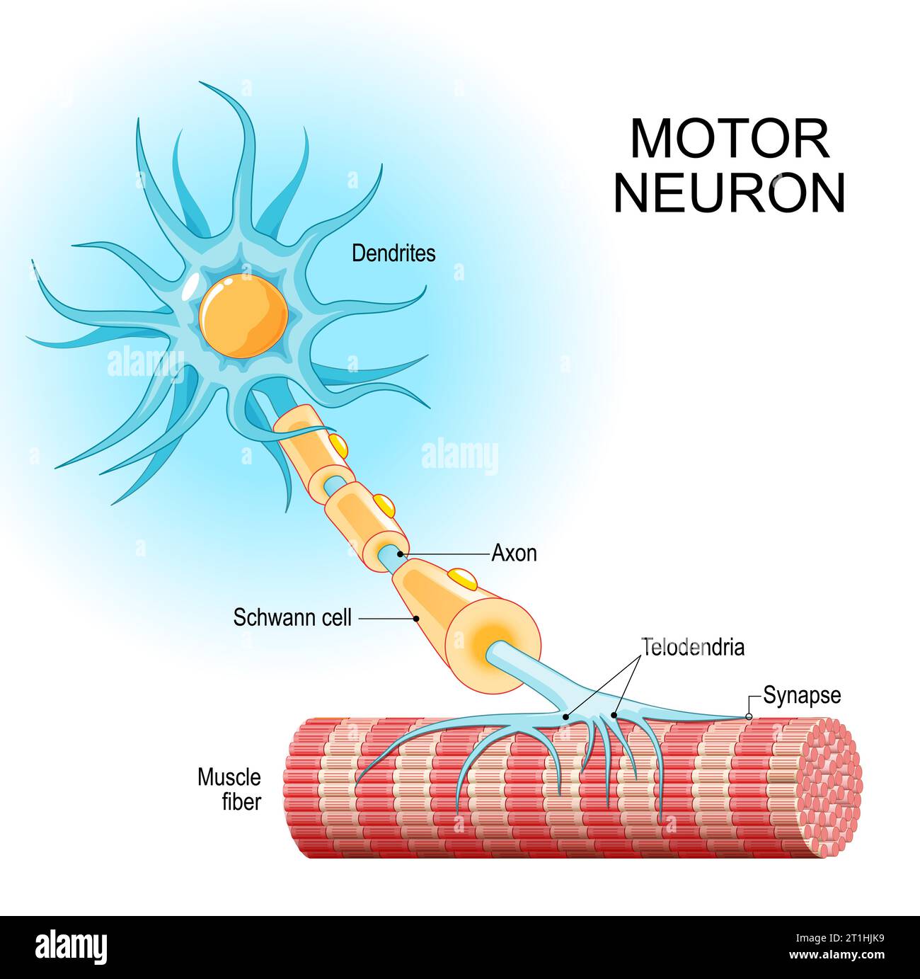
To assess muscle loss in motor neuron diseases, the number of motor units is studied. A motor unit includes a motor neuron and all the skeletal muscle fibers innervated by the neuron's axon terminals, including the neuromuscular junctions between the neuron and the fibers. Groups of motor units often work together as a motor pool to coordinate the contractions of a single muscle.

The number of motor units per muscle can change due to aging, disease, or injury.

Motor unit number estimation (MUNE) is a technique that uses electromyography to estimate the number of motor units in a muscle.

Motor unit number estimation (MUNE) methods, such as MScanFit MUNE, provide accurate results. This study uses MScanFit MUNE to compare patterns of motor unit loss in SMA, ALS, and healthy controls. Key findings include:

Patients with SMA had more severe degeneration of the hypothenar muscles compared to the thenar muscles. The authors call this: The reverse split-hand phenomenon, in reference to the split-hand phenomenon in ALS. Patients with ALS had a traditional split-hand phenomenon, with more severe involvement of the thenar muscles.

Reinnervation in SMA: Evidence of compensatory nerve sprouting in the ADM muscle has been noted in SMA but not in ALS, highlighting distinct pathophysiological mechanisms in these two diseases.

For SMA, the APB muscle has emerged as an optimal biomarker to monitor disease progression and therapeutic response due to its resistance to degeneration.

Curiously, although the authors confirmed the correlation between MScanFit MUNE neurophysiological parameters and disease severity in SMA patients, they did not find convincing correlations between MUNE neurophysiological parameters and clinical variables in ALS patients, particularly with the ALSFRS-R scale. On this issue, although several studies have reported the correlation between MUNE values ​​and the ALSFRS-R scale, others have not confirmed it.

Limitations and future directions The small cohort of AMS in the study and the lack of longitudinal data (over a period of several years) limit the conclusions. Future research should examine whether the reverse split hand phenomenon is exclusive to SMA and clarify why certain hand muscles are more vulnerable.

In this blog, I avoid studies that are not done on humans, firstly because the further away from humans and primates, the less sincere the scientific studies are. Then it is well known that the pre-clinical studies published are all extremely positive in order to attract investors (if possible private) who will finance clinical studies.

2,4-dinitrophenol (DNP)

The study examined here explores whether small doses of 2,4-dinitrophenol, a chemical used in the manufacture of pesticides and slimming drugs because it decreases the metabolic action of mitochondria, could help protect motor neurons, preserve muscle function and slow the progression of amyotrophic lateral sclerosis (ALS) in a mouse model of the disease. The scientists' guiding idea therefore seems to be to slow down the disease, by slowing down the metabolism. This seems absolutely counter-intuitive but the scientists assure that they have had good results. The effective dose of 2,4-dinitrophenol was very low (0.5–1 mg/kg, human equivalent dose 2.5–5 mg/day), making it safer for potential use in humans. enter image description here Dinitrophenol acts as a proton transporter in the mitochondrial membrane, inhibiting oxidative phosphorylation of ATP and making energy production less efficient. This is because some of the energy that is normally produced from cellular respiration is wasted as heat. This inefficiency is proportional to the dose of dinitrophenol that is absorbed. Thus, as the dose increases, energy production becomes less efficient: metabolism is then activated - more fat is burned - to compensate for the inefficiency and meet energy demands.

The researchers used hSOD1G93A mice, a common model of ALS, to test the effects of 2,4-dinitrophenol on motor skills, muscle strength, and disease progression. However, it is important to remember that most ALS patients do not have this mutation and that its notoriety is simply because it was the first mutation to be associated with ALS and that for more than 10 years (from 1993 to 2006) no other deleterious mutations were discovered in ALS.

Results

Mice treated with microdoses of 2,4-dinitrophenol (0.5–1 mg/kg) showed better motor coordination and better results on tests measuring muscle strength, compared to untreated mice. Early treatment initiation (before symptoms appeared) delayed the onset of motor decline, while late treatment initiation (after symptoms appeared) improved motor skills (which were already impaired by the disease) and slowed disease progression. Treated mice retained their ability to perform tasks such as running at 20 cm/s for longer than untreated diseased mice. In some cases, the mice regained lost motor skills, which is unusual in ALS research.

Although ALS damages the connections between nerves and muscles (neuromuscular junctions), treatment with 2,4-dinitrophenol preserved these connections. Treated mice retained a higher number of motor units (groups of muscle fibers controlled by a single neuron), indicating that motor neuron loss was reduced.

Reduced Cellular Stress

2,4-dinitrophenol reduced oxidative stress, a harmful process linked to the progression of ALS. This was demonstrated by lower levels of damaged proteins in treated muscles. While ​​slowing down an ALS patient’s metabolism seems pretty criminal, reducing cellular stress is a very good idea.

The drug also reduced inflammation and activated pathways (like Akt/mTOR) involved in muscle growth and repair.

How would 2,4-dinitrophenol work?

2,4-dinitrophenol acts on mitochondria, the energy producers of cells, by slightly lowering their membrane potential (∆Ψm). This mild “uncoupling” reduces the production of harmful reactive oxygen species (ROS), which can damage cells. 2,4-dinitrophenol may also help clear damaged mitochondria and promote the formation of healthy mitochondria, further protecting neurons and muscles.

Future Directions

While the mouse results are promising, translating these findings to humans requires more research. The study also highlighted some limitations:

Could mechanical stimulation be beneficial in ALS?

- Posted by admin in English

In recent years, research has shown that physical exercise has many benefits for the entire body, beyond muscle growth. There has been much discussion in recent years about the value of physical activity programs in the case of Alzheimer's or Parkinson's disease.

Researchers at MIT wrote about experiments that might lend credence to the idea that mechanical stimulation could one day be beneficial in ALS or other MND diseases.

Muscles release various biochemical factors, called myokines, which circulate in the bloodstream. These molecules establish a kind of dialogue between different tissues and this allows them to collectively adapt to a new environment. Myokine receptors are found in muscle, fat, liver, pancreas, bone, heart, immune system and brain cells. The location of these receptors reflects the fact that myokines have multiple functions. First, they are involved in the metabolic changes associated with exercise, as well as in the metabolic changes that follow adaptation to training. To study these effects specifically on motor neurons, the cells that transmit movement commands from the brain to muscles, MIT researchers developed a series of in vitro systems where they could precisely control and observe the effects of muscle contractions.

In 2023, Raman and his colleagues reported that they could restore mobility in mice that had suffered traumatic muscle injury by first implanting muscle tissue at the site of the injury and then exercising the new tissue by repeatedly stimulating it with light. Over time, they found that the exercised graft helped the mice regain motor function, reaching levels of activity comparable to those of healthy mice. This meant not only that the new muscle tissue had become functional, but also that there were somehow new connections between the lower motor neurons and the new muscles. In other words, the exercise was not only beneficial for the new muscle but also for the local motor neuron that develops synapses to connect to muscle fibers.

Then the group wondered: Could exercise’s purely physical impacts have a similar benefit on motor neurons?

This claim was met with some skepticism.

So the MIT team designed experiments in which the neurons were repeatedly pulled back and forth, similar to the way muscles contract and expand during exercise. enter image description here Angel Bu is the first author, while Ritu Raman is the senior author; the other authors are from MIT’s Department of Mechanical Engineering and MIT’s Koch Institute for Integrative Cancer Research. The authors matured a set of motor neurons on a gel that was a kind of carpet into which they embedded tiny magnets. They then used an external magnet to shake the carpet—and the neurons—back and forth. In this way, they made the neurons work, for 30 minutes a day.

The researchers demonstrated that the physical force generated during muscle contraction has direct mechanical effects on motor neurons, promoting growth in both a biochemical and mechanical way. And the two effects, myokine release and motor neuron work, have similar effects.

These results could have important implications for the treatment of motor neuron diseases, such as amyotrophic lateral sclerosis (ALS) or spinal muscular atrophy, where motor neurons progressively lose their function. Therapies that mechanically stimulate muscle contractions could encourage nerve growth and regeneration, potentially slowing the progression of these diseases or facilitating recovery after nerve damage.

Of course, this study is extremely preliminary, it is carried out in vitro. We cannot even talk about micro-organs. This is basic research indeed, the authors didn't talk about applications in ALS/MND diseases. Another limitation of this study is that the authors did not explore a wide variety of mechanical or biochemical stimulation protocols with different frequencies, magnitudes, and durations.

If these mechanisms were confirmed in preclinical studies with primates and then clinical studies with humans, exercise protocols could be refined to maximize the biochemical and mechanical benefits for motor neurons, potentially improving motor function or slowing disease progression in patients with motor neuron diseases.

For example, patients could benefit from muscle stimulation devices that could help maintain or even regenerate motor neuron pathways. Mechanical muscle stimulation could indeed be a way to mimic the combined biochemical and mechanical effects of muscle contractions in patients with weakened muscles, potentially helping to slow neuronal degeneration or even stimulate nerve repair.

Electrical stimulators already exist for physiotherapy and rehabilitation, and future protocols could incorporate fine-tuning to produce exercise-like contractions that not only provide muscle benefits but also encourage motor neuron activity. Given that MND patients vary greatly in progression and severity, personalized electrical stimulation programs would be essential. Clinicians could develop individualized regimens, perhaps informed by biomarkers or real-time feedback, to maximize neuron growth-promoting effects without overloading the system.

Future research could test whether electrical muscle stimulation in MND patients produces benefits similar to biochemical and mechanical stimulation observed in vitro. If effective, this could lead to new physiotherapy protocols or devices aimed at improving the quality of life and motor function of people with MND.

Checking the levels of SOD1 in CSF?

- Posted by admin in English

Until Tofersen from Biogen, ALS drugs were only slightly slowing the disease. Tofersen aims to lower the levels of a specific mutation of SOD1. This therapy is probably a lifeboat for the patients with the matching SOD1 mutation. It's certainly slowing the disease progression, but only for a tiny portion of ALS patients less than 2%.

Not all patients with the right SOD1 mutation react to Tofersen similarly.

  • People have two SOD1 genes in their cells and sometimes only one allele is mutated and the other can make up for the defective one. There are also other proteins named SOD2 and SOD3 that can assume partly the functions of SOD1.

  • In the worst case with ASO medications, ALS patients are merely exchanging a defective protein with a lower production of the same protein. This is not a great perspective, a genetic therapy that makes cells produce the correct version of SOD1 would be better. Yet genetic therapies have their share of problems, including low efficiency and increased cancer risk.

All members of this protein family, transform a byproduct of the mitochondrial electron transport chain, into hydrogen peroxide and diatomic oxygen. Yet those two chemical species are themselves quite reactive. SOD1 is located in the cytoplasm of cells, SOD2 in their mitochondria, and SOD3 is extracellular.

In that case, where there is still one functional allele, it would be nice to check SOD1 levels in the body of patients treated with Tofersen. If it's too low (because of Tofersen) the therapy renders itself ineffective. This could be done by analyzing blood, but scientists reckon that checking SOD1 in CSF would be a better idea.

The discovery of children developing SOD1 Deficiency Syndrome (ISODDES) which is characterized by injury to the motor system, suggests that a too-low SOD1 antioxidant activity may be deleterious in humans. Measuring SOD1 activity in cerebrospinal fluid (CSF) in Tofersen-treated patients is recommended but difficult due to low concentration and the presence of the isoenzyme SOD3.

To assess SOD1 activity, the scientists propose to remove SOD3 from CSF samples with antibodies and subsequently measure the SOD1 activity. enter image description here To propose this as a standard procedure for human beings is a bit weird. Repeatedly piercing the membranes that protect the spinal cord sounds like an unhealthy proposal, particularly if it's done to patients having motor neuron disease.

Rethinking antisense oligonucleotide therapies

- Posted by admin in English

A recent publication provides a detailed overview of recent advances and challenges in the development of RNA therapies for neurodegenerative diseases, particularly for amyotrophic lateral sclerosis (ALS). enter image description here Early immunotherapies targeting amyloid-β in Alzheimer's disease initially showed reductions in amyloid plaques but failed to prevent cognitive decline. The FDA recently approved lecanemab as the first disease-modifying drug for Alzheimer's disease. However, its benefits appear to be limited to the early stages of the disease, as it does not stop neurodegeneration or improve cognition. This highlights the difficulty of modifying neurodegenerative diseases, which often progress despite treatment.

Most cases of ALS are recognized as proteinopathies involving RNA dysregulation, but in others, such as those with mutations in superoxide dismutase 1 (SOD1), these abnormalities are absent, suggesting different pathogenetic pathways.

Nucleic acid-based therapies (NATs) represent an emerging treatment class that targets RNA rather than proteins. These agents act by degrading disease-associated mRNA or altering translation processes. For example, ASOs inhibit translation by disrupting ribosome assembly or degrading mRNA.

Although ASO approaches are both reasonable and scientifically sound, discernible clinical benefit has not always been observed in humans.

Three clinical trials using ASOs to reduce huntingtin (HTT) protein in Huntington’s disease have been halted because the therapies did not consistently reduce mutant HTT levels or improve clinical outcomes. A trial of one ASO, tominersen, caused unexpected worsening in patients who received higher doses. This has raised concerns about potential ASO toxicity due to off-target effects, immune responses, or loss of normal HTT function, which is essential for cellular health.

ASOs targeting toxic genes in ALS, such as TDP-43 in ALS, have shown promise in preclinical studies in extending life and restoring motor function in animal models. Yet, inhibitions of SOD1 and C9orf72 in mice show motor deficits and memory impairment over time, warning researchers of the potential risks of long-term ASO treatments. Clinical trials, such as the Phase III study of tofersen (an ASO targeting SOD1), have demonstrated a reduction in SOD1 mRNA, although clinical (i.e. visible) benefit has not been established.

Results from a Phase III trial of the ASO tofersen, which targets SOD1 mRNA in ALS, were reported in 2022. In this study, a total of 108 participants were enrolled, 60 of whom were classified in the faster progression subgroup. Approximately 7% of participants receiving tofersen experienced serious neurological adverse events, including myelitis, chemical or aseptic meningitis, and lumbar radiculopathy, but the pharmaceutical and medical community has highlighted the case of a Swedish patient who saw real benefit from his therapy.

A new study, ATLAS (NCT04856982), aims to test tofersen in asymptomatic, and some are thought to be presymptomatic, carriers of the SOD1 mutation to determine whether early intervention can delay the onset of ALS.

ASO-mediated inactivation in neurodegenerative diseases faces major challenges. By the time symptoms of ALS appear, motor neuron damage may be too advanced. Symptoms appear because compensatory mechanisms are exhausted. This is similar to Parkinson’s disease, where symptoms only appear after significant neuronal loss.

It is also likely that current ASOs are not very selective, and may reduce the production of essential proteins, causing side effects. Targeting mutations or aberrant transcripts specifically with more selective ASOs, such as those used in the PRECISION-HD trials, could help preserve healthy protein function.

Identifying sensitive biomarkers, such as elevated plasma TDP-43 or cryptic HDGFL2 levels, could help diagnose ALS before symptoms appear. This could allow for timely therapeutic interventions to slow or prevent neurodegeneration before it becomes irreversible.

In summary, RNA-based therapies such as ASOs offer hope for neurodegenerative diseases but face significant challenges.

Biotechs are weird, NP001 is back.

- Posted by admin in English

Readers of this blog probably know the story of the drug NP001. One phase I and two phase II trials were conducted by Neuraltus in ALS patients (NCT01281631 and NCT02794857). These were both 6-month studies, the usual clinical trial duration. Phase 2A was completed in 2012, and Phase 2B in 2017. Both failed. Now some people want to revive this drug, through a new biotech named Neuvivo.

What the authors of a new publication did was to try to find a subset of patients that showed some longer survival with NP001 in those old trials.

This is a widely used technique by unsuccessful companies, but it is statistically meaningless.

The authors found a subset of patients who had inflammation and a longer survival. Yet this analysis would be comically wrong if we were not speaking of dying people. enter image description here For example, the authors claim that survival of ALS patients with inflammation is 16 months longer than in the placebo arm. At 72 months there were only 3 people in the inflammation subset and two people in the placebo arm. Nobody can say anything about these numbers. If we use the same criteria, it shows that NP001 worsened the condition in all pALS with respect to the placebo branch, as starting from 72 months there were fewer survivors in the NP001 arm than in the placebo.

In addition, one could read the subset of pALS with inflammation as them having a comorbidity. If you prescribe an inflammation therapy to people having inflammation, they will improve a bit, which would be reflected in longer survival to ALS.

The proposed mechanism of action is vague (as usual, at least here there is a proposal): They said that NP001 is converted by macrophages to taurine chloramine, a regulator of inflammation. As the authors assert that persistent immune activation in patients with ALS is the cause of loss of muscle, an anti-inflammatory drug would help.

There are few publications about taurine chloramine, but, when taurine is in the presence of highly toxic hypochlorous acid, it generates the less toxic taurine chloramine. It's the result of the body's mitigation to a poison.

Any Relation Between Sleep Apnea and Amyotrophic Lateral Sclerosis?

- Posted by admin in English

The current paper is about an untested hypothesis. Scientists have proposed thousands of assumptions about the etiology of ALS, and the same is true for many other diseases. Single-authored papers are often dismissed, as are papers that make wild hypotheses without trying to test them. I mention this paper because the single author is a remarkable scientist in the field, not one of the countless hacks who write a paper on ALS today with very little knowledge of the disease and will never write about it again.

Another reason might be because it may have some relation with a drug developed by Richard B. Silverman, and P. Hande Ozdinler at Northwestern University. AKAVA Therapeutics, started last year by Silverman, is carrying out studies of the drug AKV9 (ex NU-9).

This article discusses the possible relationship between sleep, the glymphatic system, and neurodegenerative diseases. It suggests that sleep issues might exacerbate ALS disease progression by impairing the brain's waste-clearance mechanisms and compromising neuronal health, but it does not describe any experiment. It also tells that neuron health is diminished when the organism is sleep-deprived, in particular neurons experience dendritic spine loss.

Sleep is crucial for brain health because the clearance of metabolic waste and the remodeling of synapses happens during sleep. Many ALS and Parkinson's patients experience sleep disturbances. Disrupted sleep patterns, especially those associated with conditions like sleep apnea, a condition where upper airways collapse during sleep, can lead to impaired glymphatic function. The author equals poor glymphatic function with accumulation of agglomerates of misfolded proteins in cellular cytosol. I am not sure there is any evidence of this relation.

The glymphatic system is responsible for clearing waste products from the brain, primarily during sleep. It is facilitated by cerebrospinal fluid (CSF) circulation. During sleep, the glymphatic system operates optimally, removing waste products from the brain. Impaired glymphatic clearance, especially when sleep is disrupted, might be a significant contributor to protein buildup and possibly subsequent neurodegeneration.

Evidence from studies shows a faster CSF clearance during sleep, suggesting that adequate sleep is crucial for maintaining brain health. Dysfunction of this system can contribute to the accumulation of toxic proteins, such as amyloid-beta and tau, associated with neurodegenerative diseases. Yet this clearance happens in the periphery of the brain, so it's hard to see how a good clearance would improve the health of those neurons that are deep inside the brain.

Early signs of neurodegeneration in ALS include the loss of dendritic spines. A dendritic spine is a small membrane protrusion from a neuron's dendrite that typically receives input from a single axon at the synapse. Motor neurons in the spinal cord, for example, can have a dense arrangement of dendritic spines in certain regions, as these neurons need to process a large number of excitatory and inhibitory signals to regulate muscle activity effectively. Sleep plays a vital role in spine pruning and remodeling, ensuring healthy neuronal connectivity.

Synaptic connections, especially dendritic spines, are dynamic and undergo constant remodeling, particularly during sleep. This process is crucial for maintaining neuronal network stability and function. Sleep deprivation or sleep apnea disrupts spine pruning, leading to excessive or unhealthy connections, which stresses neurons and hampers brain connectivity. Experiments show that even brief sleep deprivation reduces spine elimination, resulting in abnormal spine density and neuronal hyperactivity in brain areas like the hippocampus. This disruption may have a profound impact on memory and cognitive function.

Therefore investigating the relationship between sleep, glymphatic function, and biomarkers in CSF could lead to earlier diagnosis and more effective disease monitoring.

Yet, sleep is not routinely assessed in clinical diagnostics for neurodegeneration. Integrating sleep studies into patient assessments could enhance diagnostic precision and enable targeted interventions. Moreover, understanding the molecular changes in CSF associated with sleep problems could provide valuable biomarkers for monitoring disease progression. There’s potential for developing therapies focused on improving sleep quality, as addressing hypoxia and improving glymphatic function might reduce protein buildup and protect against neuronal damage.

The importance of studying different muscle groups in ALS

- Posted by admin in English

A couple of days ago, I complained about the lack of publications on ALS metabolism in skeletal muscles, which represent only a tiny fraction of the huge number of ALS (mostly useless) publications.

Here is one article describing the evolution of the masseter muscle, one of the muscles of mastication, during the disease course in a SOD1 mice model. Why study the masseter muscle in ALS? Because it is one of the few skeletal muscles that is not affected by the disease. enter image description here The authors observed that, despite a decrease in limb motor functions, the feeding function of these mice was preserved until the late stages of the disease. Remarkably, the masseter muscle showed no reduction in muscle volume, wet weight, or muscle fiber cross-sectional area. Furthermore, no changes were observed in muscle fiber types, indicating a possible resistance of the masseter muscle to ALS-induced impairment. A potential reason for the lack of atrophy in the masseter muscle could be its higher number of muscle satellite cells compared to that of the gastrocnemius muscle. This abundance may promote the maintenance of muscle fiber nuclei, thereby contributing to muscle tissue regeneration.

What they are saying in the background is: * Something is stressing the skeletal muscles in ALS, which looks quite a reasonable assumption to me. * Maybe it would be possible to design an ALS therapy targeting muscle satellite cells. This looks less likely to me.

While the authors promote the idea that the disease may, at least partly, start in muscles, I can't help myself thinking that maybe the reason it is preserved is simply that this muscle (as for eye movements) is activated by the trigeminal nerve which is usually preserved in ALS.

It seems to me that in spinal ALS, the longer motoneurones fail early (hands, feet) and the shorter ones survive longer. Indeed in bulbar ALS, the situation is reversed, but ALS mice models attempt only to model spinal ALS. So in my opinion, the authors should have considered that the lack of wasting of mice's masseter muscle may stem from a still functioning trigeminal nerve.

Trial designs for motor neuron disease in the 21st century

- Posted by admin in English

We previously reported that Memantine was found ineffective in an ALS clinical trial for the fifth time. Several articles about persistent failure in ALS clinical trials appeared in the last issue of The Lancet Journal.

Trial designs for motor neuron disease in the 21st century

A new era of drug discovery for amyotrophic lateral sclerosis

Scientists attribute Memantine's (and the many other drugs that were tried) lack of efficacy to the "complexity of the pathophysiological mechanisms."

In simpler words, they have no idea why it failed, yet it was tested for the fifth time in ALS, so in a rational world, they should, on the contrary, have expected it to fail. In addition, no pre-clinical studies have shown any special value of Memantine in ALS.

And it's not only about Memantine, hundreds of drugs with dozens of different mechanisms of action have been trialed in ALS, as well as in other neurodegenerative diseases. For example, in the same journal issue, there are the results of the ROCK-ALS phase II clinical trial which has a purported mechanism of action entirely different from the usual suspects (glutamate excitotoxicity, impaired proteostasis, autophagy, and neuroinflammation).

Clearly, scientists know nearly nothing about these diseases, otherwise, they would concentrate their efforts on specific drugs.

It is time to reconsider century-old and unquestioned assumptions about these diseases. What makes ALS patients die? Skeletal muscle wasting which leads to respiratory failure. Efforts should concentrate in this direction. We also know that ALS is not always a death sentence, for example Stephen Hawking lived 76 years and 55 years with the disease. We know that a BMI in the 27 range helps for survivability, yet publications on ALS metabolism in skeletal muscles represent only a tiny fraction of the huge amount of ALS publications. In 2023 there were only 23 publications on this topic, versus 2507 publications on ALS.

Why this situation? Laboratories and CRO are not organized to study whole-body mechanisms in large mammals. It would cost a lot, universities and biotech prefer to work on small rodents or even worse on immortalized cell lines.

There is also the question of the time span, most studies are conducted within 6 months, or even two months, because students are used as a cheap workforce. You can't detect any statistically meaningful clinical change in neurodegenerative diseases in two months.

Studies must last at least one year and use large mammals, if possible mammals which have a corticospinal tract similar to ours, with direct connection between upper and lower motor neurons for fine control of skeletal muscles such as in hands..

A recent publication poses a question that has become increasingly pressing in the face of numerous unsuccessful trials for the treatment of amyotrophic lateral sclerosis (ALS), also known as Lou Gehrig's disease. "Why is the treatment and management of amyotrophic lateral sclerosis so difficult?"

The article suggests that when a phase II clinical trial succeeds while its phase III trial fails, it's because they have a different profile of patient population.

The authors argue that because they recruit fewer patients, the characteristics of patients enrolled in phase II trials are more homogeneous than those in phase III trials, which aim to recruit hundreds of participants. This is contrary to what statistics teaches.

I also find this hypothesis unlikely, given the constraints on patient recruitment and the sensitivity of principal investigators to the fragility of ALS diagnoses and the variability of patient phenotypes.

The authors also propose specifically that edaravone and Relyvrio (AMX0035) phase II studies yielded better results than their phase III studies. However, the phase II trials for these two drugs did not provide clear evidence of a therapeutic benefit, only showing marginal improvements in the ALSFR-R criterion, which is known to be influenced by non-medical factors such as access to better equipment. Relyvrio/AMX0035 in particuliar benefited from the intense pressure by ALS organisations. It should be noted that one of these organisations would have financially greatly benefited in a market authorization.

Furthermore, post-hoc analyses, which are often favored by drug manufacturers, are statistically unreliable. It is too easy to select favorable results from a small sample and attribute them to a common characteristic, which is why large-scale clinical trials are necessary.

In my opinion, it is time to recognize that we are on the wrong track in relying on molecular biology experts to develop treatments for neurodegenerative diseases. The current pharmacological approach is heavily influenced by our understanding of communicable diseases, where identifying the pathogen and suppressing it can lead to recovery. Molecular biology is conceptually far removed from medicine, as it ignores cellular mechanisms, tissue-level interactions, physiological systems, and the complex interplay between organs.

I believe that we would be better served by acknowledging that neurodegenerative diseases are the result of a complex interplay of factors, including a patient's medical history, lifestyle, and environmental exposures. Each patient over the age of 50 likely has a unique combination of age-related neurological diseases, with some having a history of strokes, high-intensity sports, environmental toxins, or head trauma. Some may have also had a history of substance abuse or smoking.

In summary, I believe that neurodegenerative diseases are the result of a complex interplay of factors in a patient's history, rather than a single molecular event. Therefore, funding for most molecular biology efforts should be gradually shifted to regenerative medicine, which addresses the restoration of damaged tissues and organs.


Please, help us continue to provide valuable information: