A tool for ALS or FTD gene carriers.
Amyotrophic lateral sclerosis (ALS) and frontotemporal dementia (FTD) are devastating neurodegenerative diseases. A significant number of cases are linked to a hexanucleotide repeat expansion in the C9orf72 gene, making it the most common known genetic cause of both conditions.
Genetic counseling is essential in informing families about their risk, especially for those with a family history of the disease. Currently, children of C9orf72 mutation carriers are often told they have a 50% chance of inheriting the mutation. While technically correct based on Mendelian inheritance, this figure overlooks a critical factor: age-related penetrance.
Penetrance describes the likelihood that someone carrying a disease-causing gene will develop the disease. In cases of C9orf72-related ALS/FTD, penetrance increases with age, peaking around 58 years old. This means that simply knowing you carry the mutation does not give the full picture of your personal risk.
A new study addresses this limitation by developing a more precise method for calculating risk and providing an online tool for families.
The tool is available here: https://lbbe-shiny.univ-lyon1.fr/ftd-als/
While other research has focused on identifying genetic modifiers of disease risk, this study centers on a readily available and easily measurable factor: age.
The researchers used a Bayesian approach, a statistical method that updates probabilities with new evidence. In this case, the evidence includes the individual's age and family history. By integrating age-related penetrance data, the researchers created a model to estimate the probability of carrying the C9orf72 mutation and developing ALS or FTD within a specific timeframe. This approach is especially relevant for asymptomatic relatives, such as children, siblings, grandchildren, and niblings of mutation carriers.
Importance of this work:
This research is significant because it moves beyond the simplified 50% risk figure, offering a more personalized and accurate risk assessment for individuals at risk of C9orf72-related ALS/FTD. It helps inform decisions about genetic testing and could influence lifestyle choices or participation in clinical trials. As testing for C9orf72 becomes more common, the need for nuanced interpretation of results increases. The findings are highly relevant for families affected by ALS/FTD, providing a more realistic understanding of their individual risk profiles.
Originality:
The study offers original insights beyond the basic concept. Although age-related penetrance is a known idea, this research presents a concrete, mathematically sound method to incorporate it into risk calculations. The online simulator further enhances its practical use. The novelty is in applying a Bayesian framework to refine risk estimates in C9orf72-related ALS/FTD, providing a more sophisticated and personalized approach than traditional Mendelian risk assessments.
Conclusion:
This study makes a valuable contribution to ALS/FTD genetics. By offering a more detailed and personalized risk assessment, it can improve genetic counseling, aid in clinical trial recruitment, and deepen the understanding of the disease. The online simulator makes this complex information accessible to clinicians and families, increasing its practical impact.

 The researchers specifically focused on molecules that could reduce or reverse stress granule formation, particularly those that act directly on stress granule proteins and may be useful as therapeutic agents. From the initial screening, lipoamide emerged as a novel, potent modulator of stress granules.
Once lipoamide was identified as a hit, the researchers sought to determine its effects in cells regarding specificity, potency, intracellular localization, and its effects on other cellular condensates.
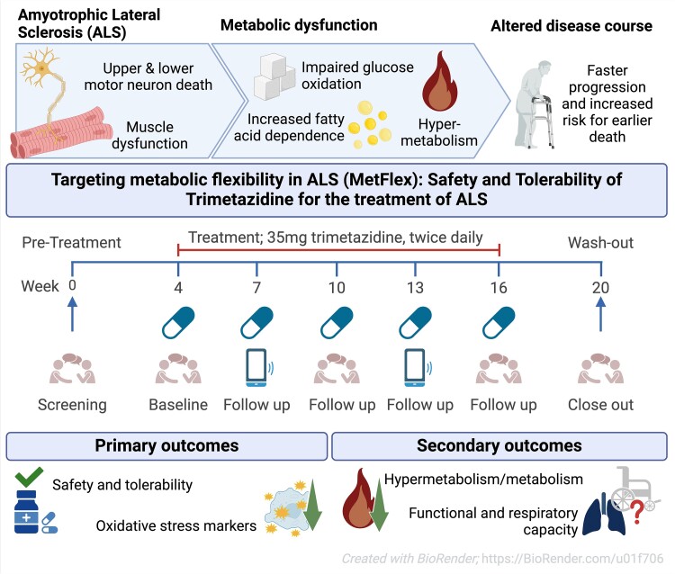 While the publication recounts that trimetazidine was beneficial for patients (this is not a phase III trial), for me the results section does not show conclusive results. For example, the results improved only during the wash-out period.
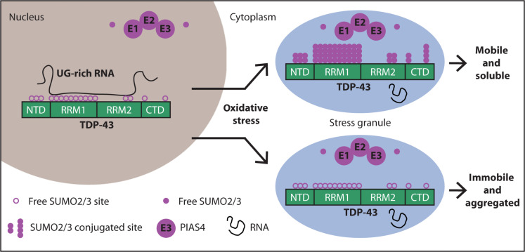 Transactive response DNA-binding protein 43 (TDP-43) is a nuclear RNA binding protein (RBP) involved in RNA metabolism.
TDP-43 has a high propensity to aggregate because of its low solubility in cells and in vitro.
The aggregation propensity of TDP-43 is increased by ALS/FTD-linked mutations and upon exposure to stress and has been observed in patients with C9orf72 hexanucleotide repeat expansion, the most common genetic cause of sporadic and familial.
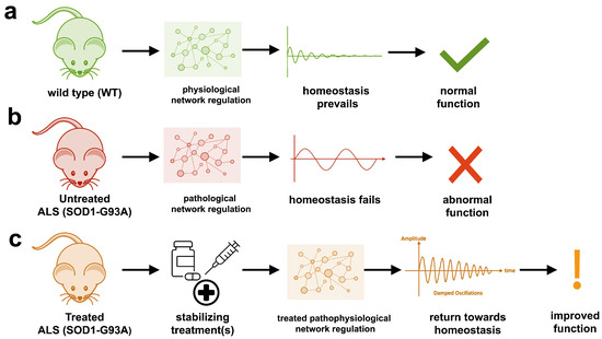 The study uses an innovative and integrative framework to model the regulatory dynamics of wild-type (WT) and SOD1-G93A ALS mice. The models are based on first-order ordinary differential equations (ODEs) that describe how the system output evolves over time. The research uses dynamic meta-analysis to synthesize experimental data from the literature and parameter optimization based on genetic algorithms to infer missing data. Indeed, to build a model, data are needed and here these are obtained from results reported in the literature on SOD1-G93A ALS mouse models.
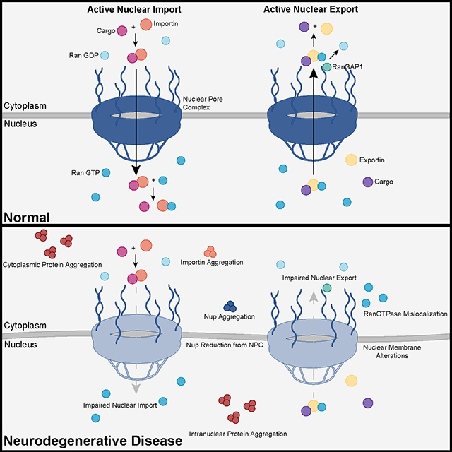 Recent advances into the underlying pathogenic mechanisms have associated mislocalization and aberrant accumulation of disease-related proteins with defective nucleocytoplasmic transport and its mediators called karyopherins.
  They measured somatic repeat expansion over time in individual neurons from donors of different ages. They found that early-phase expansions (e.g., from 40 to 80 CAG repeats) were slow and stochastic, taking decades, while later expansions (e.g., from 80 to 150 repeats) occurred more rapidly.
They then analyzed genetic markers and DNA repair mechanisms associated with repeat instability, such as those involving DNA mismatch repair (MMR) proteins (e.g., MSH3, PMS1). Variants in these genes have been shown to influence the rate of somatic instability. The progression of CAG repeat expansion is driven by errors in DNA replication, repair, and maintenance, particularly in neurons. Key mechanisms include:
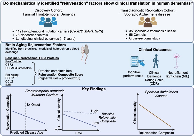 The results appear relatively reliable because the scientists found similar effects in two different types of dementia. The effects were seen across multiple measures (cognitive, functional, and biological markers).
 The endoplasmic reticulum (ER) is an important organelle in cells that is involved in protein conformation. This step occurs after protein synthesis by ribosomes and after conformation, the new protein will be sent to its final destination by the Golgi apparatus. Protein conformation requires energy, so when disease occurs, the ER may not be able to properly conform the new proteins.