Here we will discuss about two recent studies about the relation between protocatechuic acid and Alzheimer's disease.

Diet appears to be have some relation with the progression of Alzheimer's disease, maybe related to the gut microbiome. There is a clear relation between gut's microbiome and Alzheimer's disease which was formalized in Braak's stages.

The aims of the first study were to detect the main anthocyanins of blueberry extract; then to verify the protective effects of anthocyanin-rich blueberry extract on hippocampal neurons and the promotion of autophagy; and finally to investigate the main protective effects and mechanisms of protocatechuic acid, a major metabolite of blueberry extract, for promoting autophagy and thus playing a neuroprotective role.

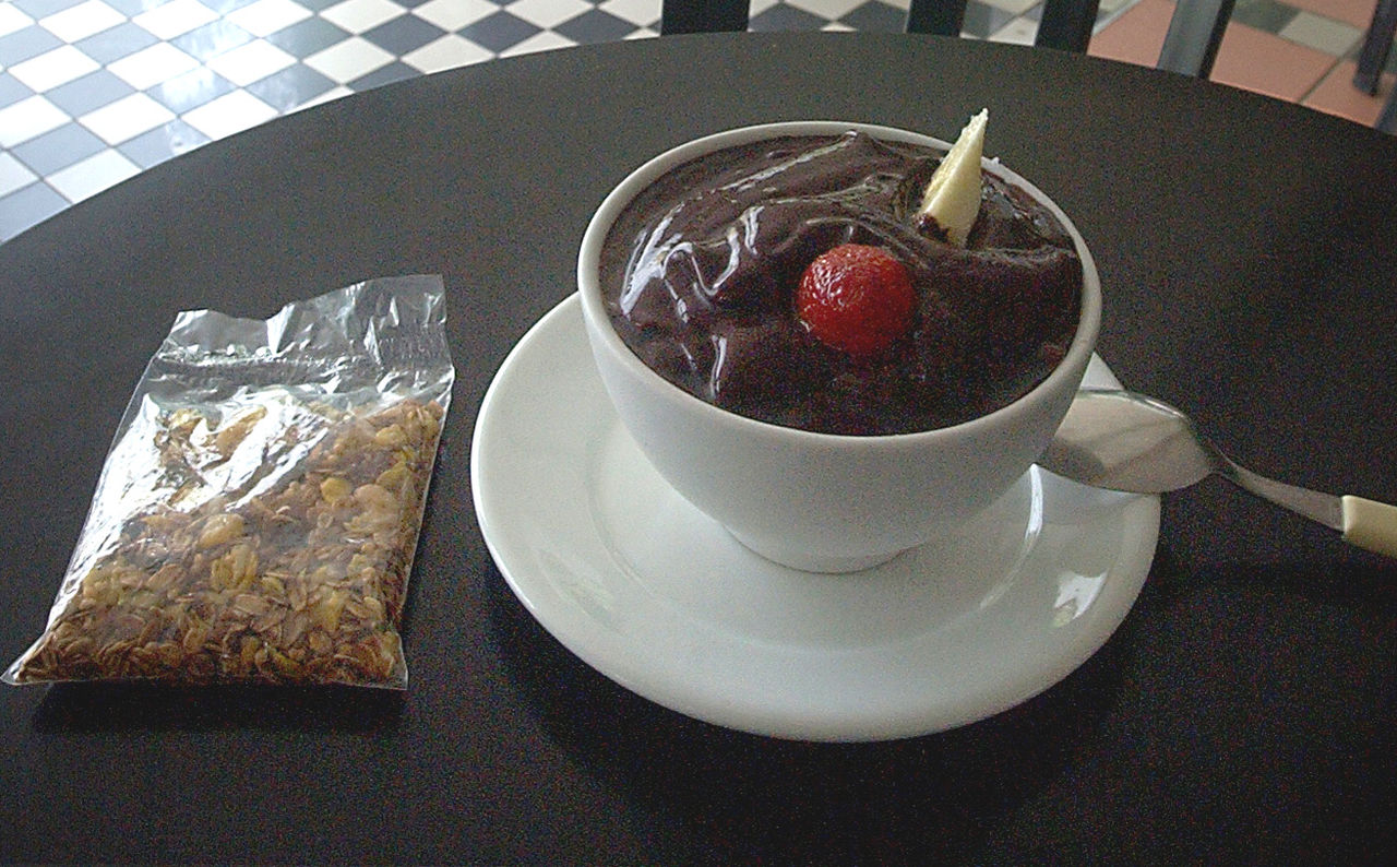
Protocatechuic acid is a type of phenolic acid increasing bringing plants resistance against fungus. It is a major metabolite of antioxidant polyphenols found in green tea. It has mixed effects on normal and cancer cells in in vitro and in vivo studies. Açaí oil, obtained from the fruit of the açaí palm (Euterpe oleracea), is rich in protocatechuic acid (630±36 mg/kg). enter image description here

Protocatechuic acid is regarded as an active component in traditional Chinese herbal medicine. For example the dried and mature fruits of the Zingiberaceae plant Alpinia oxyphylla Miq, is a choice in traditional Chinese medicine to treat Alzheimer's disease.

Recent studies have demonstrated its potent activities in modulating multiple signaling pathways associated with β-amyloid deposition, tau protein phosphorylation, chronic inflammation, oxidative stress.

In this study mice models of Alzheimer's disease (APP/PS1) were given 150 mg/kg blueberry extract daily for 16 working days. The mice were then sacrificed and morphology of neurons was observed under transmission electron microscope and autophagy-related proteins were detected.

Protocatechuic acid also promoted autophagy of neurons and the mechanism was mainly involved in increasing autophagosome degradation.

Neuron damage in morphology was reduced and the expression of autophagy-related proteins in APP/PS1 mice were promoted after blueberry extract treatment. In vitro, Aβ25-35-induced cytotoxicity, including decreased neuron viability and increased levels of lactate dehydrogenase and reactive oxygen species, was effectively reversed by protocatechuic acid.

Furthermore, by adding autophagy inducers rapamycin and autophagy inhibitors Bafilomycin A1, it was verified that degradation of autophagosomes was upregulated and autophagy was promoted by protocatechuic acid.

Yet it seems unlikely that any chemical coumpound would dramatically improve Alzheimer's disease in a few weeks and we all know that few results like that are credible. However other recent studies have hinted at similar results.

Protocatechuic acid is a dihydroxybenzoic acid with is a precursor of some insecticides and neurotransmitters like catecholamines, so it is indeed a potent and possibly dangerous drug.

In the treatment of Alzheimer's disease, it is important to develop alternative cholinesterase inhibitors with antioxidant properties that will reduce acetylcholine deficiency and free radical formation.

The aim of this other study was to investigate the effect of hydroquinone, 4-hydroxybenzoic acid, 3,5-dihydroxybenzoic acid, caffeic acid, vanillic acid and chlorogenic acid against acetylcholinesterase (AChE), partially purified from serum.

Binding of compounds with effective inhibitory potential to the AChE active site as competitive was illuminated by molecular docking.

Hydroquinone, chlorogenic acid and 4-hydroxybenzoic acid have been found to have higher inhibitory potential than others against the AChE. IC50 and KI values of the phenolic compounds against AChE were found in the range of 0.26 ± 0.01-36.34 ± 2.72 mM and 0.72 ± 0.00-29.23 ± 2.62 mM, respectively.

The effectiveness of the compounds has been associated with its structure. Consequently, the phenolic compounds, which have AChE inhibitory potential and antioxidant properties, can be considered as alternative drugs in the treatment of Alzheimer's disease.

During 2019 January I thought a TDP-43 therapy was really missing and the next month I wrote a plea to develop a TDP-43 therapy. I am not sure it had any effect, but I sent emails to nearly 250 scientists the next months. A flurry of patents have been written which indicates it is now an active research topic. Now we are at a phase where there are even clinical trials targeting TDP-43.

Reducing the TDP-43 misfolded mislocated aggregates may stop ALS progression and it could help as well in other neurodegenerative diseases.

Now I think that a part of the set of drugs needed to really recover (in addition to TDP-43 therapies) is something targeting faulty UPR (Unfolded Protein Response). Here is a list of natural products targeting the UPR signaling to tilt it towards pro-survival. Some, it will not surprise you, have been discussed since a long time on Internet forums. (source doi: 10.1007/s11010-021-04223-0)


Name of compound / Mode of action / References


  • Ginkgolide / K(GK) Reduce ER by accentuating the IRE1/XBP1 activity. / [136]

  • Elatoside C / Inhibit ER stress induced genes like GRP78, CHOP, caspase-12 and JNK, reduce apoptosis. / [137]

  • Sulforaphane (SFN) /Inhibit GRP78,CHOP and caspase-12 by activating the SIRT1 pathway.SIRT1 decrease ER induced apoptosis by deacetylating eIF2α / [138]

  • Resveratrol / Reduce ER stress mediated apoptosis by downregulating the expression of GRP78, GRP94 and CHOP and upregulating the expression of Bcl-2 and Bax. Lowers the expression of GRP78 and CHOP in doxorubicin treated H9c2 cell. / [139–141]

  • Baicalin / Target the CHOP/eNOS/NO pathway by inhibiting CHOP and thus reducing apoptosis / [142]

  • Berberine / Decrease apoptosis by decreasing the phosphorylation rate of PERK and eIF2α and downregulating the expression of ATF4 and CHOP / [143]

  • Anisodamine / Downregulate the expression GRP78, CHOP, and cleaved caspase 3 and thus reduce cell death / [144, 145]

  • Rare ginsenoside-standardized extract (RGSE) / Inhibit the overexpression of GRP78, GRP94 and CHOP as well as decrease the phosphorylation level of PERK and IRE1α / [146]

  • Panax quinquefolium saponin (PQS) Improve ventricular remodeling by downregulating the expression of GRP78, CHOP, and Bax protein as well as increasing the expression of Bcl-2 protein, thus reducing apoptosis. Also inhibits apoptosis by targeting the PERK-eIF2α- ATF4- CHOP pathway / [147–149]

  • Notoginsenoside R1 (NGR1) Protect cells from acute ER stress by delaying the onset of ER stress by decreasing the expression of GRP78, p-PERK, ATF6, IREα. Inhibit the expression of CHOP, caspase-12, and p-JNK. Scavenges free radicals, thereby increasing the activity of antioxdidases / [150]

  • Paeonol / Relieve ER stress by activating AMPK/PPARδ pathway which in turn results in down regulation GRP78, eIF2α as well as lower ROS overproduction / [151]

  • Tournefolic acid B / Accentuate the phosphorylation of P13K and AKT as well as it downregulates the expression CHOP, caspase-12 thus inhibiting apoptosis during ER stress via PI3K/AKT pathways / [152]

  • Crocetin / Impair the function of nuclear factor erythroid-2 related factor 2 (Nrf2)/heme oxygenase-1 signaling. Loss of Nrf2 activity was in turn shown to attenuate the expression of ER stress associated proteins / [153, 154]

  • Salvianolic acid B / Exert its cardioprotective role by improving cellular survival and reducing ER stress mediated apoptosis / [155, 156]

  • Flavonoids of astragalus (TFA) / Restores the mRNA and protein level of ER chaperone calumenin, rescues the interaction between SERCA2 and calumenin thus restoring ER homeostasis / [157]

  • Curcumin and masoprocol / Rescue Protein disulfde isomerase (PDI). Reduces the ROS generated ER stress by increasing the expression of GRP98 and inhibiting the activation of caspase-12 / [158]

  • SP600125 / Ameliorate the expression of CHOP in cardiomyocytes, reduce apoptosis [159] Panax Notoginseng Saponins (PNS) Protects cardiomyocytes against ER stress mediated mitochondrial injury by augmenting the autophagic response / [160]

  • Inonotus obliquus (IO) / Protects heart against Myocardial I/R injury by activating SIRT1 which in turn inhibits ER stress induced apoptosis - [161]

  • Fuziline Imparts its cardioprotective role by attenuating isoproterenol induced ER stress by targeting the PERK/eIF2α/ATF4/CHOP signaling axis / [162]

  • Protocatechualdehyde Imparts its anti-apoptotic role during oxygen–glucose deprivation/reoxygenation (OGD/R) mediated myocardial ischemic injury via targeting the PERK/ATF6α/IRE1α signaling molecules / [163]

  • Beta carotene / Exhibits its cardioprotective role in advanced glycation end products (AGEs)-induced cardiomyocyte apoptosis during diabetic cardiomyopathy by decreasing hyperactive ER stress molecules CHOP, ATF4 and GRP78 / [164]

  • Qishen granule (QSG) / Imparts its cardioprotective role during myocardial ischemia by augmenting the inositol requiring enzyme 1 (IRE-1)-αBcrystallin (CRYAB) signaling pathway thereby decreasing cardiac apoptosis / [165]

Contact the author

Advertisement

This book retraces the main achievements of ALS research over the last 30 years, presents the drugs under clinical trial, as well as ongoing research on future treatments likely to be able stop the disease in a few years and to provide a complete cure in a decade or two.


[136]. Wang S, Wang Z, Fan Q, Guo J, Galli G, Du G, Wang X, Xiao W (2016) Ginkgolide K protects the heart against endoplasmic reticulum stress injury by activating the inositol-requiring enzyme 1α/X box-binding protein-1 pathway. Br J Pharmacol 173(15):2402–2418. https://doi.org/10.1111/bph.13516

[137]. Wang M, Meng XB, Yu YL, Sun GB, Xu XD, Zhang XP, Dong X, Ye JX, Xu HB, Sun YF, Sun XB (2014) Elatoside C protects against hypoxia/reoxygenation-induced apoptosis in H9c2 cardiomyocytes through the reduction of endoplasmic reticulum stress partially depending on STAT3 activation. Apoptosis 19(12):1727–1735. https://doi.org/10.1007/s10495-014-1039-3

[138]. Li YP, Wang SL, Liu B, Tang L, Kuang RR, Wang XB, Zhao C, Song XD, Cao XM, Wu X, Yang PZ, Wang LZ, Chen AH (2016) Sulforaphane prevents rat cardiomyocytes from hypoxia/reoxygenation injury in vitro via activating SIRT1 and subsequently inhibiting ER stress. Acta Pharmacol Sin 37(3):344–353. https:// doi.org/10.1038/aps.2015.130

[139]. Lin Y, Zhu J, Zhang X, Wang J, Xiao W, Li B, Jin L, Lian J, Zhou L, Liu J (2016) Inhibition of cardiomyocytes hypertrophy by resveratrol is associated with amelioration of endoplasmic reticulum stress. Cell Physiol Biochem 39(2):780–789. https:// doi.org/10.1159/000447788

[140]. Hubbard BP, Sinclair DA (2014) Small molecule SIRT1 activators for the treatment of aging and age-related diseases. Trends Pharmacol Sci 35:146–154. https://doi.org/10.1016/j.tips.2013. 12.004

[141]. Lou Y, Wang Z, Xu Y, Zhou P, Cao J, Li Y, Chen Y, Sun J, Fu L (2015) Resveratrol prevents doxorubicin-induced cardiotoxicity in H9c2 cells through the inhibition of endoplasmic reticulum stress and the activation of the Sirt1 pathway. Int J Mol Med 36(3):873–880. https://doi.org/10.3892/ijmm.2015.2291

[142]. Shen M, Wang L, Yang G, Gao L, Wang B, Guo X, Zeng C, Xu Y, Shen L, Cheng K, Xia Y, Li X, Wang H, Fan L, Wang X (2014) Baicalin protects the cardiomyocytes from ER stressinduced apoptosis: inhibition of CHOP through induction of endothelial nitric oxide synthase. PLoS ONE 9(2):e88389. https://doi.org/10.1371/journal.pone.0088389

[143]. Zhao GL, Yu LM, Gao WL, Duan WX, Jiang B, Liu XD, Zhang B, Liu ZH, Zhai ME, Jin ZX, Yu SQ, Wang Y (2016) Berberine protects rat heart from ischemia/reperfusion injury via activating JAK2/STAT3 signaling and attenuating endoplasmic reticulum stress. Acta Pharmacol Sin 37(3):354–367. https://doi.org/10. 1038/aps.2015.136

[144]. Jia LJ, Chen W, Shen H, Ji D, Zhao XM, Liu XH (2008) Efects of anisodamine on microcirculation of the asystole rats during the cardiopulmonary resuscitation. Zhongguo Wei Zhong Bing Ji Jiu Yi Xue 20(12):737–739

[145]. Yin XL, Shen H, Zhang W, Yang Y (2011) Inhibition of endoplasm reticulum stress by anisodamine protects against myocardial injury after cardiac arrest and resuscitation in rats. Am J Chin Med 39(5):853–866. https://doi.org/10.1142/s0192415x1 1009251

[146]. Wang L-C, Zhang W-S, Liu Q, Li J, Alolga R, Liu K, Liu B-L, Li P, Qi L-W (2015) A standardized notoginseng extract exerts cardioprotection by attenuating apoptosis under endoplasmic reticulum stress conditions. J Funct Foods 16:20–27. https://doi. org/10.1016/j.jf.2015.04.018

[147]. Liu M, Wang XR, Wang C, Song DD, Liu XH, Shi DZ (2013) Panax quinquefolium saponin attenuates ventricular remodeling after acute myocardial infarction by inhibiting chop-mediated apoptosis. Shock 40(4):339–344. https://doi.org/10.1097/shk. 0b013e3182a3f9e5

[148]. Liu M, Wang XR, Wang C, Song DD, Liu XH, Shi DZ (2012) Panax quinquefolium saponins reduce myocardial hypoxia-reoxygenation injury by inhibiting excessive endoplasmic reticulum stress. Shock 37(2):228–233. https://doi.org/10.1097/shk.0b013 e3182a3f9e5

[149]. Liu M, Xue M, Wang XR, Tao TQ, Xu FF, Liu XH, Shi DZ (2015) Panax quinquefolium saponin attenuates cardiomyocyte apoptosis induced by thapsigargin through inhibition of endoplasmic reticulum stress. J Geriatr Cardiol 12(5):540–546

[150]. Yu Y, Sun G, Luo Y, Wang M, Chen R, Zhang J, Ai Q, Xing N, Sun X (2016) Cardioprotective efects of Notoginsenoside R1 against ischemia/reperfusion injuries by regulating oxidative stress- and endoplasmic reticulum stress- related signaling pathways. Sci Rep 6:21730. https://doi.org/10.1038/srep21730

[151]. Choy KW, Mustafa MR, Lau YS, Liu J, Murugan D, Lau CW, Wang L, Zhao L, Huang Y (2016) Paeonol protects against endoplasmic reticulum stress-induced endothelial dysfunction via AMPK/PPARdelta signaling pathway. Biochem Pharmacol 116:51–62. https://doi.org/10.1016/j.bcp.2016.07.013

[152]. Yu Y, Xing N, Xu X, Zhu Y, Wang S, Sun G, Sun X (2019) Tournefolic acid B, derived from Clinopodium chinense (Benth.) Kuntze, protects against myocardial ischemia/reperfusion injury by inhibiting endoplasmic reticulum stress-regulated apoptosis via PI3K/AKT pathways. Phytomedicine 52:178–186. https:// doi.org/10.1016/j.phymed.2018.09.168

[153]. Yang M, Mao G, Ouyang L, Shi C, Hu P, Huang S (2020) Crocetin alleviates myocardial ischemia/reperfusion injury by regulating infammation and the unfolded protein response. Mol Med Rep 21(2):641–648. https://doi.org/10.3892/mmr.2019.10891

[154]. Wang X, Yuan B, Cheng B, Liu Y, Zhang B, Wang X, Lin X, Yang B, Gong G (2019) Crocin alleviates myocardial ischemia/ reperfusion-induced endoplasmic reticulum stress via regulation of miR-34a/Sirt1/Nrf2 pathway. Shock 51:123–130. https://doi. org/10.1097/shk.0000000000001116

[155]. Xu L, Deng Y, Feng L, Li D, Chen X, Ma C, Liu X, Yin J, Yang M, Teng F, Wu W, Guan S, Jiang B, Guo D (2011) Cardioprotection of salvianolic acid B through inhibition of apoptosis network. PLoS ONE 6(9):e24036. https://doi.org/10.1371/journ al.pone.0024036

[156]. Chen R, Sun G, Yang L, Wang J, Sun X (2016) Salvianolic acid B protects against doxorubicin induced cardiac dysfunction via inhibition of ER stress mediated cardiomyocyte apoptosis. Toxicol Res (Camb) 5(5):1335–1345. https://doi.org/10.1039/c6tx0 0111d

[157]. Zhou X, Xin Q, Wang Y, Zhao Y, Chai H, Huang X, Tao X, Zhao M (2016) Total favonoids of astragalus plays a cardioprotective role in viral myocarditis. Acta Cardiol Sin 32(1):81–88. https:// doi.org/10.6515/acs20150424h

[158]. Pal R, Cristan EA, Schnittker K, Narayan M (2010) Rescue of ER oxidoreductase function through polyphenolic phytochemical intervention: implications for subcellular trafc and neurodegenerative disorders. Biochem Biophys Res Commun 392:567–571. https://doi.org/10.1016/j.bbrc.2010.01.071

[159]. Cheng WP, Wang BW, Shyu KG (2009) Regulation of GADD153 induced by mechanical stress in cardiomyocytes. Eur J Clin Invest 39:960–971. https://doi.org/10.1111/j.1365-2362.2009. 02193.x

[160]. Chen J, Li L, Bai X, Xiao L, Shangguan J, Zhang W, Zhang X, Wang S, Liu G (2021) Inhibition of autophagy prevents panax notoginseng saponins (PNS) protection on cardiac myocytes against endoplasmic reticulum (ER) stress-induced mitochondrial injury, Ca2+ homeostasis and associated apoptosis. Front Pharmacol 12:620812. https://doi.org/10.3389/fphar.2021. 620812

[161]. Wu Y, Cui H, Zhang Y, Yu P, Li Y, Wu D, Xue Y, Fu W (2021) Inonotus obliquus extract alleviates myocardial ischemia/reperfusion injury by suppressing endoplasmic reticulum stress. Mol Med Rep 23(1):77. https://doi.org/10.3892/mmr.2020.11716

[162]. Fan CL, Yao ZH, Ye MN, Fu LL, Zhu GN, Dai Y, Yao XS (2020) Fuziline alleviates isoproterenol-induced myocardial injury by inhibiting ROS-triggered endoplasmic reticulum stress via PERK/eIF2α/ATF4/Chop pathway. J Cell Mol Med 24(2):1332– 1344. https://doi.org/10.1111/jcmm.14803

[163]. Wan YJ, Wang YH, Guo Q, Jiang Y, Tu PF, Zeng KW (2021) Protocatechualdehyde protects oxygen-glucose deprivation/ reoxygenation-induced myocardial injury via inhibiting PERK/ ATF6α/IRE1α pathway. Eur J Pharmacol 891:173723. https:// doi.org/10.1016/j.ejphar.2020.173723

[164]. Zhao G, Zhang X, Wang H, Chen Z (2020) Beta carotene protects H9c2 cardiomyocytes from advanced glycation end productinduced endoplasmic reticulum stress, apoptosis, and autophagy via the PI3K/Akt/mTOR signaling pathway. Ann Transl Med 8(10):647

[165]. Zhang Q, Shi J, Guo D, Wang Q, Yang X, Lu W, Sun X, He H, Li N, Wang Y, Li C, Wang W (2020) Qishen Granule alleviates endoplasmic reticulum stress-induced myocardial apoptosis through IRE-1-CRYAB pathway in myocardial ischemia. J Ethnopharmacol 252:112573. https://doi.org/10.1016/j.jep.2020. 112573
In most neurodegenerative diseases there are contradictory explanations of where the disease strikes and how it progresses. For a majority of scientists it begins somewhere in the brain, while for a minority it appears in guts, mucous membranes or muscles.

Aggregation of alpha-synuclein into inclusion bodies, termed Lewy pathology, is a defining feature of Parkinson's disease (PD) and Dementia with Lewy bodies (LBD), a less agressive form of the Parkinson's disease.

The Braak staging system is based on the idea that Lewy pathology starts in the olfactory bulb and enteric nervous system, and invades the brain via the vagus nerve. Alternative hypotheses propose that Lewy pathology does not start in the gut but inside the central nervous system (CNS), or possibly via an isolated olfactory entry route followed by intra-cerebral propagation.

A potential problem with both the Braak and DLB consortium staging systems is that relatively little emphasis is put on the magnitude of Lewy pathology in a given region. Assignment of a case to a specific category or stage is based mainly on the spatial distribution of pathology. Thus, two patients who display Lewy pathology in the same anatomical regions, but who have quite different levels of pathology in those regions, will nevertheless be labeled with the same Braak or DLB consortium stage.

A recent paper analyzed 124 Lewy pathology-positive post mortem cases in a population-based sample of subjects aged over 85 years (Raunio et al., 2019). The authors showed that approximately two-thirds of the cases conformed to a caudo-rostral pattern with relatively more pathology in the brainstem than in the telencephalon.

The remaining one-third showed an amygdala-centered pattern with a peak of pathology in the “center of the brain”, including the amygdala, entorhinal cortex, and SN, and then relatively less pathology in the lower brainstem, spinal intermediolateral column (IML), and also in neocortical regions.

Here, the authors use two existing post mortem datasets to explore the possibility that clinical body-first and brain-first subtypes are equivalent to the caudo-rostral and amygdala-centered patterns of Lewy pathology seen at post mortem.

In summary, caudo-rostral and amygdala-centered profiles of Lewy pathology seem to be the two most common patterns seen in post mortem studies. In cases with a low global burden of Lewy pathology, thought to be early prodromal disease, the patterns are particularly clear. Therefore, it seems likely that patients with Lewy body disorders most commonly evolve from one of these two initial conditions.

Using in vivo, multi-modal imaging, it was recently shown that patients with prodromal and de novo PD can be divided into two clusters compatible with brain-first and body-first etiology.

In this paper the authors argue that body-first patients, in whom the sympathetic system degenerates before the dopamine system, are equivalent to post mortem cases with a caudo-rostral distribution of Lewy pathology. In early caudo-rostral cases, the autonomic structures display more pathology than the substantia nigra and limbic structures.

In contrast, brain-first patients show dopaminergic degeneration before sympathetic denervation. The scientists remarked that these patients are equivalent to post mortem cases with an amygdala-centered distribution of Lewy pathology, where limbic structures and the nigra show more pathology than do the autonomic structures.

This hypothesis could be tested in future studies, especially once a PET tracer targeting alpha-synuclein becomes available.

This line of reasoning suggests that the gut-first hypothesis posed by Braak and colleagues is only valid for that part of LBD patients, which the authors refer to as the body-first subtype.

Importantly, if the Lewy pathology in the remaining part of patients truly originates inside the brain or olfactory bulb and generally shows a top-down direction of propagation, it will be much more difficult to identify brain-first LBD at the prodromal stage.

These patients will generally not develop the most recognizable prodromal symptoms such as RBD and autonomic dysfunction until after parkinsonism or dementia have manifested.

If correct, this further emphasizes the importance of developing robust biomarkers in body fluids to detect brain-first LBD at an early stage, where neuroprotective treatments will be most effective.

Read the original article on Pubmed

Tau protein belongs to an heterogeneous group of proteins that are associated with neurodegenerative diseases. While scientists have hoped that each protein would be associated with only one disease, actually most of those diseases cases display several harmful proteins aggregates at the same time.

These aggregates are misfolded, mislocated often partial proteins in the cytosol of neurons, including in mitochondria. Most experimental therapies that target those proteins have shown excellent pre-clinical results.

Previous studies have demonstrated that mitochondrial dysfunction is a key pathogenetic event in ALS. Interestingly, studies in Alzheimer's disease (AD) post-mortem brain and animal models link alterations in mitochondrial function to interactions between hyperphosphorylated tau and dynamin-related protein 1 (DRP1), the GTPase involved in mitochondrial fission.

Recent evidence suggest that tau may be involved in ALS pathogenesis, therefore, we sought to determine whether hyperphosphorylated tau may lead to mitochondrial fragmentation and dysfunction in ALS and whether reducing tau may provide a novel therapeutic approach.

The findings of the authors demonstrated that pTau-S396 is mis-localized to synapses in post-mortem motor cortex (mCTX) across ALS subtypes. Additionally, the treatment with ALS synaptoneurosomes (SNs), enriched in pTau-S396, increased oxidative stress, induced mitochondrial fragmentation, and altered mitochondrial connectivity without affecting cell survival in vitro.

Furthermore, pTau-S396 interacted with DRP1, and similar to pTau-S396, DRP1 accumulated in SNs across ALS subtypes, suggesting increases in mitochondrial fragmentation in ALS.

Reducing tau levels with QC-01-175, a selective tau degrader, prevented ALS SNs-induced mitochondrial fragmentation and oxidative stress in vitro. QC-01-175 is sometimes used in cerebral malaria, a severe complication of malaria.

Collectively, their findings suggest that increases in pTau-S396 may lead to mitochondrial fragmentation and oxidative stress in ALS and decreasing tau may provide a novel strategy to mitigate mitochondrial dysfunction in ALS.

pTau-S396 mis-localizes to synapses in ALS. ALS synaptoneurosomes (SNs), enriched in pTau-S396, increase oxidative stress and induce mitochondrial fragmentation in vitro. pTau-S396 interacts with the pro-fission GTPase DRP1 in ALS.

Reducing tau with a selective degrader, QC-01-175, mitigates ALS SNs-induced mitochondrial fragmentation and increases in oxidative stress in vitro.

Read the original article on Pubmed

Contact the author

Advertisement


This book retraces the main achievements of ALS research over the last 30 years, presents the drugs under clinical trial, as well as ongoing research on future treatments likely to be able stop the disease in a few years and to provide a complete cure in a decade or two.

The CMAP idealizes the summation of a group of almost simultaneous action potentials from several muscle fibers in the same area. These are usually evoked by stimulation of the peripheral motor nerve.

Scientists wanted to determine which compound muscle action potential (CMAP) scan-derived electrophysiological markers are most sensitive for monitoring disease progression in amyotrophic lateral sclerosis (ALS), and whether they hold value for clinical trials.

Theye used four independent patient cohorts to assess longitudinal patterns of a comprehensive set of electrophysiological markers including their association with the ALS functional rating scale (ALSFRS-R).

The scientists recorded 225 thenar CMAP scan in 65 patients.

Electrophysiological markers showed extensive variation in their longitudinal trajectories. Expressed as standard deviations per month, motor unit number estimation (MUNE) values declined by 0.09, D50, a measure that quantifies CMAP scan discontinuities, declined by 0.09 and maximum CMAP by 0.05.

ALSFRS-R declined fastest, however the between-patient variability was larger compared to electrophysiological markers, resulting in larger sample sizes. MUNE reduced the sample size by 19.1% (n = 388 vs n = 314) for a 6-month study compared to the ALSFRS-R.

Conclusions: CMAP scan-derived markers show promise in monitoring disease progression in ALS patients, where MUNE may be its most suitable derivate.

Yet this study is weird, as it is known that electrophysiology (an very indirect measure of lower motor neuron activity done on muscle surface) is not accurate as it relies much on interpretation by the practitioner. In addition comparing electrophysiological markers to the unreliable ALSFRS-R looks like a very bad idea.

Read the original article on Pubmed

Deep Brain Stimulation (DBS) of the subthalamic nucleus or globus pallidus internus is used to treat the motor symptoms of Parkinson's disease.

The former can worsen impulsive and compulsive behaviors after controlling for the reduction of dopaminergic medications. However, the effect of pallidal DBS on such behaviors in PD patients is less clear.

The authors hypothesized that greater stimulation spread to the pallidum with prefrontal connectivity would reduce motor impulsivity.

Seven Parkinson's patients with stable globus pallidus internus DBS settings for 3 months, disease duration of 13 ± 1.3 years, and Montreal Cognitive Assessment of 26.8 ± 1.1 each had two stimulation settings defined based on reconstructions of lead placement and volume of tissue activation targeting either a dorsal or ventral position along the DBS electrode but still within the globus pallidus internus.

Subjects performed a stop signal reaction time task with the DBS turned off vs. on in each of the defined stimulation settings, which was correlated with the degree of stimulation effect on pallidal subregions.

Results: A shorter distance between the volume of tissue activation and the right prefrontally-connected GPi correlated with less impulsivity on the stop signal reaction time task (r = 0.69, p < 0.05). Greater volume of tissue activation overlap with the non-prefrontally-connected globus pallidus internus was associated with increased impulsivity.

Conclusion: These data can be leveraged to optimize DBS programming in PD patients with problematic impulsivity or in other disorders involving impulsive behaviors such as substance use disorders.disorders involving impulsive behaviors such as substance use disorders.

Read the original article on Pubmed

" Background and purpose: The aim of this study was to investigate the role of the autonomic nervous system in Amyotrophic Lateral Sclerosis using a multimodal approach. NMS were studied with the NMS assessment scale for Parkinson's disease and an autonomic subscale was calculated. Cardioautonomic innervation at rest and while standing was assessed by different parameters of heart rate variability. NMS in general were more frequent in Amyotrophic Lateral Sclerosis patients and correlated inversely with the Amyotrophic Lateral Sclerosis functional rating scale whereas the autonomic subscore of NMSScale did not differ between both groups and was not related to functional impairment. Cardioautonomic assessment solely revealed an increased heart rate at rest in Amyotrophic Lateral Sclerosis patients, whereas the other HRV parameters did not differ from controls. Using a multimodal approach the authors found evidence for a rather mild cardio-sympathetic overactivity in Amyotrophic Lateral Sclerosis patients. Overall, autonomic dysfunction seems to be subtle and is not related to the functional state of Amyotrophic Lateral Sclerosis patients.

Read the original article on Pubmed

Scientists and pharmaceutical industry have an interest in ultra-segmenting each disease despite the obvious overlap both clinically and at molecular level of most neurodegenerative diseases.

The motor unit number estimation (MUNE) method, reflects motor unit loss in motor neuron diseases, yet there is more and more doctors using it in other contexts.

The aim of this study was to evaluate the involvement of a peripheral motor neuron in Parkinson Disease using the motor unit number estimation method, which reflects motor unit loss in motor neuron diseases.

Multipoint incremental MUNE method was calculated in abductor pollicis brevis and abductor digiti minimi in forty one patients with Parkinson disease and forty five healthy volunteers.

From the analysis, the MUNE of APB was lower in Parkinson disease than in the control group, especially in the sub-group aged 60 years or older. MUNE was negatively correlated with the age of patientsfor APB, but not with the duration of the disease and advancement of Parkinson disease.

The loss of motor units in sporadic Parkinson's disease revealed by multipoint incremental MUNE method is considered a sign of lower motor neuron involvement, however, loss of motor neurons is slight and does not manifest equally in all muscles .

Read the original article on Pubmed

Positron emission tomography (PET) amyloid imaging has become an important part of the diagnostic workup for patients with primary progressive aphasia (PPA) and uncertain underlying pathology.

Connected speech is a linguistic term that is quite a synonym of "conversation". Analysis of connected speech shows sound changes affecting linguistic units.

Here, scientists employ a semi-automated analysis of connected speech (CS) with a twofold objective.

  • First, to determine if quantitative connected speech features can help select primary progressive aphasia (PPA) patients with a higher probability of a positive PET amyloid imaging result.
  • Second, to examine the relevant group differences from a clinical perspective.

117 connected speech samples from a well-characterised cohort of PPA patients who underwent PET amyloid imaging were collected. Expert consensus established PET amyloid status for each patient, and 40% of the sample was amyloid positive.

The scientists found that leave-one-out cross-validation yields 77% classification accuracy (sensitivity: 74%, specificity: 79%).

Their results seem to confirm the potential of connected speech analysis as a screening tool. Discriminant connected speech features from lexical, syntactic, pragmatic, and semantic domains are discussed.

Read the original article on Pubmed

Parkinson's disease (PD) is a neurodegenerative disorder characterized by inclusions of aggregated α-synuclein (α-SYN) in the cytoplasmic region of neurons. Clinical evidence suggests that stimulation of pro-inflammatory cytokines leads to neuroinflammation in the affected brain regions.

The JAK-STAT signaling pathway is a chain of interactions between proteins in a cell, and is involved in processes such as immunity, cell division, cell death and tumour formation. Since the JAK-STAT pathway plays a major role in many fundamental processes, such as apoptosis and inflammation, dysfunctional proteins in the pathway may lead to a number of diseases. Yet it is rarely associated by scientists to Parkinson's disease. This review take the opposite point of view: The JAK-STAT signaling pathway is involved in Parkinson's disease.

The JAK-STAT pathway communicates information from chemical signals outside of a cell to the cell nucleus, resulting in the activation of genes through a process called transcription. There are three key parts of JAK-STAT signalling:

  • Janus kinases (JAKs),
  • signal transducer and activator of transcription proteins (STATs),
  • receptors (which bind the chemical signals).

Upon neuroinflammation, the Janus Kinase/Signal Transducers and Activators of Transcription (JAK/STAT) signaling pathway, and other transcription factors such as nuclear factor κB (NF-κB), NOD-, LRR- and pyrin domain-containing protein 3 (NLRP3), mammalian target of rapamycin (mTOR), and toll-like receptors (TLRs) are upregulated and induce the microglial activation, contributing to Parkinson's disease via dopaminergic neuron autophagy.

Aberrant activation or phosphorylation of the components of JAK/STAT signaling pathway has been implicated in increased transcription of the inflammation-associated genes and many neurodegenerative disorders such as Parkinson's disease.

Interferon gamma (IFN-γ), and interleukine (IL)-6 are two of the most potent activators of the JAK/STAT pathway, and it was shown to be elevated in PD.

Stimulation of microglial cell with aggregated α-SYN results in production of nitric oxide (NO), tumor necrosis factor (TNF)-α, and IL-1β in PD.

Dysregulation of the JAK/STAT in PD and its involvement in various inflammatory pathways make it a promising PD therapy approach. So far, a variety of synthetic or natural small-molecule JAK inhibitors (Jakinibs) have been found promising in managing a spectrum of ailments, many of which are in preclinical research or clinical trials.

Janus kinase inhibitors, also known as JAK inhibitors or jakinibs, are a type of medication that functions by inhibiting the activity of one or more of the Janus kinase family of enzymes (JAK1, JAK2, JAK3, TYK2), thereby interfering with the JAK-STAT signaling pathway.

These inhibitors have therapeutic application in the treatment of cancer and inflammatory diseases such as rheumatoid arthritis. JAK3 inhibitors are attractive as a possible treatment of various autoimmune diseases since its functions is mainly restricted to lymphocytes.

Herein, scientists provided a perspective on the function of the JAK/STAT signaling pathway in Parkinson's disease progression and gathered data that describe the rationale evidence on the potential application of Jakinibs to improve neuroinflammation in Parkinson's disease.

Read the original article on Pubmed


Please, help us continue to provide valuable information: