ALS reversals and hydrogen for Parkinson's disease

- Posted by admin in English

After weeks after weeks of uninteresting publications in neurodegenerative diseases, two publications, at last, are a bit above the lot. enter image description here

Plateaus in ALS

The first article is about genetic analysis of cases of ALS reversal, or maybe it's not reversals but long duration plateaus in the disease course. As usual with the use of "reversal" word, Dr. Bedlack is involved.

He and his team found that in approximately 1% of patients, there is a plateau phase where the disease stops progressing for a long time. Studying those patients might teach new information. Bedlack and colleagues think they found that those patients have a different genetic code in areas related to insulin growth factor (IGF-1).

IGF-1 was tested in ALS a long time ago, and some interesting results were obtained. It was at a time were the criteria to judge if a clinical trial succeeded were much more stringent than today. Maybe it would be interesting to revisit these trials.

there is also the possibility that an IGF-1 therapy works with 1% of patients, but not with other patients.

Mitigating hydroxyl radicals and excess iron in Parkinson's disease

The other article is about the role of Iron in Parkinson's disease. Its writing style is not academic, yet the authors claim they were able to improve the state of disease in three Parkinson's patients by making them inhale (for one and a half hours!) a gas mixture containing hydrogen. They say hydrogen could dissolve in blood, reach the brain (it is a tiny molecule), and chelate unmetabolized iron.

The authors explain that when there are too many iron ions in the brain for ferritin to mitigate its oxidative effect, the iron ions are released into brain cells and mitochondria. Hydroxyl radicals (HO) with strong oxidizing power are produced, resulting in cellular and mitochondrial damage. In the views of the authors, hydrogen reacts with hydroxyl radicals resulting in water, so the toxicity of hydroxyl radicals is mitigated.

Yet it's not clear to me, why iron ion deposition results in hydroxyl radicals and what happens to iron after hydrogen reacts with hydroxyl radicals.

Iron toxicity in neurodegenerative diseases and aging has been suspected for a long time, yet all chelating drugs that were tried were unable to significantly change the course of the disease.

Excessive consumption of certain foods is not harmless.

- Posted by admin in English

On forums dedicated to ALS (Charcot/Lou Gehrig's disease), there are often messages from people asking if their symptoms are reminiscent of those of ALS. In general, the authors of these messages experience fasciculations, which when you don't know, is relatively disturbing (fasciculation anxiety syndrome).

Fasciculations are discrete, rapid, repetitive, painless, and localized muscle contractions of the limbs that often occur in isolation or can be associated with muscle cramps. When my uncle was affected by ALS, I briefly saw certain muscles in the thigh twitch, which was distressing. I suppose it was a psychological effect. In any case, fasciculations are not specific to ALS and should not be confused with the phenomenon of clonus. Skeletal muscle cramps are a distinct phenomenon characterized by sudden, involuntary, painful muscle contractions lasting from seconds to minutes and relieved by voluntary extension of the limbs.

The cramp-fasciculations syndrome, which combines these two phenomena, is a benign and usually short-lived disorder without the development of muscle weakness or atrophy. The syndrome is rare, affecting less than 1% of the population and more women than men.

Muscle cramps, fasciculations, and myokymia result from hyperexcitability of peripheral nerves. Muscle contractions and spasms occur in hypothyroid myopathy, and pregnancy can reveal various subclinical neuromuscular disorders, including amyotrophic lateral sclerosis (ALS). It can also be due to ingesting various foods or dietary supplements.

A recent study reports cases of transient diet-related cramp-fasciculations syndrome linked to excessive ingestion of monosodium glutamate or white lupin seeds. These cases illustrate the health hazards of some popular dietary practices.

The first group of patients experienced acute headaches, flushing, muscle stiffness, and fasciculations after consuming umami-flavored foods containing high concentrations of monosodium glutamate. Monosodium glutamate has been used for over 100 years to flavor foods. Monosodium glutamate is harmless for human consumption as a flavor enhancer. However, it is prudent to limit its consumption to a few grams. enter image description here Source: Jean-Claude ECHARDOUR

The second group of patients consuming foods derived from lupin seeds developed acute cholinergic toxicity, the cramp-fasciculations syndrome, and, with chronic consumption, significant, self-limiting, but incompletely reversible deficits of the upper and lower motor neurons! White lupin is mainly used as an appetizer, but it is toxic, to prepare it it must be soaked in cold salted water, after cooking, for a week, changing the water twice a day. They are sold canned, vacuum-packed, or in brine.

While the symptoms appear to have been short-lived in both cases, this is not always the case. The medical literature describes a case, where a 28-year-old woman who had consumed 3 grams of lupin seeds per month for 8 years, presented symptoms very similar to ALS. Twenty months after stopping lupin seed ingestion (probably L. albus), she was neurologically stable but had pyramidal signs, weakness, and amyotrophy in all four extremities. Fasciculation was no longer present, dysarthria had improved, and dysphagia had resolved.

A link between a protein called IL-11 and aging in mice.

- Posted by admin in English

A new publication presents work on the link between a protein called IL-11 and aging in mice.

These studies identify IL11 as a key inflammatory factor and therapeutic target for mammalian health. What is new is that this effect is positive even for elderly subjects and for both sexes. enter image description here IL11 belongs to the IL6 family of cytokines, which bind to alpha receptors and the gp130 coreceptor to initiate intracellular signaling via JAK/STAT. This family is evolutionarily ancient, with homologs found in ascidians and fish.

The authors found that IL-11 levels increase with age and that inhibition of IL-11 provides several benefits, including improved metabolism, muscle function, and duration of life. Inhibition also appears to reduce age-related cancers.

Researchers believe that inhibiting IL-11 could be a promising approach to extending human lifespan and health, especially since drugs targeting IL-11 are already being tested for safety in the framework of clinical trials.

Initially perceived as anti-fibrotic, anti-inflammatory, and pro-regenerative, IL11 could be pro-fibrotic, pro-inflammatory, and anti-regenerative. Two misinterpretations of previous studies have shaped this incorrect understanding.

Identified in 1990 as a factor secreted by bone marrow cells, IL11 appeared to be synergistic with IL3 for the formation of megakaryocytes and the increase in platelets. This belief was validated by studies using recombinant human IL11 (rhIL11), leading to its approval by the FDA in 1998 to treat thrombocytopenia. However, as early as 1997, studies suggested that IL11 had primary functions other than hematopoiesis.

The putative role of IL11 as a hematopoietic factor has led to the development of rhIL11 as an antifibrotic, anti-inflammatory, and pro-regenerative in murine models. Clinical trials have been carried out on various diseases, but without conclusive success, suggesting a lack of effectiveness or significant toxicities.

In 2016, IL11 was shown to activate fibrogenic proteins in human fibroblasts, causing a disease: Fibrosis. This finding contradicted 20 years of data showing beneficial effects. rhIL11 appeared to inhibit the function of endogenous IL11 in mice, explaining the beneficial effects observed in previous studies. The authors of these studies did not understand that the observed effects reflected rhIL11-mediated inhibition of endogenous IL11.

From the beginning of IL11 studies, it was observed that IL11 is upregulated in tissues of aged rodents. This discovery spurred a six-year (2017-2023) study of IL11 in terms of lifespan and health. During this period, it became clear that IL11 is part of the senescence-associated secretory phenotype and can directly stimulate senescence in lung fibroblasts and epithelial cells.

Serum levels of IL11 are increased in very elderly people. Healthspan studies have identified IL11 as an inflammatory factor responsible for ERK/mTOR-mediated sarcopenia, metabolic dysfunction, and frailty in aged mice while showing that IL11 inhibition increases the mouse's health duration.

Chronic inflammation is an important feature of aging, intimately linked to senescence and implicated in the pathogenesis of age-related frailty, metabolic dysfunction, and multimorbidity. Studies in invertebrates have shown that innate immune signaling, including Jak-Stat signaling in fly adipose tissue, can impair metabolism and lifespan. The relative contributions of canonical (JAK–STAT3) and noncanonical (MEK–ERK) IL-11 signaling, alone or in combination, to aging phenotypes remain to be determined.

Inhibition of ERK or mTOR or activation of AMPK by trametinib, rapamycin, or metformin, respectively, increases lifespan in model organisms and some advocate the use of these drugs in humans. However, sometimes these agents have detrimental, effects on health and inflammation.

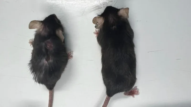
As mice age, IL11 gradually appears in the liver, skeletal muscle, and fat. to stimulate an ERK/AMPK/mTORC1 axis of cellular, tissue, and organism aging pathologies. In aged mice, the deletion of Il11 protects against metabolic multimorbidity, sarcopenia, and frailty. Administering anti-IL11 treatment to aged mice for six months reactivates an age-repressed white fat loss program, reverses metabolic dysfunction, restores muscle function, and reduces frailty. In all cases studied, IL11 inhibition lowers epigenetic age, reduces telomere attrition, and preserves mitochondrial function.

The metabolic effects observed with IL-11 inhibition in aged mice resemble those of young mice with white adipose-specific Raptor deletion. White adipose tissue (fat) and brown adipose tissue are the two main types of adipose tissue in mammals. The authors speculate that inhibition of IL-11 prevents mTORC1 activation in fat, thereby decreasing the amount of white adipose tissue. However, the authors did not identify the mechanism leading to weight loss with IL-11 inhibition, which in further studies could inform some of the present findings.

Beyond metabolism, IL-11 inhibition ameliorated deterministic features of aging common in vertebrates (such as frailty and sarcopenia), demonstrating generic anti-aging benefits at the organismal level. Intriguingly, some of the beneficial effects of germline Il11ra1 or Il11 deletion, notably on muscle and fat, were apparent even in young mice. The authors did not see any specificity of this effect to certain tissues.

Inhibition of IL-11 increased lifespan in both male and female mice. The extent of the increase in lifespan remains unclear, but current data suggest that anti-IL-11 treatment given late in life increases median lifespan by more than 20% in both sexes. In these experiments, anti-IL-11 was injected into mice at 75 weeks (human equivalent to approximately 55 years).

Treatment with IL-11 is therefore effective in prolonging lifespan, as is the case for rapamycin. The mortality of mice in the elderly is often linked to cancer and the end-of-life autopsy data carried out by the authors support the idea that inhibition of IL-11 considerably reduces cancers linked to 'age. Of note, IL-11 is often linked to tumor onset and immune evasion. Clinical trials of anti-IL-11 in combination with immunotherapy to treat cancer are already planned.

As a first step toward creating a drug, the authors designed a high-affinity humanized neutralizing IL11 antibody.

It should be noted that some of the authors have strong links with companies that have acquired the patents resulting from this work or financed this work.

Also, the green and yellow vegetables and fruits, such as leafy greens, herbs, broccoli, peas, green bell peppers, and squash, are relatively rich sources of the oxycarotenoids lutein and zeaxanthin which may prevent the overexpression of IL-11 and ERK signaling

The death of motor neurons is one of the main pathological hallmarks of ALS, and the disease often starts at a small muscle and propagates to other muscles. Muscle denervation appears during the early stages of ALS pathogenesis, and it can be observed by electromyography. This denervation is the result of motor neuron degeneration, probably with a series of pathogenetic factors converging to create a toxic microenvironment. Yet some scientists are not so sure motor neurons die, they think they might simply enter a sort of frozen state to mitigate a stressful situation. This article belongs to a tiny circle as it tells that it's possible to partially reverse the disease and it presents a good mechanism of action, whereas most articles are extremely vague about ALS etiology.

Indeed, it is known that plastic events, such as synaptic plasticity, axonal sprouting, and morphological changes, within the spared motor neuron population can be responsible for compensatory adaptation after the loss of function caused by the neurotoxic removal of a spinal motor neuron subset. These spontaneous plastic changes are known to take place also in ALS models, but their ability to sustain motor function is transient and incapable of counteracting disease progression. Therefore, a therapeutic approach to manage the disease (but not cure it) should be capable of both improving plastic changes and supporting neuroprotection to slow down motor neuron degeneration.

In the authors' view, the use of a simplified in vivo model of motor neuron degeneration would help in the step-by-step dissection of ALS pathogenesis.

The authors used a specific toxin, CTB-Sap, to selectively kill certain motor neurons in the spinal cord by injecting a compound into muscles. CTB-Sap is a compound made by combining cholera toxin-B (which binds to neurons) with saporin (a toxin that kills cells). When injected into muscles, this compound is taken up by the synapses of the lower motor neurons that control those muscles. After the motor neurons take up CTB-Sap, it travels backward (retrograde) along the neuron to the cell body in the spinal cord. The saporin then kills the neuron.

It is a valuable tool for studying compensatory plastic changes, including synaptic plasticity, axonal sprouting, and other morphological and functional adaptations. The authors think that in ALS animal models, when motor neuron degeneration occurs progressively, the remaining cells may try to compensate for the motor deficits. It's when the progressive loss of motor neurons exceeds the compensatory capacity of the surviving cells, that the first signs of the disease appear.

Despite intensive research, it remains poorly understood why motor neurons are specifically targeted in ALS. As motor neurons and the skeletal muscle they control consume enormous amounts of energy, mitochondria use aerobic respiration to generate adenosine triphosphate (ATP), which is used throughout the cell as a source of chemical energy. enter image description here Mitochondria are sort of microbes symbiotes of cells and like other microbes, they can divide or even fusion depending on the needs of the host cell. Several genes encode fission and fusion proteins: MFN1, MFN2, OPA1, DRP1 (Dynamin-Related Protein 1): This protein is essential for mitochondrial fission. MFF (Mitochondrial Fission Factor).

Several publications have associated abnormal mitochondrial dynamics with excessive mitochondrial fission predominantly mediated by the hyperactivation of the dynamin-related protein 1 (DRP1). This cytosolic GTPase, is recruited to the outer mitochondrial membrane, where it assembles into a ring-like structure around the mitochondria, causing constriction and subsequent division. High levels of DRP1 trigger mitochondrial damage which causes insufficient ATP production, indeed fission and fusion events consume a lot of energy.

To prove that an abnormal fission (division) of mitochondria causes a motor neuron disease, it's necessary to show that in such a case inhibiting mitochondria restores some muscle function. Previous studies have proven that spontaneous motor recovery is possible sometimes after toxin administration. Yet these kinds of plastic changes are not enough to counteract the functional effects of the progressive motoneuron degeneration. The authors wanted to use a mitochondrial division inhibitor to prove that it's the mitochondrial division that is a cause of motor neuron disease.

Mdivi-1, a cell-permeable quinazolinone, is an inhibitor of DRP1, so it is capable of inhibiting the fission process by directly decreasing the GTPase enzymatic activity of DRP1. This results in neuroprotection in animal models of Alzheimer’s disease, Parkinson’s disease, and multiple sclerosis. In an attempt to determine the therapeutic impact of Mdivi-1 after motor neuron loss, the scientists used the already established mouse CTB-Sap model which is characterized by up-regulation of DRP1, together with increased mitochondrial fission. They wanted to investigate if the administration of Mdivi-1 could be neuroprotective on damaged or stressed motor neurons, and whether it may promote spinal cord (SC) plasticity.

The drug was administered to the mouse model, a localized removal of spinal motor neurons was induced by injection of CTB-Sap in the calf muscle and it moved from the muscle to the lower motor neuron retrogradly. This simple model of selective motor neuron depletion allows the scientists to focus on the functional and molecular mechanisms of neuroplastic changes upon motor neuron removal.

As expected, a few days after CTB-Sap injection in the right calf muscle, all animals started to display an evident decline in the motor activity of the right back leg that reached a maximum during the first two weeks after the lesion. The observation of limb motion during free exploration of an open field revealed frequent curling of toes, loss of support, and foot-dragging. Motor deficits were accompanied (and caused by) the partial loss of motor neurons innervating the calf muscle and located in the lumbar region of the spinal cord, and the muscle denervation is confirmed by the presence of spontaneous electromyography activity in anesthetized mice.

This functional decline was followed by a spontaneous partial recovery during the experimental period and, as the scientists hypothesized, Mdivi-1 treatment was capable of reducing the early back leg deficit despite the presence of a slightly toxic effect of the drug, as demonstrated by the loss of body weight. The grid walk test confirmed the beneficial effects of treatment in the preservation of motor performance, although a spontaneous recovery (but slower) was seen also in untreated animals.

The beneficial effects of the Mdivi-1 drug were probably limited to some aspects of the motor activity, such as motor coordination, as suggested by clinical scoring and grid walk test results, whereas gait analysis was not able to efficiently reveal the effects of treatment.

However, the observed effects of treatment on motor coordination cannot be explained only by its action on the affected muscle, and a detailed mechanistic study of mitochondrial dynamics should include, for instance, the spinal cord, cerebellum, motor cortex, basal ganglia, and also some general aspects of metabolism.

The phenomena of motor neurons attempting to form new connections and adapt to new conditions in the tissue microenvironment in response to tissue damage or neuronal loss have been well documented in the literature. This process may increase in soma size and dendritic complexity of surviving motor neurons, which might be attributable to their active hunt for new synapses and increased synaptic efficacy. Therefore, the observed increase in motor neurons’ size only in Mdivi-1-treated mice may be proof of neuronal adaptation, promoted by the known activity of the drug onto mitochondrial dynamics, and likely involving motor neuron itself but also the whole sensorimotor spinal cord circuitry and supraspinal pathways.

The results of the present study have confirmed that the CTB-Sap model is a valid tool for research in motor neuron diseases, proving that compensatory plastic changes may take place after the removal of a spinal motor neuron subset. Moreover, it seems likely that treating this animal model with a drug known to inhibit mitochondrial fission may increase this intrinsic plastic capability and protect motor neurons from degeneration.

Some people advocate for Parkinson's patients to consume mannitol. Many individuals affected by the disease began consuming daily oral mannitol. Self-reported outcomes included an improved sense of smell, a reduction in the dose of PD medications, and general improvement in well-being. A clinical trial was done at Hadassah Medical Center in Israel by David Arkadir and colleagues.

The lobby group CliniCrowd was probably instrumental in this decision.

The study lasted 36 weeks and included four dose escalations of oral mannitol (manufacturer Roquette, France) or dextrose as a placebo from 2.5 g to a maximal dose of 18 g per day. The COVID-19 pandemic in 2020 dramatically slowed the recruitment rate in the 3rd year of the clinical trial and led to the decision to earlier trial termination. They did not observe a clear reduction in Parkinson's symptoms. It is possible that a longer exposure would enable clinically to demonstrate disease modification. Anyway, such a high dose of mannitol is not without innuendoes, mannitol is hypertonic, it forces water to be excreted from cells. Yet the mechanism by which mannitol reduces α-synuclein accumulation in PD models was still unknown.

In 2022 another study showed that glycation agents (sugars) can ameliorate α-synuclein folding. Glycation is increased in the brains of hyperglycemic patients. Alpha-synuclein (αSN), a central player in the etiology of Parkinson’s disease, can be glycated so reducing αSN fibril formation. The best glycating agents were unfortunately toxic, but one agent was mildly efficacious while not toxic: Mannose.

Mannitol, while usually derived from fructose, can also be derived from a mannose by reduction. In the human body, mannose residues are used to assemble N-glycans by adding them to a dolichol phosphate (Dol-P) core in the Endoplasmic Reticulum (ER) of cells.

There's a growing body of research suggesting a connection between abnormal N-glycans and neurodegenerative diseases like Alzheimer's and Parkinson's. N-glycans are sugar chains attached to proteins in a specific way, and changes in their structure or abundance seem to play a role in these diseases. This means the sugar chains are either attached differently, have different structures, or are present in abnormal amounts.

N-glycans can influence how proteins fold, interact with other molecules, and get transported within cells. In a recent publication, scientists analyzed neurons in iPSC midbrain cultures derived from patients with Parkinson's disease and they discovered the disruption of a metabolic pathway, the hexosamine pathway. The hexosamine pathway is important for protein synthesis, transport, and folding in the neuron's endoplasmic reticulum. enter image description here The hexosamine pathway produces N-linked glycans, essential molecules that support protein folding in the endoplasmic reticulum.

The hexosamine pathway (a biological pathway is the way a molecule is created from components) uses glucose and uridine-5’-triphosphate to generate N-linked glycans for protein folding in the endoplasmic reticulum. In Parkinson's midbrain cultures, however, this N-glycosylation process was interrupted, causing protein misfolding and accumulation of α-synuclein.

Accelerating glucose flux through the hexosamine pathway rescued hydrolase function and reduced pathological α-synuclein.

So as a non-scientist, I can conclude this post by saying that there is some rationality in using mannitol in Parkinson's disease, while it might not be the silver bullet people are waiting for.


Please, help us continue to provide valuable information: