La dérégulation du fer a été impliquée dans de multiples maladies neurodégénératives, y compris la maladie de Parkinson et la sclérose latérale amyotrophique (SLA). Les cellules constituant la microglie se trouvent fréquemment chargées de fer dans les régions cérébrales touchées, mais la façon dont l'accumulation de fer influence la physiologie de la microglie et contribue à la neurodégénérescence est mal comprise.

Dans une nouvelle publication, les auteurs montrent qu'une tri-culture de cellules de microglie dérivée de cellules souches pluripotentes humaines est très sensible au fer et sensible à la ferroptose, une forme de mort cellulaire dépendante du fer. La microglie est une population de cellules gliales — des macrophages que l'on retrouve dans le système nerveux central et qui en forme la principale défense immunitaire active grâce à ses capacités phagocytaires. Les cellules gliales sont les cellules qui forment l'environnement des neurones. Elles jouent un rôle de soutien et de protection du tissu nerveux en apportant les nutriments et l'oxygène, en éliminant les cellules mortes et en combattant les pathogènes.

Les cultures in vitro d'astrocytes et de microglies sont des outils puissants pour étudier les voies moléculaires spécifiques impliquées dans la neuroinflammation. Cependant, afin de mieux comprendre l'influence de la diaphonie cellulaire sur la neuroinflammation, de nouveaux modèles de culture multicellulaires sont nécessaires. En effet, les interactions entre les neurones, les astrocytes et la microglie influencent de manière critique les réponses neuro-inflammatoires à l'insulte dans le système nerveux central. La « tri-culture » composée à la fois de neurones, d'astrocytes et de microglie imite plus fidèlement les réponses neuro-inflammatoires in vivo que les mono-cultures standard.

Parmi les trois types de cellules, la microglie a eu la réponse transcriptionnelle la plus forte à la dérégulation du fer, et les scientifiques ont identifié un sous-ensemble de microglie avec une signature transcriptomique distincte associée à la ferroptose qui est enrichie dans la moelle épinière SLA post-mortem et la microglie du mésencéphale du patient PD post-mortem.

L'élimination de la microglie du système de tri-culture a considérablement retardé la neurotoxicité induite par le fer.

Dans la maladie, l'absorption microgliale de fer peut initialement être protectrice, mais, lorsque les cellules succombent à la ferroptose, elles entrent dans un état cellulaire neurotoxique qui entraîne des lésions et elles meurent en masse.

Pour élucider les mécanismes régulant la réponse du fer dans la microglie, les scientifiques ont effectué un criblage CRISPR à l'échelle du génome et identifié de nouveaux régulateurs de la ferroptose, y compris le gène de trafic de vésicule SEC24B. enter image description here 
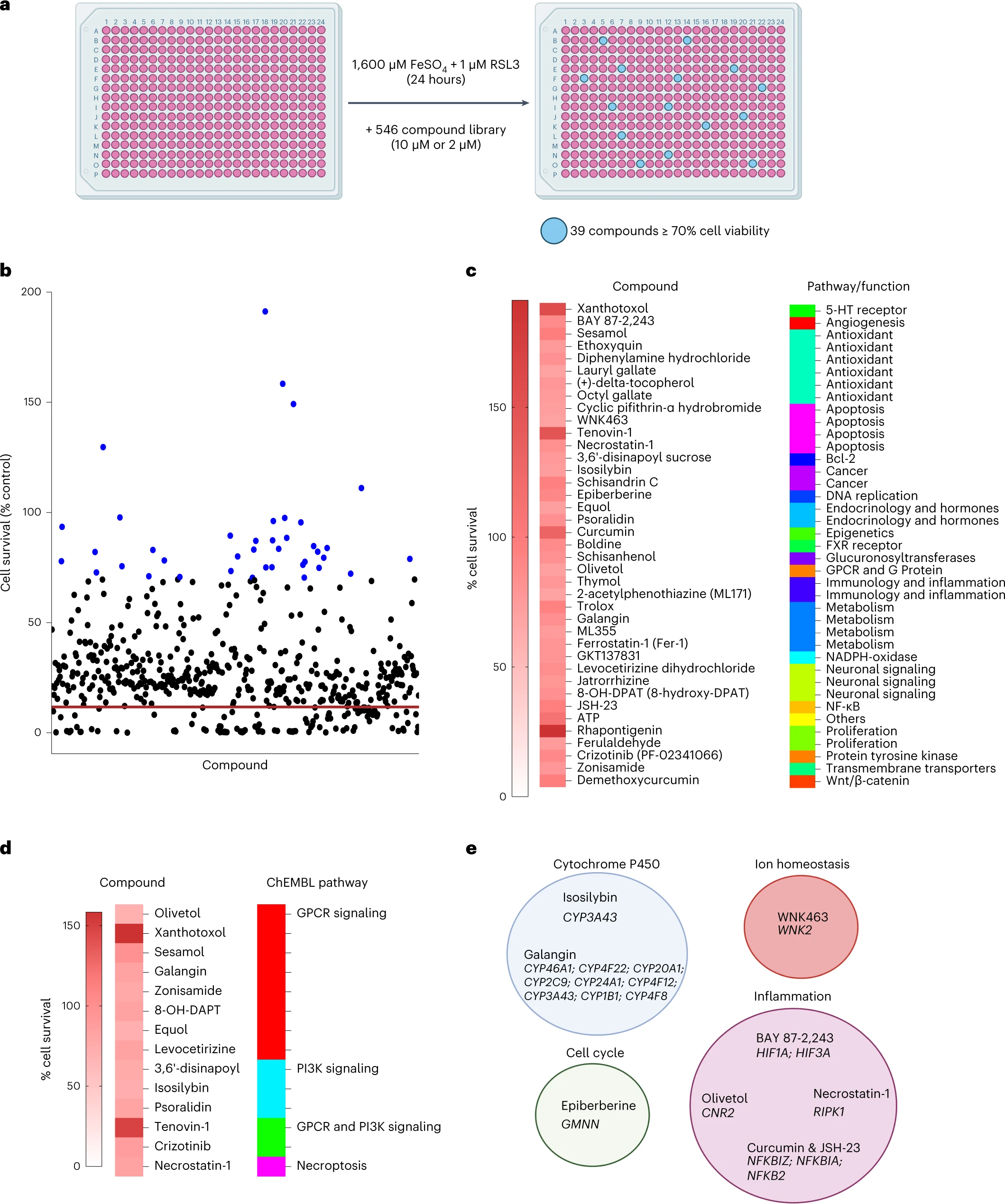
Enfin, les auteurs ont effectué un criblage de petites molécules pour identifier les inhibiteurs de la ferroptose de la microglie. Sur les 546 composés, ils ont trouvé 39 composés qui inhibaient la ferroptose dans la microglie. Parmi ceux-ci Rhapontigenin, Xanthotoxol, Tenovin-1, Curcumin, ATP ou encore sésamol. La rhapontigénine est un stilbénoïde. Il peut être isolé de la vigne du Japon (Vitis coignetiae) ou du Gnetum cleistostachyum. Il montre une action sur les cellules cancéreuses de la prostate. Il a été démontré qu'il inhibe le cytochrome humain P450 1A1, une enzyme impliquée dans la biotransformation d'un certain nombre de composés cancérigènes et immunotoxiques. Le xanthotoxol est une furanocoumarine. C'est l'un des principes actifs majeurs de Cnidium monnieri. Cnidium monnieri (L.) Cuss. est l'une des plantes médicinales traditionnelles les plus largement utilisées et ses fruits ont été utilisés pour traiter diverses maladies en Chine, au Vietnam et au Japon. Le sésamol est un composé organique naturel qui entre dans la composition des graines de sésame et de l'huile de sésame, aux propriétés anti-inflammatoires, antioxydantes, antidépressives et neuroprotectrices.

Malgré le fait que la ferroptose a été impliquée dans de nombreux troubles, on ne connait aucun traitement efficace pour atténuer la ferroptose. Les chélateurs du fer sont une approche potentielle, mais beaucoup od'entre eux peuvent perturber les fonctions redox homéostatiques. Cependant, les études précliniques existantes utilisant des inhibiteurs de la peroxydation lipidique, tels que lip-1, et les données présentées dans cette étude fournissent une justification solide pour le développement de thérapies ciblant la ferroptose. Plusieurs composés ciblant la peroxydation lipidique et le stress oxydatif sont d'ailleurs en cours d'essais cliniques, notamment le dérivé de la vitamine E vatiquinone, l'acide linoléique deutéré et les activateurs de la voie antioxydante NRF2.

How iron can drive neurodegeneration.

- Posted by admin in English

Iron dysregulation has been implicated in multiple neurodegenerative diseases, including Parkinson's disease and amyotrophic lateral sclerosis (ALS).

Cells making up microglia are frequently found loaded with iron in affected brain regions, but how iron accumulation influences microglia physiology and contributes to neurodegeneration is poorly understood.

In a new publication, authors show that a tri-culture of microglia cells derived from human pluripotent stem cells is highly iron-sensitive and susceptible to ferroptosis, a form of iron-dependent cell death.

Microglia is a population of glial cells, macrophages that are found in the central nervous system and which form the main active immune defense thanks to their phagocytic abilities. Glial cells are the cells that form the environment of neurons. They play a role in supporting and protecting nervous tissue by providing nutrients and oxygen, eliminating dead cells and fighting pathogens.

In vitro cultures of astrocytes and microglia are powerful tools to study the specific molecular pathways involved in neuroinflammation. However, in order to better understand the influence of cell crosstalk on neuroinflammation, new multicellular culture models are needed. Indeed, interactions between neurons, astrocytes and microglia critically influence neuroinflammatory responses to insult in the central nervous system. The "tri-culture" composed of both neurons, astrocytes and microglia more closely mimics neuro-inflammatory responses in vivo than standard mono-cultures.

Of the three cell types, microglia had the strongest transcriptional response to iron dysregulation, and scientists identified a subset of microglia with a distinct transcriptomic signature associated with ferroptosis that is enriched in the spinal cord Postmortem ALS and midbrain microglia from postmortem PD patient.

Removal of microglia from the tri-culture system significantly delayed iron-induced neurotoxicity.

In the disease, microglial iron uptake may initially be protective, but when cells succumb to ferroptosis, they enter a neurotoxic cellular state that leads to damage and they die en masse.

To elucidate the mechanisms regulating the iron response in microglia, scientists performed a genome-wide CRISPR screen and identified novel regulators of ferroptosis, including the vesicle trafficking gene SEC24B. enter image description here Finally, the authors performed a small molecule screen to identify inhibitors of microglia ferroptosis. Of the 546 compounds, they found 39 compounds that inhibited ferroptosis in microglia. Among these Rhapontigenin, Xanthotoxol, Tenovin-1, Curcumin, ATP or sesamol.

Rhapontigenin is a stilbenoid. It can be isolated from Japanese grapevine (Vitis coignetiae) or Gnetum cleistostachyum. It shows an action on prostate cancer cells. It has been shown to inhibit human cytochrome P450 1A1, an enzyme involved in the biotransformation of a number of carcinogenic and immunotoxic compounds. Xanthotoxol is a furanocoumarin. It is one of the major active principles of Cnidium monnieri. Cnidium monnieri (L.) Cuss. is one of the most widely used traditional herbal medicines and its fruits have been used to treat various diseases in China, Vietnam and Japan. Sesamol is a natural organic compound that is part of the composition of sesame seeds and sesame oil, with anti-inflammatory, antioxidant, antidepressant and neuroprotective properties.

Despite the fact that ferroptosis has been implicated in many disorders, no effective treatment is known to alleviate ferroptosis. Iron chelators are a potential approach, but many of them can disrupt homeostatic redox functions. However, the existing preclinical studies using lipid peroxidation inhibitors, such as lip-1, and the data presented in this study provide strong rationale for the development of therapies targeting ferroptosis. Several compounds targeting lipid peroxidation and oxidative stress are also in clinical trials, including the vitamin E derivative vatiquinone, deuterated linoleic acid and activators of the antioxidant pathway NRF2.

Read the original article on Pubmed

I am not sure of the value of this study, yet as it is well known that a bile acid (TUDCA) may have benefits in ALS, so every story making a connection between bile acids and ALS may be of interest.

Recent studies suggest that the bile acid metabolism is associated with cognitive function.

Cognitive impairments and behavioral abnormalities in 35% of patients with amyotrophic lateral sclerosis (ALS) have been reported. However, the underlying mechanisms have been poorly understood. Mutations in the C9orf72 gene explain the association between ALS and frontotemporal dementia. About 5%–15% of Western ALS patients satisfy the diagnostic criteria for frontotemporal dementia. An intriguing fact is that this C9orf72 mutation barely occurs in Chinese ALS patients, yet about 40% of Chinese ALS patients exhibited CI and that 30% had behavioral abnormalities.

In the current study, the authors explored the role of gut microbiota in cognitive impairment of ALS patients. They collected fecal samples from 35 ALS patients and 35 healthy controls. The scientists analyzed these samples by using 16S rRNA gene sequencing as well as both untargeted and targeted (bile acids) metabolite mapping between patients with cognitive impairment and patients with normal cognition.

They found altered gut microbial communities and a lower ratio of Firmicutes/ Bacteroidetes in the cognitive impairment group, compared with the normal cognition group. In addition, the untargeted metabolite mapping revealed that 26 and 17 metabolites significantly increased and decreased, respectively, in the cognitive impairment group, compared with the normal cognition group.

These metabolites were mapped to the metabolic pathways associated with bile acids. They further found that cholic acid and chenodeoxycholic acid were significantly lower in the cognitive impairment group than in the normal cognition group. Chenodeoxycholic acid and cholic acid are the two primary bile acids in humans.

As primary bile acids move from the small intestine to the colon, they are converted to secondary bile acides (including TUDCA) by the biotransformation of the resident microbial community. So bile acids changes may be associated with a microbiome change in ALS patients.

Bile acids are essential products of cholesterol metabolism. Apolipoprotein E (Apo-E) is a protein involved in the metabolism of fats in the body of mammals. A subtype is implicated in Alzheimer's disease and cardiovascular disease. Similarly a defect in lipid (cholesterol) metabolism may induce cognitive changes. But this is still highly hypothetical.

In conclusion, the authors found that the gut microbiota and its metabolome profile differed between ALS patients with and without cognitive impairment and that the altered bile acid profile in fecal samples was significantly associated with cognitive impairment in ALS patients.

These results need to be replicated in larger studies in the future.

We all know about TUDCA, a bile acid which has been used medicinally for centuries, is used in combination with sodium phenylbutyrate, in treatment of amyotrophic lateral sclerosis (ALS). TUDCA is also used in retinal degenerative disorders and is studied in Parkinson's disease.

There is evidence that there are differences in the serum levels of bile acids in males and females and their risk of developing Alzheimer's disease.

The authors of a new publication on medRxiv previously reported that serum bile acids are associated with Alzheimer's disease. It remains unclear, however, how changes in serum bile acids may relate to the development of Alzheimer's disease in a sex-dependent manner.

They analyzed 33 bile acids in the sera of 4219 samples from 1180 subjects in a Alzheimer's disease cohort and they examined the associations between bile acids and mild cognitive impairment (MCI) progression and clinical markers. enter image description here

The scientists found that significant alterations in bile acids profiles occurred at an early stage of MCI and were associated with the onset and progression of MCI. These changes were more dramatic in men than in women. Bile acids markers improved the ability to diagnose MCI and predict its progression. enter image description here

Many studies hint at a role of cholesterol in neurodegenerative diseases. For example protein clearance ability is age-related, regulated by brain cholesterol. Bile acids have metabolic actions in the body resembling those of hormones in triglyceride metabolism, glucose metabolism, and liver growth.

Half of the amount of cholesterol produced by the body is used for bile acid synthesis. Bile acid sequestrants are used to lower the level of LDL cholesterol circulating in the blood. As many aged people are prescribed bile acid sequestrants it could be worth to investigate the link between this kind of medication and the apparition of some neurodegenerative diseases.

Read the original article on medRxiv

This article is a bit unusual as it claims a relationship between several genes in ALS (Lou Gerigh disease), including APP which is associated with Alzheimer's disease. On the overall it says that C9 ALS is a problem of ribosome quality control (RQC) which leads to cellular stress response. It suggests that C9, FUS, TDP-43 mutations influence mTORC2 protein which in turn alters the translation mechanism. When the translation mechanism is altered, cellular stress response is activated and protein production nearly stops, rendering the cell non-functional. It's the start of the disease.

ALS is a muscle wasting disease characterized by degeneration of lower motor neurons and axons and loss of upper motor neurons and their corticospinal tracts. FTD is a progressive neuronal atrophy with neuronal loss in the frontal and temporal cortices and associated behavioral and personality changes and impairment of language skills. Advances in human genetics have identified multiple genetic mutations commonly associated with ALS and FTD, revealing that these two diseases are related and may represent a continuum of a broad neurodegenerative disorder. C9orf72 mutation is present in approximately 40% of familial ALS and 8-10 % of sporadic ALS. It is currently the most common demonstrated mutation related to ALS - far more common than SOD1.

The research on association of C9ORF72 with ALS or FTD is relatively recent and its mechanisms are not clear. This obviously impairs the proposal of new drugs. Gene expression or translation is the process in which ribosomes in the cytoplasm or endoplasmic reticulum synthesize proteins after the process of transcription of DNA to RNA in the cell's nucleus. Yet during translation elongation, ribosomes may slowdown or even stall for various reasons. The polypeptide later folds into an active protein and is sent to its final location to perform its functions in the cell.

RAN translation, is an irregular mode of mRNA translation that can occur in eukaryotic cells. RAN translation produces a variety of dipeptide repeat proteins (DPR) by translation of expanded hexanucleotide repeats present in an intron of the C9orf72 gene. The expansion of the hexanucleotide repeats and thus accumulation of dipeptide repeat proteins are thought to cause cellular toxicity that leads to neurodegeneration in ALS disease.

Previous studies of protein quality control have focused on how proteins were handled after translation. However, rproblems with proteostasis are prevalent even with translating nascent peptide chains still associated with ribosomes, necessitating ribosome-associated quality control (RQC) mechanisms.

Mutations in other genes that are commonly linked to ALS/FTD have also shed lights on disease pathogenesis such as TDP-43 and FUS. Other genes linked to ALS/FTD include VCP, a member of the AAA ATPase family with established function in the recycling and degradation of ubiquitinated proteins, and genes with functions in protein clearance or maintenance of protein homeostasis. In addition, upregulation of APP, a protein whose aberrant processing or metabolism having been implicated in Alzheimer’s disease (AD), was observed at early stages of ALS and FTD, presumably as a compensatory response to neuronal damage or impairment of axonal transport. However, the relationships among the various ALS/FTD genes remain underexplored.

It is therefore important to understand cellular mechanisms underlying the quality control of poly(GR). Previous studies of protein quality control have focused on how proteins were handled after translation, e.g., by chaperone-mediated refolding, or proteasome- and lysosome-mediated degradation. However, recent studies reveal that problems with proteostasis are prevalent even with translating nascent peptide chains still associated with ribosomes, necessitating ribosome-associated quality control (RQC) mechanisms.

In the case of poly(GR), it was shown that its translation was frequently stalled, presumably due to positively charged arginine residues interacting with negatively-charged residues lining the exit tunnel of 60S ribosome. Stalled poly(GR) translation activates the RQC process, the inadequacy of which can lead to the accumulation of aberrant, C-terminally modified (CAT-tailed) poly(GR) species that can perturb proteostasis and contribute to poly(GR) accumulation and neuromuscular degeneration.

In this study, the scientists from USA and China set out to test whether the other ALS/FTD associated genes may participate in the quality control of poly(GR). Strikingly, they discovered that overexpression of APP, FUS, and TDP-43 restrains poly(GR) protein expression. Mechanistically, APP, FUS and TDP-43 act through the mTORC2/AKT/VCP axis to regulate the RQC of poly(GR) translation. Inhibition of the mTORC2/AKT/VCP axis could restore poly(GR) protein expression attenuated by APP, FUS, or TDP-43. Their data strongly implicate the mTORC2/AKT/VCP axis as a major regulator of protein quality control in ALS/FTD.

Their data support the working model that mutated APP, FUS, and TDP-43 are upstream regulators of the mTORC2/AKT/VCP axis, which regulates the RQC of poly(GR) during its translation stalling. Moreover, they suggest that APP, FUS, and TDP-43 can also induce repression of global translation when ribosome stalling is persistent.

APP acts through the mTORC2/AKT signaling axis to regulate the RQC of C9-ALS/FTD-associated poly(GR) translation. The involvement of APP in ALS has previously been studied in the context of ALS, and APP or its metabolite was found to exacerbate ALS-related phenotypes in the SOD1-G93A mouse model. This new results suggest that APP can activate mTORC2/AKT signaling to alleviate stalled translation of poly(GR) and restrain the expression of aberrant poly(GR) translation products, at least at the initial stage. It is possible that in ALS/FTD setting, APP is upregulated as a protective response in response to neuronal damage at an early stage of disease as previously suggested

It is difficult to not think about the controverse about the role of amyloid plaques found in the brains of Alzheimer's disease patients. Amyloid beta is a fragment from the larger amyloid-beta precursor protein (APP) a transmembrane protein that penetrates the neuron's membrane. APP is critical to neuron growth, survival, and post-injury repair. While the authors write only about ALS, chronic upregulation of APP may contribute not only to ALS, but also also to Alzheimer's disease due to the accumulation of APP metabolites, the stalled translation of APP itself, or the prolonged activation of stress response pathways by APP may lead to the depression of global translation.

Indeed the authors found that integrated stress response as indicated by eIF2α phosphorylation was heightened in transgenic flies expressing poly(GR). This is presumably caused by the ribosome stalling occurring during poly(GR) translation.

A similar situation may occur with TDP-43 and FUS. In fact, both mutations in TDP-43 and FUS genes have been shown to associate with stalled ribosomes, and in the case of TDP-43, its association with stalled ribosomes provides neuroprotection function in the face of sublethal stress.

Intriguingly, the authors showed that the a portion of APP (APP-C99) is sufficient to activate the mTORC2/AKT axis and regulate GR80 translation, whereas the Aβ-42 portion of APP was without effect. This finding resonates with recent revelation of aberrant APP-C99 as the etiological driver of Alzheimer’s disease.

Remarkably, the translation of this portion of APP is also frequently stalled, the inadequate RQC of which can generate aberrant translation products that precipitate hallmarks of Alzheimer’s disease. It is therefore fascinating that overexpression one stalled translation product (APP-C99) would abrogate the stalled translation of another portion (GR-80).

Future studies will investigate at the biochemical level how APP/APP-C99, FUS, and TDP-43 signal to the mTORC2/AKT/VCP axis to regulate the RQC of stalled poly(GR) translation, whether endogenous stalled peptides that serve as RQC substrate(s) may also targeted by this pathway, and how this signaling process may be targeted for therapeutic purposes.

Forecasting individual progression trajectories in Alzheimer's disease.

- Posted by admin in English

The anticipation of progression of Alzheimer's disease is crucial for evaluations of secondary prevention measures thought to modify the disease trajectory. However, it is difficult to forecast the natural progression of Alzheimer's disease, notably because several functions decline at different ages and different rates in different patients. enter image description here Disease modeling uses computational and statistical methods to address this question. These models learn the variability of disease progression from observational longitudinal cohort data and can then predict the progression of patients from their historical data. They require various clinical or biomarker assessments at one or several time points as input.

European authors evaluate in Nature communications Alzheimer's disease Course Map, a statistical model predicting the progression of neuropsychological assessments and imaging biomarkers for a patient from current medical and radiological data at early disease stages.

Alzheimer's disease Course Map is a spatiotemporal atlas of Alzheimer's disease progression. It summarizes the variability in the progression of a series of neuropsychological assessments, the propagation of hypometabolism and cortical thinning across brain regions and the deformation of the shape of the hippocampus. The analysis of these variations highlights strong genetic determinants for the progression, like possible compensatory mechanisms at play during disease progression. AD Course Map also predicts the patient's cognitive decline with a better accuracy than the 56 methods benchmarked in the open challenge TADPOLE. Finally, AD Course Map is used to simulate cohorts of virtual patients developing Alzheimer's disease.

The scientists from France, Germany and Italy propose a software tool using a disease progression model for participant selection in clinical trials. The goal is to enrich the selected population of participants likely to display progression during the trial, a concept called prognostic enrichment.

The scientists tested the method on more than 96,000 cases, with a pool of more than 4,600 patients from four continents.

They measured the accuracy of the method for selecting participants displaying a progression of clinical endpoints during a hypothetical trial.

They show that enriching the population with the predicted progressors decreases the required sample size by 38% to 50%, depending on trial duration, outcome, and targeted disease stage, from asymptomatic individuals at risk of Alzheimer's disease to subjects with early and mild Alzheimer's disease.

In conclusion this method introduces no biases regarding sex or geographic locations and is robust to missing data. It performs best at the earliest stages of disease and is therefore highly suitable for use in prevention trials.

Read the original article on Pubmed

Chez les patients atteints de la maladie de Parkinson, on pense depuis les travaux de Heiko Braak, que la pathologie de l'α-synucléine se propage au cerveau via le nerf vague. Le lien entre le microbiote intestinal et la maladie de Parkinson a été exploré dans diverses études. Un indice est que les patients sont souvent constipés. entrez la description de l'image ici

Récemment, l'appendicectomie a été associée à un risque plus faible de maladie de Parkinson, probablement en raison du rôle de l'appendice dans la modification du microbiome intestinal. Cependant, le microbiote fécal chez les patients sans antécédent d'appendicectomie peut présenter des variations interindividuelles considérables qui rendent difficile toute conclusion.

Les auteurs d'un nouvel article publié dans Nature/Scientific reports, ont cherché à élucider si le microbiote intestinal affectait le développement de la maladie de Parkinson dans une cohorte d'appendicectomie.

Ces scientifiques japonais ont analysé la composition microbienne fécale d'une vingtaine de patients atteints de la maladie de Parkinson et de témoins sains avec et sans antécédent d'appendicectomie. 10 sujets avaient la maladie de Parkinson, tandis que 10 autres constituaient le groupe contrôle. Chacun de ces deux groupes a été subdivisé en deux groupes de 5 sujets, l'un pour les sujets n'ayant pas subi d'appendicectomie, l'autre groupe constitué de ceux ayant subi cette opération.

L'abondance de microbes de la famille des entérobactéries était plus élevée dans les échantillons de matières fécales de patients atteints de la maladie de Parkinson que dans les échantillons prélevés sur des témoins sains. Il convient de noter que les entérobactéries, telles que Escherichia coli et Salmonella, produisent des amyloïdes bactériens appelés curli. Par conséquent, les entérobactéries peuvent induire une pathologie de Parkinson.

Les microbes des groupes Proteobacteria, Gammaproteobacteria, Enterobacteriales et Enterobacteriaceae étaient plus enrichis chez les patients parkinsoniens que chez les témoins sains. Les scientifiques ont découvert que le genre Serratia, de l'ordre des entérobactéries, avait une abondance plus élevée dans les échantillons fécaux de patients atteints de maladie de Parkinson que dans les témoins sains.

On pensait que Serratia était une bactérie environnementale inoffensive jusqu'à ce que l'espèce la plus commune du genre, S. marcescens, soit découverte comme étant un pathogène opportuniste. Chez l'homme, S. marcescens est principalement associé à des infections nosocomiales ou nosocomiales, mais il peut également provoquer des infections des voies urinaires, une pneumonie et une endocardite. S. marcescens se trouve fréquemment dans les douches, les cuvettes de toilettes et autour des carreaux humides sous forme de biofilm rosâtre à rouge.

Certaines études ont démontré que le microbiote intestinal des patients atteints de maladie de Parkinson a une faible abondance de Prevotella, tandis que d'autres études ont montré des résultats contradictoires. Il est possible que l'abondance des Prevotellaceae dans le microbiote intestinal soit corrélée à la sévérité de la constipation, et non à la maladie de Parkinson.

Les individus du groupe en bonne santé avaient les indices de masse corporelle les plus élevés. Une réduction du ratio Firmicutes/Bacteroidetes chez les patients atteints de maladie de Parkinson a été observée entre les groupes maladie de Parkinson et contrôle dans une étude précédente.

De plus, il y avait une différence phylogénétique significative entre les patients atteints de la maladie de Parkinson et les témoins sains ayant subi une appendicectomie. Il y avait une différence phylogénétique significative entre les patients atteints de la maladie de Parkinson et les témoins sains qui avaient subi une appendicectomie. Ces résultats suggèrent la corrélation entre le microbiote intestinal et la maladie de Parkinson chez les patients ayant subi une appendicectomie.

Read the original article on Pubmed

In patients with Parkinson's disease, it is believed since the work of Heiko Braak, that α-synuclein pathology spreads to the brain via the vagus nerve. The link between gut microbiota and Parkinson's disease has been explored in various studies. One clue is that patients are often constipated. enter image description here

Recently, appendectomy has been associated with a lower risk of Parkinson's disease, possibly due to the appendix's role in altering the gut microbiome. However, the faecal microbiota in patients without a history of appendectomy may show considerable interindividual variation which makes any conclusion difficult.

The authors of a new paper published in Nature/Scientific reports, sought to elucidate whether gut microbiota affects the development of Parkinson's disease in an appendectomy cohort.

The Japanese scientists analyzed the fecal microbial composition in about twenty patients with Parkinson's disease and healthy controls with and without a history of appendectomy. 10 subjects had Parkinson's disease, while 10 others constituted the control group. Each of these two groups was subdivided into two groups of 5 subjects, one for subjects who had not undergone appendectomy, the other group consisting of those who had undergone this operation.

The abundance of microbes from the Enterobacteriaceae family was higher in fecal matter samples from patients with Parkinson's disease than in samples taken from healthy controls. It should be noted that Enterobacteriaceae, such as Escherichia coli and Salmonella, produce bacterial amyloids called curli. Therefore, Enterobacteriaceae can induce PD α-synuclein pathology.

Microbes from the Proteobacteria, Gammaproteobacteria, Enterobacteriales, and Enterobacteriaceae groups were more enriched in PD patients than in healthy controls. The scientists found that the genus Serratia, of the order Enterobacteriales, had a higher abundance in fecal samples from patients with PD than from healthy controls.

Serratia was thought to be a harmless environmental bacterium until the most common species of the genus, S. marcescens, was discovered to be an opportunistic pathogen. In humans, S. marcescens is primarily associated with nosocomial or nosocomial infections, but can also cause urinary tract infections, pneumonia, and endocarditis. S. marcescens is frequently found in showers, toilet bowls, and around wet tiles as a pinkish to red biofilm.

Some studies have demonstrated that the gut microbiota of patients with PD has a low abundance of Prevotella, while other studies have shown conflicting results. It is possible that the abundance of Prevotellaceae in the gut microbiota may be correlated with the severity of constipation, and not with PD.

Individuals in the healthy group had the highest body mass indices. A reduction in the Firmicutes/Bacteroidetes ratio in patients with PD was observed between the PD and control groups in a previous study.

Moreover, there was a significant phylogenetic difference between patients with Parkinson's disease and healthy controls who had undergone appendectomy. There was a significant phylogenetic difference between patients with Parkinson's disease and healthy controls who had undergone appendectomy. These results suggest the correlation between gut microbiota and Parkinson's disease in patients who have undergone appendectomy.

Read the original article on Pubmed

Alzheimer's disease is characterized by the accumulation of amyloid β peptides and impaired glucose metabolism in the brain. Traditionally when there is impaired glucose metabolism the focus is on:

  • Sugar
  • Insulin
  • And cholesterol

Yet there are other biological entities at work in this case. Osteocalcin, an homonal protein, is secreted by osteoblasts. Osteoblasts are cells that synthesize bone. enter image description here 
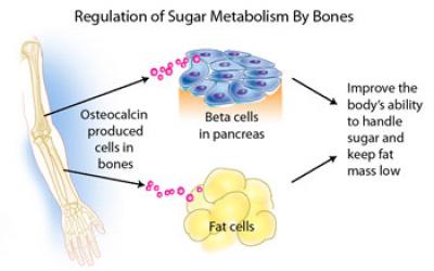
Patients with type 2 diabetes mellitus (T2DM) and/or cardiovascular disease (CVD), conditions of hyperinsulinaemia, have lower levels of osteocalcin and bone remodelling, and increased rates of fragility fractures.

In this study, daily intraperitoneal injection of osteocalcin for 4 weeks ameliorated the anxiety-like behaviors and cognitive dysfunctions in the APP/PS1 transgenic Alzheimer's disease mice model enter image description here Aβ burden in the hippocampus and cortex of Alzheimer's disease mice was ameliorated by osteocalcin. Besides, osteocalcin improved the neural network function of the brain, mainly in the enhanced power of high gamma band in the medial prefrontal cortex of Alzheimer's disease mice.

The proliferation of astrocytes in the hippocampus in Alzheimer's disease mice was also inhibited by osteocalcin Furthermore, OCN enhanced glycolysis in astrocytes and microglia.

Such an effect was abolished when the receptor of osteocalcin (Gpr158) was knockdown in astrocytes.

So osteocalcin maybe a novel therapeutic factor for Alzheimer's disease potentially through reducing Aβ burden and upregulation of glycolysis in neuroglia.

It should be noted that osteocalcin production is accelerated in acute stress response (like in physical effort), which stimulates osteocalcin release from bone within minutes humans. Vitamin D deficiency reduces osteocalcin synthesis and favours osteocyte apoptosis while its synthesis depends on vitamin K avilability.

Read the original article on Pubmed

La consommation d'un régime riche en graisses provoque diverses maladies métaboliques, notamment le syndrome métabolique et le diabète de type 2 en développant une résistance à l'insuline et même en diminuant la production d'insuline.

Par exemple un bon moyen d'induire un syndrome de Parkinson chez les rats est de leur donner un régime alimentaire très riche en graisse. On a parfois qualifié la maladie d'Alzheimer comme étant diabète de type 3.

Un mystère est celui de la SLA (maladie de Charcot) où on a souvent une résistance à l'insuline, mais très rarement un diabète en comorbidité. Il semble aussi que l'existence protéines mal repliées dans le cytoplasme des malades soit liée à un stress du réticulum endoplasmique, un organe des cellules dont la fonction est justement de replier les protéines nouvellement produites par les ribosomes, avant leur expédition vers leur destination dans l'appareil de Golgi.

Une exposition à long terme aux acides gras saturés (acides gras saturés) dans les cellules β pancréatiques provoque une désensibilisation et une altération de la sécrétion d'insuline.

Par exemple, la consommation pendant trois mois de régime alimentaire très riche en graisse contenant de la graisse de porc et de l'huile de tournesol (80 % de graisse) réduit chez la souris la teneur en insuline des îlots pancréatiques (50 %), l'ARNm de la proinsuline (35 %), la biosynthèse et la sécrétion de l'insuline en réponse au glucose (50 %) , et l'oxydation du glucose.

Selon des recherches antérieures, WFS1 (syndrome de Wolfram 1) est impliqué dans la synthèse et la libération d'insuline, ainsi que dans la préservation de la masse des cellules β pancréatiques. Ce gène Wfs1 a été identifié pour la première fois par Wolfram et Wagener (1983) chez des patients atteints du syndrome de Wolfram (c.-à-dire un diabète sucré et une atrophie du nerf optique).

Le gène WFS1 exprime une glycoprotéine dans le réticulum endoplasmique (réticulum endoplasmique) des cellules β pancréatiques, du cœur, du placenta, des poumons et du cerveau.

Les acides gras saturés comme le palmitate induisent un stress réticulum endoplasmique. Des études ont montré qu'il existe une relation mutuelle entre le stress oxydatif et le stress réticulum endoplasmique. On l'acide trouve dans l'huile de palme, mais aussi dans toutes les graisses et huiles animales (beurre, fromage, lait et viande) ou végétales.

Pendant le stress du réticulum endoplasmique, l'expression de WFS1 augmente pour inhiber la signalisation de ce stress et ainsi empêcher l'apoptose (l'un des processus aboutissant à la mort de la cellule).

Etant donné le rôle de la protéine WFS1 dans le maintien de l'homéostasie du réticulum endoplasmique, on s'attend à ce que l'expression de cette protéine soit augmentée dans le réticulum endoplasmique des cellules β, et sa translocation vers le cytoplasme est réduite et conduit à une diminution du GSIS des îlots pancréatiques et teneur en insuline.

Bien que de nombreuses études aient étudié les effets de chaque régime alimentaire très riche en graisse et l'implication de WFS1 dans la synthèse et la sécrétion d'insuline, aucune étude n'a examiné l'interaction de régime alimentaire très riche en graisse et WFS1 en relation avec la synthèse et la sécrétion d'insuline et donc l'homéostasie du glucose.

Après le sevrage, les rats ont été divisés en six groupes et nourris avec un régime alimentaire normal et régime alimentaire très riche en graisse (30%) pendant 20 semaines, puis de l'acide 4-phényl butyrique (4-PBA, un inhibiteur du stress réticulum endoplasmique) a été administré. On notera qu'il s'agit d'une des deux composantes de l'AMX0035.

Après avoir effectué un test de tolérance au glucose, les animaux ont été disséqués et leurs pancréas ont été prélevés pour extraire l'réticulum endoplasmique, l'isolement des îlots et l'évaluation du GSIS. De plus, les biomarqueurs du stress du réticulum endoplasmique pancréatique.

Ce régime alimentaire très riche en graisse a diminué les niveaux de protéine pancréatique WFS1 et de GSH, et augmenté l'activité de la catalase pancréatique. En conséquence, il a augmenté les niveaux de protéines BIP, CHOP et WFS1 dans le RE extrait du pancréas. De plus, le régime alimentaire très riche en graisse a provoqué une intolérance au glucose et a diminué la teneur en GSIS et en insuline des îlots.

Cependant, l'administration de 4-PBA a restauré les niveaux antérieurs. Il semble donc bien que la consommation de régime alimentaire très riche en graisse en induisant un stress réticulum endoplasmique pancréatique ait altéré les niveaux d'expression de WFS1, réduit la teneur en GSIS et en insuline des îlots et finalement altéré l'homéostasie du glucose.

L'administration de 4-PBA ne me semble pas être une solution au problème du stress du réticulum endoplasmique, par contre c'est certainement un moyen de pallier à une conséquence dramatique de ce stress: La mort cellulaire, ce qui dans le cas de la SLA intervient très certainement sur les cellules qui consomment le plus d'énergie comme les muscles squelettiques et les neurones moteurs.

Lire l'article original sur Pubmed


Please, help us continue to provide valuable information: