SUMO (Small Ubiquitin-like Modifier) proteins are a family of small proteins that are attached to and detached from other proteins in cells to modify their function. This process is called SUMOylation. SUMOylation is to signal to other cellular mechanisms that the protein attached must be processed. There are at least 4 SUMO isoforms in humans; SUMO-1, SUMO-2, SUMO-3, and SUMO-4. SUMO proteins are involved in a variety of cellular processes, such as nuclear transport, transcriptional regulation, apoptosis, and protein stability. enter image description here Transactive response DNA-binding protein 43 (TDP-43) is a nuclear RNA binding protein (RBP) involved in RNA metabolism. TDP-43 has a high propensity to aggregate because of its low solubility in cells and in vitro. The aggregation propensity of TDP-43 is increased by ALS/FTD-linked mutations and upon exposure to stress and has been observed in patients with C9orf72 hexanucleotide repeat expansion, the most common genetic cause of sporadic and familial.

Stress conditions trigger the accumulation of TDP-43 in cytosolic stress granules. The role of stress granules in modulating TDP-43 aggregation is ambiguous. Functionally, SUMO2/3-ylation has been shown to maintain stress granules in a dynamic state: SUMOylation inhibition impairs stress granule disassembly, but the underlying mechanism is still unknown.

In this paper, the authors report that upon oxidative stress (induced by sodium arsenite), TDP-43 becomes modified by SUMO2/3 protein chains and moves from the nucleus to cytoplasmic stress granules. This conclusion is probably also valid for other stress conditions, so this study is of great interest. When researchers blocked SUMOylation, TDP-43 became less mobile within the cell, stress granules took longer to disassemble, and TDP-43 was more likely to form insoluble aggregates. Yet this is not a study on humans, the authors used various immortal cell lines, motor neuron cells, and Caenorhabditis elegans worms. Furthermore, the authors used genetic therapies to infect cells to express or repress PIAS4. This is easy to do in vitro but most probably hard to achieve in a seriously ill patient.

Because TDP-43 aggregation is central to familial and sporadic ALS, approaches aimed at preventing TDP-43 aggregation hold promise for future treatments. How cells control TDP-43 aggregation is poorly understood. Modifiers of TDP-43 solubility include molecular chaperones and posttranslational modifications. Besides ubiquitination, which is a key posttranslational modification required to clear aggregation-prone proteins, phosphorylation of TDP-43 is emerging as a protective response to counteract its misfolding. Phosphorylation of TDP-43 decreases its assembly into condensates and suppresses TDP-43 aggregation and toxicity.

Under normal growth conditions, cells prefer to modify proteins with SUMO1, while during cellular stress, SUMO2 and SUMO3 are usually conjugated in the form of SUMO2/3 chains (referred to as SUMO2/3-ylation). TDP-43 when SUMO1-ylated stays in the nucleus, does not aggregate in the cytoplasm, and its splicing activity is modified.

Using experiments in cells, the authors show that conjugation of TDP-43 with SUMO2/3 coincides with stress granule assembly. Pharmacological inhibition of TDP-43 SUMO2/3-ylation triggers TDP-43 aggregation inside stress granules.

E3 SUMO-protein ligase PIAS4 is one of several protein inhibitors of activated STAT (PIAS) proteins. PIAS proteins act as transcriptional co-regulators with at least 60 different proteins to either activate or repress transcription. The transcription factors STAT, NF-κB, p73, and p53 are among the many proteins that PIAS interacts with. PIAS4 has been shown to recruit proteins to the site of the DNA damage and promote repair.

The authors found that PIAS4 helps attach SUMO2/3 to TDP-43. - PIAS4-mediated SUMO2/3-ylation increases the solubility of TDP-43 and prevents its aggregation in the cytoplasm. - Depleting PIAS4 leads to TDP-43 aggregation In motor neurons from the human spinal cord in familial ALS cases with TDP-43 and C9orf72 mutations, reduced cytoplasmic PIAS4 correlates with increased TDP-43 aggregates.

RNA binding appears to compete with SUMOylation: When cells are not subjected to stress, TDP-43 is mainly localized inside the nucleus, binding with high affinity to RNA. When TDP-43 is bound to RNA, it's less likely to be SUMOylated. When cellular RNA levels are low, there's increased SUMO2/3 modification This suggests SUMOylation may be a protective mechanism when TDP-43 isn't bound to RNA

The authors conclude that modification with SUMO2/3 chains maintains the solubility of RNA-free TDP-43 during stress.

There are many studies on reducing TDP-43 aggregates in ALS, but this one looks much more sophisticated than the previous ones. Yet this is mostly an in-vitro study. Long pre-clinical studies must be conducted on mammals to verify if a simple and safe agonist of PIAS4 (which does not exist today) could improve the health of ALS patients.

Modelling ALS: Dynamic Regulatory Instability

- Posted by admin in English

A few years ago I tried to model the course of ALS using SBML, a systems biology tool now abandoned by scientists.

At the time there was no model of ALS, but since then there have been very interesting attempts, for example, this one:enter link description here

However, few scientists and almost no doctors use models based on differential equations today, we live in an era where statistical models are all the rage. In addition, biology as it is practiced is essentially qualitative, which makes it not very suitable for modeling and which attracts the mockery of "soft science", because by nature it is not capable of making consistent predictions. One only has to look at the colossal failure rate of clinical trials to be convinced of this.

The publication reviewed in this post focuses on understanding the regulatory dynamics of amyotrophic lateral sclerosis (ALS) using the widely used SOD1-G93A transgenic mouse model.

ALS is a multifactorial disease, and previous studies have often focused on isolated aspects rather than its complex and interconnected nature.

The study suggests that ALS regulation may be hypervigilant, meaning that the system overcorrects in response to stress, leading to damaging oscillatory behavior that contributes to disease progression. enter image description here 
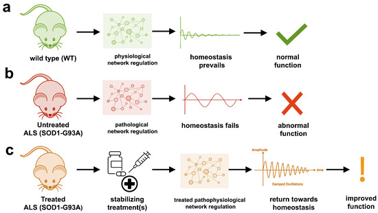
The study uses an innovative and integrative framework to model the regulatory dynamics of wild-type (WT) and SOD1-G93A ALS mice. The models are based on first-order ordinary differential equations (ODEs) that describe how the system output evolves over time. The research uses dynamic meta-analysis to synthesize experimental data from the literature and parameter optimization based on genetic algorithms to infer missing data. Indeed, to build a model, data are needed and here these are obtained from results reported in the literature on SOD1-G93A ALS mouse models.

The study shows that SOD1-G93A mice with ALS exhibit unstable physiological regulation, characterized by oscillatory behavior due to hypervigilant regulation. This instability intensifies near disease onset and worsens with progression.

Computational models of the physiological dynamics of wild-type (WT) and transgenic SOD1-G93A mice were constructed: a WT mouse model to simulate normal homeostasis and a SOD1-G93A ALS model to simulate the dynamics of ALS pathology and their response to treatments in silico. The model simulates the functional molecular mechanisms of apoptosis, metal chelation, energetics, excitotoxicity, inflammation, oxidative stress, and proteomics using data curated from published SOD1-G93A mouse experiments. Time-course measures of disease progression (rotarod, grip strength, body weight) were used to validate the results from the literature.

The health of untreated SOD1-G93A ALS mouse models cannot be maintained due to oscillatory instability. The onset and magnitude of homeostatic instability corresponded with disease onset and progression. Oscillations are associated with high feedback gain due to hypervigilant regulation.

Multiple virtual treatments combined were able to stabilize the dynamics of SOD1-G93A ALS mice to near-normal WT homeostasis. However, treatment timing and effect size are critical for stabilization to match therapeutic success. The most common unidirectional stabilizing treatment was pro-proteomics, while the most common bidirectional stabilizing treatment was energy consumption and anti-apoptosis. The authors cite anti-apoptosis factors such as caspase-9 inhibitor, caspase-3 inhibitor, Bax inhibitor, and Bcl-2 homolog BCL-XL.

A major drawback is that this type of ALS only affects less than 2% of ALS cases and if it is still largely overrepresented in the literature, it is because the association of this type of mutation with ALS is historically the first to have been discovered during the boom in genetics and for almost 15 years no other association has been found.

Another important point is that we do not know what mechanisms cause this disease, or even if it is a single disease. Models of this type therefore make many assumptions and their results are less robust than they appear.

The study highlights the multifactorial nature of ALS, which involves various molecular mechanisms, such as apoptosis, bioenergetics, excitotoxicity, inflammation, oxidative stress, and proteomics. These mechanisms are interconnected, and their dysregulation contributes to disease progression.

The study also explores the potential of combination therapies to stabilize the regulatory dynamics of ALS. Previous studies focusing on single therapeutic targets have often been inadequate, but combination treatments have shown success in the management of other complex diseases such as cancer, COVID-19, and HIV. The study suggests that precisely timed combination therapies targeting multiple pathways may be necessary to achieve meaningful results in ALS.

Code and data are not available at the indicated Github address.

The authors acknowledge that first-order feedback models may not fully capture the complexity of biological systems, including phenomena such as bistability and hysteresis. But for now, we are still in the prehistory of biological systems modeling, the field needs to be developed before focusing on its imperfections.

I fear that this publication will go way over the heads of health professionals working in the field of ALS.

Differences in disease treatment between countries are evidence that medicine is not an exact science (if there ever was one). For example, it has been shown in certain cancers that crossing a state border can offer a better chance of survival. The article that is the subject of this post takes us to China in Taizhou, in the Zhejiang province.

It seems that in Asia (China, Japan) we talk about Parkinson's disease with dementia, as distinct from Lewy body disease. This dementia is managed with donepezil, which is not done in the West where this drug is rather used for Alzheimer's disease.

Donepezil is one of those drugs with unpleasant side effects that sometimes lead to patients abandoning them.

Murine NGF (nerve growth factor) has been licensed in China since 2003. It appears to improve patient outcomes for several nervous system diseases. This is important because few drugs can treat nervous system diseases. Unfortunately, research and clinical use outside of China are limited.

Doctors in Taizhou wanted to investigate the clinical efficacy of donepezil combined with nerve growth factor (NGF) in the treatment of Parkinson's disease (PD) dementia and its potential impact on serum adiponectin (APN) and soluble tumor necrosis factor receptor-1 (sTNFR-1) levels.

Clinical data from 140 PD patients treated at Taizhou People's Hospital from March 2021 to December 2023 were retrospectively analyzed. Patients were grouped according to the treatment received. Patients receiving donepezil alone (n = 68) were in the Donepezil group, and patients treated with a combination of donepezil and NGF (n = 72) were assigned to the Donepezil and NGF group.

The overall efficacy of the combination therapy was superior to that of donepezil alone treatment. enter image description here The authors focused on adiponectin, an adipocytokine, i.e. a molecule produced by adipose tissue, which is involved, among other things, in the regulation of lipid and glucose metabolism. Adiponectin modulates inflammatory cascades by modifying the action and production of inflammatory cytokines, but the link between adiponectin and Parkinson's disease is not obvious unless we consider that Parkinson's disease is due to a metabolic disorder. The relationship with the soluble tumor necrosis factor receptor (sTNFR) is even less obvious. Nothing in the article explains why these two molecules were studied.

The serum APN levels after treatment in the donepezil and NGF group were significantly higher than in the donepezil alone group, while the sTNFR-1 level was significantly lower. There was no significant difference in the incidence of adverse events between the two groups.

In conclusion, the combined treatment regimen of donepezil and NGF is more effective than donepezil monotherapy in improving cognitive function, neurological function, and severity of the condition in patients with Parkinson's disease with dementia, and is associated with suppression of the inflammatory response without a significant increase in the incidence of adverse events. Hopefully, these studies will be considered in the Western world.

Yet another theory about the Alzheimer's disease

- Posted by admin in English

Here is a paper that outlines a new theory on the cause of Alzheimer's disease with implications for Parkinson's disease as well as ALS.

This is just speculation, based on almost only one fact: The expression of many genes is involved in this disease, so it would imply a global deregulation of the cellular machinery. Unfortunately, as usual in biology, this is a purely qualitative theory and, therefore, susceptible to many possibly contradictory interpretations. However, it is a theory that sees many neurodegenerative diseases as belonging to a spectrum rather than as distinct diseases. I endorse this point of view. enter image description here Alzheimer's disease research has produced many hypotheses over the years, including cholinergic, inflammatory, viral, mitochondrial, tau, and amyloid. However, none of these hypotheses have led to treatments that can stop or reverse the disease. This leads to a search for new theories to explain these failures. But this may be because interventions occur too late in the disease progression, with brain damage irreparable and compensatory mechanisms saturated.

Most publications ignore physiology, such as the importance of drainage in the cerebral lymphatic channels that have been discovered in recent years. This publication is no exception to this unfortunate trend, it is a discussion of the functioning of a cell in general, not even a brain cell like a neuron or an astrocyte, and the theory is even mostly not specific to humans or mammals, which still leaves one very skeptical. In this publication, the authors suggest that a disrupted nucleocytoplasmic transport system, linked to the formation of stress granules (SG), plays a central role. Cellular stress itself can have multiple causes independent of each other. There is no clear explanation why a general blockage of the cell would specifically lead to the appearance of beta-amyloid in Alzheimer's disease, nor that of alpha-synuclein in Parkinson's disease or TDP-43 in ALS.

In this model, cellular stress triggers SGs, which disrupt the movement of molecules between the nucleus and the cytoplasm, affecting RNA transport, chromatin accessibility, and alternative splicing. These changes lead to synaptic dysfunction, metabolic disorders, protein processing defects, and ultimately cell death. When this process propagates to brain regions, it results in clinical Alzheimer's disease.

The authors present a multistep mechanism linking SGs, NCT dysfunction, and amyloid propagation: * SG formation disrupts nucleocytoplasmic transport, altering gene expression and RNA localization. * Aβ clearance is decreased due to impaired lysosomal function, reduced proteostasis, and disrupted Aβ export. * Aβ production may increase via impaired APP processing. * Seeding and spreading of Aβ aggregates are facilitated by exosome dysregulation and chaperone sequestration. * Glial activation and BBB dysfunction further enhance Aβ diffusion in the brain.

The mention of ALS, FTD, and other conditions with similar transport disruptions strengthens the model's plausibility by showing how dysfunction of nucleocytoplasmic transport is implicated in multiple diseases.

Eukaryotic cells regulate the movement of molecules between the nucleus and the cytoplasm through nuclear pore complexes (NPCs), which are composed of nucleoporins. This transport is controlled by importins, exportins, and the protein Ran, which provides the energy for molecular movement.

Stress granules (SGs) are nonmembranous cytoplasmic structures that form in response to cellular stress, typically through phosphorylation of eukaryotic initiation factor 2 (eIF2α). During transient stress, SGs help cells recover, but during chronic stress, such as in Alzheimer's disease (AD), SGs abnormally persist and sequester key molecules, disrupting transcription and nucleocytoplasmic transport.

Disruption of nucleocytoplasmic transport in Alzheimer's disease (AD) was first reported in 2006, when cytoplasmic accumulation of nuclear transport factor 2 (NTF2) was observed in hippocampal neurons, even before the formation of neurofibrillary tangles (NFTs). This suggests that dysfunction of the transport system occurs early in the progression of AD. Analysis of gene expression data shows similar transport-related disruptions in tangle-bearing and non-tangle-bearing neurons.

Similar disruptions are observed in ALS, FTD, Huntington's disease, and even in non-neurological diseases such as cancer and heart failure. However, the specific transport disruptions vary by disease, likely due to different patterns of SG sequestration.

Some neurons maintain normal expression of the transport system and show enrichment in translational and neuronal function pathways, while others, with altered expression of the transport system, display stress-related pathways and deficits in mitochondrial function and metabolism. These findings are consistent with in vitro studies, suggesting that AD progresses along a continuum at the cellular level, ultimately leading to widespread neuronal dysfunction and clinical symptoms.

Conclusion The text suggests a causal role for SGs and transport dysfunction in AD, but much of the available supporting evidence comes from in vitro studies or studies of related diseases (e.g., ALS, FTD). The available direct in vivo evidence demonstrating SG-mediated pathology in AD patients is still limited.

Although the text discusses tau tangles and Aβ, their role appears secondary to SGs. Since amyloid and tau pathology remain at the core of AD research and therapeutic efforts, their relative downplaying constitutes a potential weakness.

The proposed model is primarily based on molecular and cellular studies, with little reference to clinical data.

Karyopherins and cellular transport disruption in neurodegeneration

- Posted by admin in English

Usually, I strive to find articles that have some interest in patients. This means eliminating the countless articles about cell culture that bring little new knowledge in basic science and have zero interest in pre-clinical research.

Yet here is one about cell culture that may retain some interest. It's about a type of karyopherin that transports protein molecules from the cell's cytoplasm to the nucleus. It does so by binding to specific recognition sequences, called nuclear localization sequences (NLS).

Upon stress, several karyopherin proteins stop shuttling between the nucleus and the cytoplasm and are sequestered in stress granules. Everyone interested in ALS knows this is an important topic in neurodegenerative diseases. Neurodegenerative proteinopathies are characterized by progressive cell loss that is preceded by the mislocalization and aberrant accumulation of proteins prone to aggregation in the cell's cytoplasm. Despite their different physiological functions, disease-related proteins include tau, α-synuclein, TAR DNA binding protein-43, fused in sarcoma, and mutant huntingtin. enter image description here 
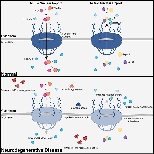
Recent advances into the underlying pathogenic mechanisms have associated mislocalization and aberrant accumulation of disease-related proteins with defective nucleocytoplasmic transport and its mediators called karyopherins. These studies identified karyopherin abnormalities in amyotrophic lateral sclerosis, frontotemporal dementia, Alzheimer’s disease, and synucleinopathies including Parkinson’s disease and dementia with Lewy bodies, that range from altered expression levels to the subcellular mislocalization and aggregation of karyopherin α and β proteins.

In addition to their classical function in nuclear import and export, karyopherins can also act as chaperones by shielding aggregation-prone proteins against misfolding, accumulation, and irreversible phase-transition into insoluble aggregates. Karyopherin abnormalities can, therefore, be both the cause and consequence of protein mislocalization and aggregate formation in degenerative proteinopathies.

A new study suggests that the upregulation of two karyopherins might improve cell health in C9orf72.

Hexanucleotide repeat expansions in C9orf72 are the primary genetic mutation associated with amyotrophic lateral sclerosis (ALS) and frontotemporal dementia (FTD), collectively referred to as C9-ALS/FTD. The resulting dipeptide repeat (DPR) proteins, such as poly(proline-arginine) (polyPR), generated from G4C2 repeat expansions, have been shown to be highly toxic.

To investigate the cellular localization of PR20, a polyPR protein, it was labeled with fluorescein isothiocyanate (FITC). Notably, several cell lines survived PR20 treatment by sequestering it in the cytoplasm. However, treatment with JQ-1 or Ivermectin (Iver) translocated PR20 into the nucleus, leading to cell death.

Mechanistically, the interaction between KPNA2/KPNB1 and PR20 in the cytoplasm prevented PR20 from entering the cell nucleus. Genetic silencing of KPNA2/KPNB1 converted PR20-resistant cells into PR20-sensitive cells. Specifically, JQ1 treatment decreased KPNA2/KPNB1 protein levels, allowing PR20 to enter the nucleus.

Conversely, overexpressing KPNA2 or KPNB1 effectively blocked cell death induced by co-treatment with JQ-1 and PR20. The authors' findings suggest that the upregulation of KPNA2/KPNB1 protects cells from PR20 toxicity.

Pharmacological targeting of karyopherins represents a promising new strategy for therapeutic intervention in neurodegenerative diseases. Yet by the time patients are diagnosed, a significant number of neurons are already lost, while the remaining functional ones are doomed to degenerate over time due to the lack of efficient treatments. So, it's important not to exaggerate the benefits of drugs in those diseases.

Several compounds targeting karyopherins are already in clinical trials to treat specific forms of cancer and viral infections, so it might be less costly to retarget these drugs toward neurodegenerative diseases than it is for entirely new drugs.


Please, help us continue to provide valuable information: