How the gut microbiome influences insulin resistance.

- Posted by admin in English

Alzheimer's disease, Parkinson's disease, ALS, and FT Dementia share many biomarkers and comorbidities. One of them is insulin resistance which is found in half of patients.

Indeed insulin resistance is also a consequence of diabetes and aging. Scientists from Japan explore in a recent article how the gut microbiome influences insulin resistance. enter image description here Previous studies have explored the role of gut microbiota in metabolizing nutrients in insulin resistance. This research aims to uncover the mechanisms underlying this relationship using a multi-omics approach. The study analyzes data from 306 individuals without diabetes, focusing on insulin resistance as defined by HOMA-insulin resistance scores.

The researchers used various techniques, including metabolomics, metagenomics, transcriptomics, and clinical data, to profile how the gut microbiome contributes to insulin resistance.

They found that certain carbohydrates in the feces, particularly those accessible to the host, are elevated in individuals with insulin resistance. These carbohydrates are linked to microbial carbohydrate metabolism and host inflammatory cytokines. Specific gut bacteria are associated with insulin resistance and insulin sensitivity, each displaying distinct carbohydrate metabolism patterns. In a mouse model, bacteria linked to insulin sensitivity demonstrate the potential to improve insulin resistance traits.

The study also involves analyzing metabolic syndrome (MetS) and its associations with fecal and plasma metabolites. Using human fecal cultures, the researchers discovered that Bacteroidales, a type of gut bacteria linked to insulin sensitivity, have a unique metabolic profile. These bacteria are efficient consumers of certain carbohydrates, affecting the production of fermentation products.

To explore causality, the researchers test the effects of seven candidate bacteria associated with insulin sensitivity on mice fed a high-fat diet. Several strains, notably Alistipes indistinctus, show promising results in reducing postprandial blood glucose levels and improving insulin resistance. These strains also impact body mass, lipid accumulation, and glucose intolerance.

Mechanistically, the researchers find that A. indistinctus administration reduces carbohydrate oxidation in mice, possibly due to decreased host-accessible carbohydrates in the intestine. This is supported by altered caecal metabolites, including reduced monosaccharides like fructose.

In conclusion, the study employs a comprehensive multi-omics strategy to investigate the relationship between gut microbiota and insulin resistance. It identifies specific bacteria associated with insulin resistance and sensitivity and highlights the potential of A. indistinctus in ameliorating insulin resistance in mice. However, further research is needed to understand the precise mechanisms and potential therapeutic implications of these findings.

Radiation therapy for Alzheimer's disease

- Posted by admin in English

Hormesis is a characteristic of many biological processes, namely a biphasic or triphasic response to exposure to increasing amounts of a substance or condition. Within the hormetic zone, the biological response to low exposures to toxins and other stressors is generally favorable.

Hormesis has been observed in a number of cases in humans and animals exposed to chronic low doses of ionizing radiation. enter image description here A new publication by Korean researchers discusses the current state of treatment for Alzheimer's disease, focusing on pharmacological and non-pharmacological approaches. It mentions that only four drugs (donepezil, rivastigmine, galantamine, and memantine) have been available to Alzheimer's patients. Yet, the improvement in neurological function is almost 0%, and it only slows the rate of cognitive deterioration. A new drug called aducanumab, targeting Aβ plaques, has been approved by the U.S. FDA, but its efficacy and accessibility are debated due to unclear clinical results and high costs.

An alternative approach, low-dose radiation therapy has gained attention after a case report described significant improvement in a patient with advanced Alzheimer's disease who underwent computed tomography (CT) scans of the brain five times. Recently, several preclinical studies based on mouse models revealed a significant reduction in Aβ plaques with low-dose radiation therapy, and low-dose radiation therapy induced the upregulation of pre-and post-synaptic molecules in the brains of Alzheimer's disease mouse models.

For example, a recent study showed that low-dose radiation therapy seems to reduce the levels of pro-inflammatory cytokines in animal models of Alzheimer's disease. Therefore, several pilot studies or clinical trials investigating the effect of low-dose radiation therapy on humans have been launched. Most recently, researchers at Virginia Commonwealth University (VCU) published positive pilot study results showing that four of five patients diagnosed with early Alzheimer's disease experienced improved or stable cognition after treatment with low-dose radiation therapy.

The Korean scientists describe an ongoing clinical trial aiming to explore low-dose radiation therapy as a treatment for Alzheimer's disease. The trial involves evaluating the safety and effectiveness of this therapy and determining appropriate dose schedules. The clinical trial is multicenter and randomized, with patients being divided into different treatment groups.

The study involves rigorous screening tests, including neurological examinations, cognitive function tests, and various imaging and laboratory tests. Eligible patients will receive low-dose radiation therapy in either 6 fractions of 4 cGy or 6 fractions of 50 cGy, while a control group will receive sham irradiation.

The clinical trial aims to evaluate cognitive improvement of at least 5% as an effective response. This would be a huge improvement over existing medications for Alzheimer's disease.

Brain implant to vocalize thoughts

- Posted by admin in English

For ALS patients, failing to access a cure, it is important to overcome certain important problems. Alas a chair that can be controlled by gaze, telemanipulation arms, intelligent ventilation, and a device for vocalizing by thought, will not be marketed for another decade or two.

The press echoed two recent articles about devices for vocalizing through thought. One acquires the data via a deep cranial implant, the other via a surface cranial implant. enter image description here I analyze below the first article because the data and programs to implement this experiment are publicly available. Which is apparently not the case for the second.

I asked one of the authors if it was possible to use his program with an EEG helmet. We'll see what his answer will be, but I anticipate that the results of this course of action will be very disappointing.

The study focused on understanding how facial movement and speech production are organized in the motor cortex at the level of individual neurons. Neural activity was recorded from microelectrode arrays implanted in the brain of a participant with amyotrophic lateral sclerosis (ALS) who had limited facial movements and an ability to vocalize but not produce intelligible speech.

The results indicated neural solid agreement on various facial movements in a region of the brain called area 6v, and this activity was very distinct for different movements. In contrast, area 44, traditionally associated with speech production, appears (in this experiment) to contain little information about facial actions or speech.

The ventral premotor cortex, which is located in the middle of the upper part of the brain has been involved in motor vocabularies in both speech and manual gestures. A recent prospective fMRI study demonstrated adaptation effects in the ventral premotor cortex to repeating syllables.

Broca's area, is a region in the frontal lobe of the dominant hemisphere, usually the left, of the brain with functions linked to speech production.

The researchers developed a decoder using a recurrent neural network (RNN) to translate neural activity into speech. The participant attempted to speak sentences and the RNN decoded the predicted words in real-time, achieving a word error rate of 9.1% for a vocabulary of 50 words and 23.8% for a vocabulary of 125 000 words. This demonstrated the feasibility of decoding speech attempts using neural signals.

The neural representation of speech sounds in the brain was analyzed, showing that the activity patterns reflected the articulatory features of the phonemes. This suggests that even after years of paralysis, the detailed articulatory code of phonemes remains preserved in the brain.

The study also discussed design considerations for improving the accuracy of speech brain-computer interfaces (BCIs), including vocabulary size, number of electrodes used, and size of the training data set. The researchers noted that while their results were promising, there was still room for further optimization and improvements in the technology.

Overall, the study presented a proof of concept for a speech BCI that could potentially enable people with severe motor impairments to communicate more effectively by translating their intended speech into text from neural signals.

Grafting muscles to heal from ALS?

- Posted by admin in English

Scientists tell that ALS is a disease striking upper motor neurons, which in a cascade effect, affects the lower motor neurons and muscles. Yet this does not match the patient's experience. enter image description here For them, symptoms often start with weakness, unreliability, and thinning of the thumb or calf, and with time the disease progresses to other (skeletal) muscles.

Scientific approaches are focussing on healing upper motor neurons (with a complete lack of success, which hints that we understand nearly nothing in this area) or replacing them (there are still no clinical studies).

For most scientists, it would be meaningless to try to replace muscles in ALS patients, as the upper motor neurons are dead (despite evidence they are not) new muscles would never be usable and therefore waste and die quickly.

And anyway grafting muscles on neuromuscular junctions is an extraordinarily difficult problem.

Yet there are contrarians: Scientists have recently unveiled an approach to address the devastating effects of amyotrophic lateral sclerosis (ALS), on muscle weakness. Their research introduces a novel technique involving a combination of grafted replacement motor neurons and optical nerve stimulation.

By employing a two-pronged strategy—grafting modified motor neurons and using light-based nerve stimulation—the researchers managed to rejuvenate muscle function, marking a tiny step toward devising effective therapies for ALS.

Led by Dr. Barney Bryson, the team of scientists had shown potential in restoring muscle denervation in a mouse model by utilizing optical nerve stimulation. This method hinged on grafting replacement motor neurons that were engineered to be light-sensitive, enabling their activation through an external light source.

In this latest study, the initial objective was to ensure the survival of donor motor neurons during the grafting process, overcoming potential immune system rejection. After determining that conventional immunosuppressive drugs were not viable for ALS mice, the researchers experimented with a specific antibody known as H57-597, which successfully prevented graft rejection and began reconnecting nerves to target muscles. Despite these positive outcomes, the force generated by muscle contractions was relatively weak, prompting the team to further refine their strategy.

Recognizing that neuromuscular junctions—the connection points between nerves and muscles—are influenced by regular stimulation, the scientists introduced a wireless optical stimulation system to enforce consistent muscle contractions for an hour each day. Astonishingly, after 21 days of this optical stimulation training, the mice exhibited an astonishing 13-fold enhancement in muscle contraction force. That would be nearly three years of rehabilitation therapy in humans.

These findings carry immense significance, demonstrating that even in the advanced stages of ALS, affected muscles remain amenable to reinnervation by healthy replacement motor neurons.

The strategy suggests the feasibility of a treatment that could potentially be universally applied to all ALS patients regardless of the kind of ALS they have (familial or sporadic).

More significantly, it tells that the model of ALS progression which was derived from stroke ~150 years ago and mostly never questioned since all these decades, is simply wrong.

However, significant challenges remain before this approach can transition to clinical use. Subsequent studies are necessary to confirm the efficacy of the grafting procedure with human motor neurons and to evaluate whether it can genuinely enhance patients' quality of life. Furthermore, the technique's applicability to other forms of MND, particularly those with longer life expectancies, must be investigated to ascertain its long-term effectiveness.

In conclusion, the study's senior author, Linda Greensmith, emphasizes that the findings underline the robustness of replacing motor neurons to reinnervate muscles even in advanced ALS stages. Should this approach successfully translate to ALS patients, it holds the potential to revolutionize treatment by employing a single type of motor neuron for various muscles, simplifying the therapy and making it more widely accessible. The introduction of this approach could be a game-changer in the ongoing battle against ALS and other related neuromuscular conditions.

Parkinson's disease can start as an autoimmune disease

- Posted by admin in English

Constipation and sialorrhea (impaired swallowing) have been considered to be important factors of neurodegenerative diseases (Parkinson's disease, but it is also common in ALS, Alzheimer, and autistic disorders), although the exact mechanism is still controversial. Constipation and dementia have similar epidemiological characteristics. A study found that people with Alzheimer's disease and constipation decline nearly 3 times quicker than people with Alzheimer's disease but no constipation. enter image description here Constipation is present in approximately 70% of Parkinson's disease patients, is 3-fold more prevalent in Parkinson's disease patients than healthy controls, and occurs as early as 20 years prior to the onset of motor symptoms. Inflammatory bowel disease patients who receive anti-tumor necrosis factor alpha (TNF-α) therapy exhibit a 78%–100% reduction in Parkinson's disease incidence compared with those who do not receive such therapy.

Constipation is often neglected by scientists and doctors who attribute it to poor hygiène, poor diet, lower water intake, comorbidities (stroke, diabetes), and brain health degradation. In consequence, constipation symptoms are treated with pharmacologic drugs, while underlying causes are ignored and therefore not treated.

Most studies on constipation in dementia patients focus on the population with α-synucleinopathies [Parkinson’s disease dementia, dementia with Lewy bodies]. Studies have shown that total truncal vagotomy, but not selective vagotomy, was associated with a lower prevalence of Parkinson's disease which hints at propagation of the disease from the gut to the brain via the vagus nerve.

20 years ago Heiko Braak hypothesized that in Parkinson's disease, a pathogen reaching the gut initiates pathology that spreads to the CNS. The main portals for the central delivery of α-synuclein are thought to be the olfactory bulb and vagus nerve.

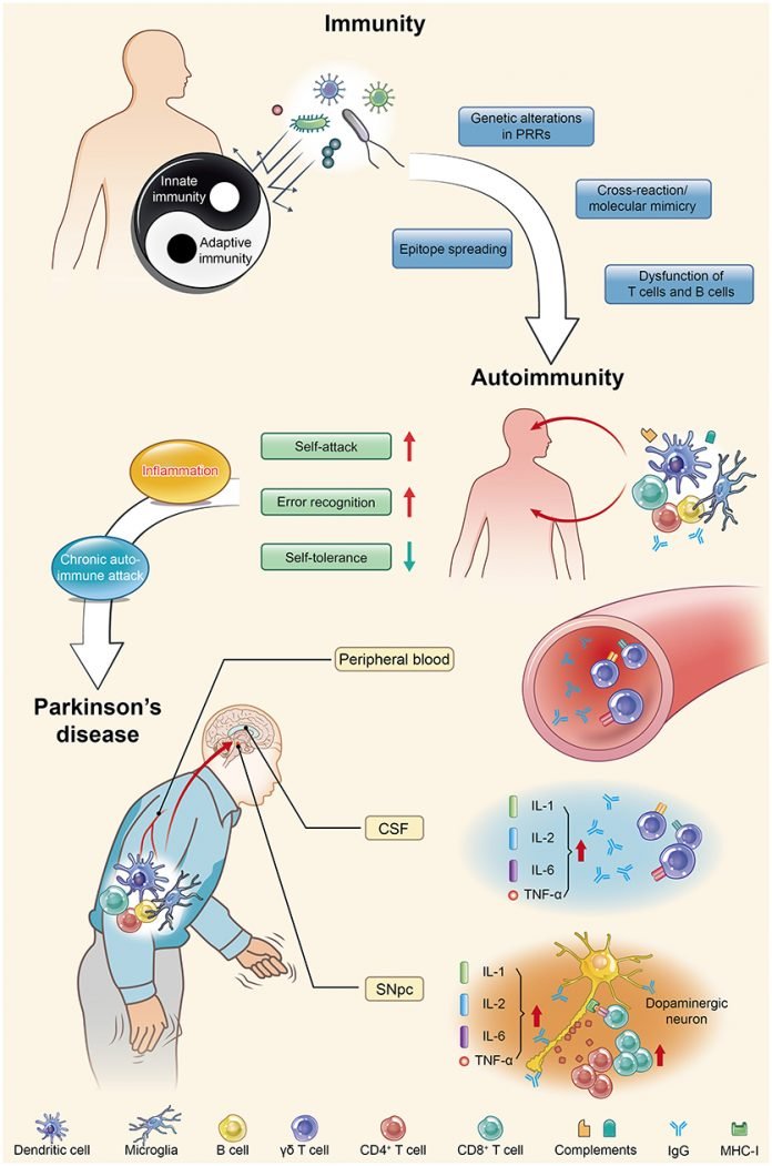
Peripheral inflammation is likely implicated in PD pathogenesis because high levels of pro-inflammatory cytokines (e.g., TNF-α, interleukin [IL]-1β, IL-6, and interferon [IFN]γ) are found in PD patients and correlate negatively with disease duration. This is true also in ALS, or Alzheimer's disease.

PD patients also possess circulating T cells, mostly CD4+ subtypes, which recognize specific α-syn-derived neo-epitopes. T cells are one of the important types of white blood cells in the immune system and play a central role in the adaptive immune response. This immune response is restricted to patients carrying a specific HLA haplotype (B∗07:02 C∗07:02 DRB5∗01 DRB1∗15:01 DQA1∗01:02 DQB1∗06:02).

There are several immune cell types. White blood cells include three main subtypes; granulocytes, lymphocytes, and monocytes.

  • CD8+ T cells are cytotoxic lymphocytes, they kill virus-infected cells, as well as cancer cells. CD8+ T cells are also able to use small signaling proteins, known as cytokines, to recruit other types of cells when mounting an immune response.

  • CD4+ T cells, function as "helper cells". They activate memory B cells and cytotoxic T cells, which leads to a larger immune response. They also secrete cytokines.

  • Regulatory T cells provide a mechanism of tolerance, which prevents immune cells from inappropriately reacting against their own cells. Alas, these same regulatory T cells can also be co-opted by cancer cells to prevent the recognition of, and an immune response against, tumor cells.

A different kind of lymphocyte, Memory B cells circulate in the bloodstream in a quiescent state, sometimes for decades. Their function is to memorize the characteristics of the antigen that activated their parent B cell during an initial infection such that if the memory B cell later encounters the same antigen, it triggers an accelerated and robust secondary immune response.

In each vertebrate cell, protein molecules are continually synthesized and degraded. Some small parts of degraded proteins (peptides) are sent to the surface of the cell where MHC molecules on the cell surface keep them and display them at the intention of the host's leukocytes.

These peptides are specific to one host, so if a leukocyte encounters a cell that displays peptides that are known to be from pathogens, they kill the cell. For most cell types this is not a threat to the host's health as many cell types are quickly renewed, yet this is damaging some cell types such as neurons which reproduce slowly or not at all (motorneurons).

These small peptides displayed by MHC molecules at the surface of vertebrate cells are called epitopes. MHC molecules constitute a very wide class of molecules with many types and sub-types. In humans, MHC molecules are called HLA molecules.

It's known that α-synuclein, the molecule which causes Parkinson's disease, is recognized by CD4+ T cells. One epitope, α-syn32-46 (a part of the α-synuclein molecule), binds with a strong affinity to the HLA-DRB1∗15:01 allele which is implicated in autoimmune diseases. This may not be an accident but on the contrary some anti-microbial mechanism. A parallel could be drawn with beta-amyloids in Alzheimer's disease. enter image description here Scientists just reported that making a mouse model with leucocytes that react to a subset of α-synuclein molecule, triggers intestinal inflammation, leading to transient constipation and weight loss, yet with no detectable effects in the central nervous system (CNS). In other words, far from defending the body, the leucocyte of this mouse model are attacking their own body cells. Given that this auto-immune artificially created disease implicated a part of the molecule incriminated in Parkinson's disease and creates effects found in Parkinson's patients, it is reasonable to suspect it has some similarity with human disease.

More precisely the scientists created a mouse strain lacking major histocompatibility complex class II (MHCII) and expressing the human HLA-DRB1∗15:01 allele. This was to model PD patients who possess circulating T cells that recognize specific α-synuclein (α-syn)-derived epitopes.

Next, the authors had to make the mice's immune system to react α-synuclein derived epitopes. They used a classic strategy which is to administrate the peptide along with some immunologic adjuvant. Vaccines commonly use such immunologic adjuvants. The authors used myelin oligodendrocyte glycoprotein peptide (MOG)35–55 plus complete Freund’s adjuvant (CFA) emulsion combined with i.v. administration of Bordetella pertussis toxin. The idea between this complex assemblage is that when leucocytes will recognize the adjuvant as hostile, CD4+ T cells will associate it with α-synuclein derived epitopes. So when CD8+ T cells will encounter cells with such epitopes, they will kill them.

This triggered intestinal inflammation and loss of enteric neurons in the submucosal plexus (SP) of the small intestine, leading to transient constipation and weight loss, with no detectable effects in the CNS. Bulk RNA sequencing (RNA-seq) of the gut reveals that α-syn32-46 immunizations induce innate and adaptive immune responses and IFN signaling. Single-cell RNA-seq (scRNA-seq) of immune cells showed altered gene signatures in CD4+ TH1 and TH17 lamina propria lymphocytes from complete Freund’s adjuvant (CFA)/α-syn32-46-immunized mice, characteristic of antigen-experienced tissue-resident memory (TRM) cells found in mucosal barriers during infection and chronic inflammation.

Depletion of CD4+, but not CD8+, T cells partially rescued enteric neurodegeneration, which confirms the hypothesized mechanism where CD4+ are responsible for immunization. In human inflammatory bowel disease, CD4+ CD69+ CD103+ TRM cells are proposed to accumulate in the gut and trigger intestinal inflammation.57,62 A similar CD4+ TRM population (CD161+ CCR5+) produces proinflammatory cytokines in Crohn’s disease.

Thus, interactions of α-syn32-46 with the HLA-DRB1∗15:01 allele are critical for the induction of enteric features resembling those seen in prodromal Parkinson's disease, suggesting that additional hits may be required for the development of CNS symptoms.
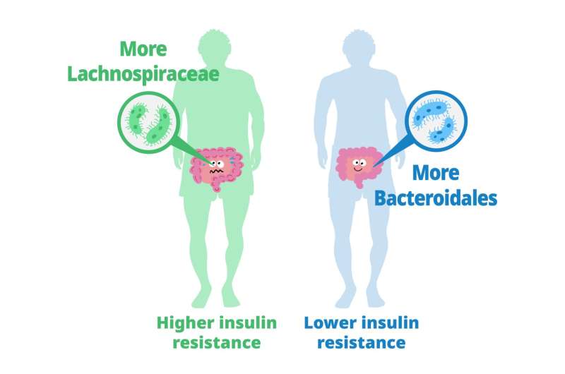
La sclérose latérale amyotrophique (dite aussi maladie de Charcot) est une maladie incurable qui provoque une faiblesse musculaire dans tout le corps, rendant les mouvements et la respiration de plus en plus difficiles jusqu'au décès. La maladie commence souvent par un problème apparemment musculaire dans le pouce ou le mollet et cette faiblesse s'étend progressivement au reste des muscles du corps que l'on contrôle consciemment (muscles dit "squelettiques").

On a trouvé de multiples causes génétiques à la SLA dite familiale, mais celle-ci ne représente qu'un faible pourcentage de l'ensemble des cas. Par contre dans la plupart des cas on trouve dans les cytoplasme des neurones moteurs, des amas d'une protéine nommée TDP-43. Ces amas souvent comporte seulement des fragments de cette protéine qui de plus est dépliée, c'est à dire non fonctionnelle.

On ne sait pas si TDP-43, qui est indispensable à la survie, est une cause de la maladie ou une conséquence d'événements tels qu'une infection virale. De tels interrogations existent aussi dans le cas de la maladie d'Alzheimer ou de Parkinson.

Ce qui est sur c'est que les mutations de cette protéine sont souvent délétères, ce qui n'étonnera personne, et qu'un surcroît ou un défaut de TDP-43 est lui aussi délétère.

Une autre controverse concerne aussi le sens de propagation de la maladie, se propage t-elle depuis les extrémitées vers le cerveau (ce qui correspond à l'expérience des malades) ou au contraire, est-ce une maladie qui nait dans le cerveau et se propage aux extrémitées (hypothèse majoritaire chez les scientifiques et dérivée de ce que l'on observe après un AVC dans la zone motrice du cerveau). Les scientifiques partisans de chacune des deux opinions ont à de multiples reprise prouvé que "l'autre" hypothèse était fausse, ce qui n'est guère informatif.

Une nouvelle étude par des scientifiques Japonais tend à dire que les deux hypothèses sont vrais mais ne sont pas assez précises dans leur formulation.

Les commandes de mouvement initiées par des neurones de la zone motrice du cortex cérébral sont transmises aux neurones moteurs supérieurs de la moelle épinière via le tractus corticospinal. Dans la moelle épinière ces commandes sont transmises aux neurones moteurs inférieurs qui à l'autre extrémité, sont reliés aux muscles.

Les scientifiques Japonais ont supposé que la protéine (TDP-43) se propage anormalement dans ces circuits neuronaux. Jusqu'à présent, on pensait que TDP-43 était cantonné à l'intérieur des neurones et des cellules non-neuronales. enter image description here 
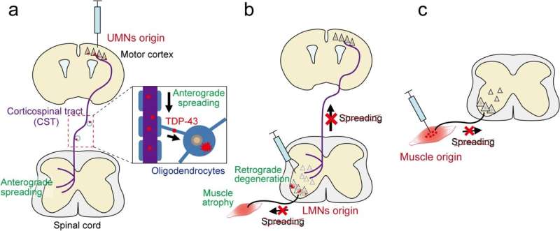
Les auteurs ont établi de nouveaux modèles de SLA de souris qui induisaient initialement des inclusions de TDP-43 mutant dans des types neuronaux ou cellulaires spécifiques dans les circuits moteurs, et ils ont étudié si le TDP-43 et les processus pathologiques pertinents se propageaient à travers les connexions neuronales ou cellulaires.

Les auteurs ont d'abord développé des modèles de SLA qui induisaient principalement des inclusions de TDP-43 dans les neurones corticospinaux, les motoneurones spinaux ou le muscle squelettique des membres antérieurs, en utilisant le virus adéno-associé (AAV) exprimant le mutant TDP-43.

Ils ont ensuite examiné comment le TDP-43 depuis des motoneurones spécifiques, ce propage à d'autres neurones moteurs entrainant la progression de la maladie. Les données ont révélé que le mutant TDP-43 se propageait à travers les connexions neurogliales de manière antérograde dans la voie corticospinale, alors qu'il présentait différentes propriétés dégénératives rétrogrades dans les circuits spinaux. Cela suggère que le TDP-43 pathogène peut induire des mécanismes antéro- et rétrogrades distincts de dégénérescence du système moteur dans la SLA.

  • Les auteurs ont constaté que le TDP-43 induit dans les neurones corticospinaux était transporté le long des axones de manière antérograde et transféré aux oligodendrocytes le long du tractus corticospinal (CST), coïncidant avec une légère dégénérescence des axones. Lorsque le TDP-43 anormal a été induit dans le cortex cérébral, le cortex cérébral et les axones subissent une dégénérescence et que les oligodendrocytes du tractus corticospinal adoptaient une morphologie réactive. Mais, bien que le TDP-43 soit apparu dans le tractus corticospinal, il ne s'est pas propagé à la moelle épinière. Les oligodendrocytes sont des cellules non neuronales qui assistent les neurones pour faciliter la transmission du signal neuronal, en enveloppant les axones d'une couche protectrice appelée myéline. C'est à dire en langage simple qu'on a bien une progression de la maladie depuis le cerveau vers la moelle épinière, mais l'atteinte est légère.

  • En revanche, le TDP-43 introduit dans les motoneurones inférieurs ne s'est pas propagé de manière rétrograde aux neurones corticaux ou aux motoneurones supérieurs. Cependant, il a induit une perte extrême en peu de temps des motoneurones inférieurs voisins. La dégénérescence intraspinale a en outre entraîné une atrophie musculaire sévère. De plus, il a été constaté que non seulement les motoneurones, mais également d'autres interneurones de la moelle épinière qui communiquent avec la zone environnante, étaient induits à mourir. De plus, parallèlement à la perte de motoneurones, les muscles se sont également atrophiés à un degré élevé, provoquant des troubles du mouvement. Mais le TDP-43 muté lui-même ne s'est pas propagé aux neurones moteurs supérieurs, au cortex cérébral ni aux muscles. Ce n'est donc pas une progression rétrograde mais plutôt latérale.

  • Enfin, le TDP-43 induit dans les muscles squelettiques ne s'est pas propagé rétrogradement aux neurones inférieurs spinaux. L'induction de TDP-43 aberrant dans le muscle a augmenté le nombre de fibres régénérantes, mais n'a pas provoqué d'atrophie musculaire ni de dyskinésie. De plus, aucune propagation du TDP-43 à la moelle épinière n'a été observée

enter image description here 
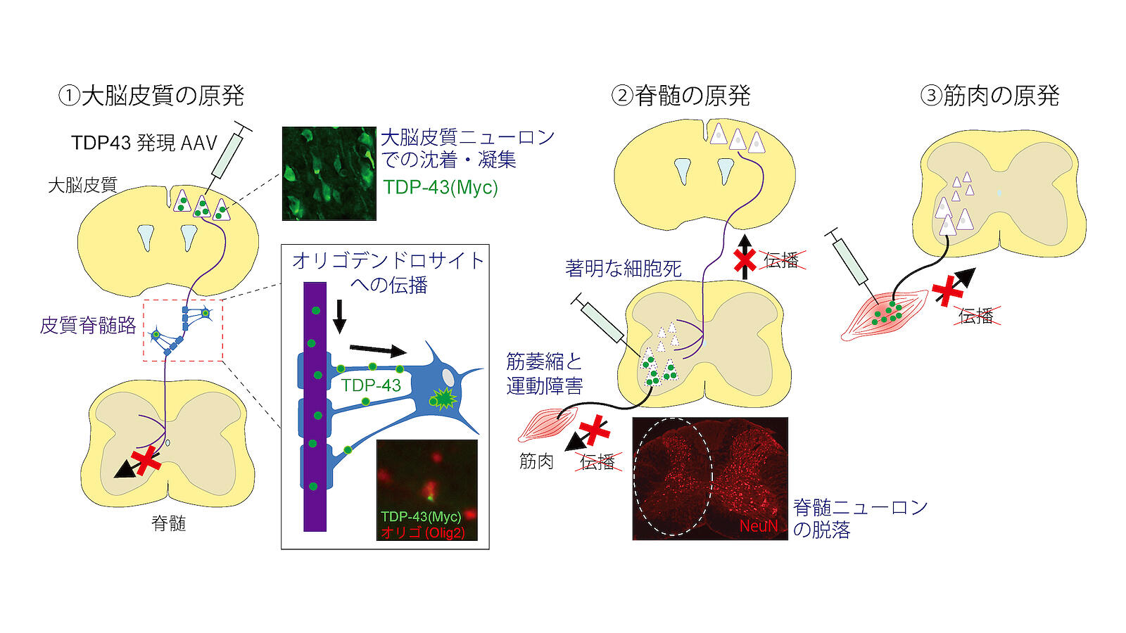
Tsuboguchi et al., Acta Neuropathologica 2023.

La maladie de Parkinson est la deuxième maladie neurodégénérative la plus courante, caractérisée par des tremblements, des mouvements ralentis, de la rigidité et des troubles cognitifs. La maladie de Parkinson affecte les neurones dopaminergiques de la substantia nigra du mésencéphale.

Une caractéristique particulière de la maladie de Parkinson est l'accumulation d'α-synucléine dans les neurones dans le cadre d'inclusions protéiques, les corps de Lewy. Comme les patients atteints d'autres maladies neurodégénératives, les patients atteints de la maladie de Parkinsons ont des biomarquers et des symptomes qu'on associe habituellement à d'autres maladies.

Il est à noter que 60% des malades d'Alzheimer ont des dépôts d'α-synucléine dans l'amygdale, et que certains patients atteints de maladie de Parkinson ont une accumulation d'Aβ dans le cerveau. Cela suggère que des voies spécifiques menant au développement de la maladie de Parkinson ou de la maladie d'Alzheimer convergent, provoquant l'apparition de signes communs.

Le rôle du cholestérol dans la maladie de Parkinson reste controversé. Un HDL sérique plus élevé est associé à une fonction cognitive plus faible chez les femmes atteintes de MP.

Une étude de radeaux lipidiques isolés du cortex frontal de sujets atteints de maladie de Parkinson à un stade précoce a montré une diminution des acides gras polyinsaturés sans modification du contenu en cholestérol et en sphingomyéline.

L'α-synucléine comprend deux domaines de liaison au cholestérol et le cholestérol membranaire affecte son agrégation. Théoriquement, la synucléine peut perturber l'intégrité du radeau lipidique, en interagissant avec le cholestérol.

Les statines inhibent l'agrégation de l'α-synucléine dans une culture neuronale et l'ajout de cholestérol exogène augmente l'agrégation de l'α-synucléine.

La privation de nourriture (contrairement à ce qui est souvent répété) provoque l'agrégation de l'α-synucléine et l'apoptose, qui est associée au stress du réticulum endoplasmique, suivie d'une augmentation de la synthèse du cholestérol. Dans la maladie de Parkinson, la concentration de certains oxystérols augmente dans le cerveau en réponse à la surproduction d'espèces réactives de l'oxygène. Une alimentation riche en cholestérol réduit le rapport 24 OHC/27 OHC dans le cerveau et augmente le niveau d'α-synucléine dans la substantia nigra sans altérer le taux de cholestérol cérébral.

Plusieurs oxystérols sont associés à des maladies liées à l'âge telles que les maladies cardiovasculaires, les maladies oculaires (cataracte, dégénérescence maculaire liée à l'âge), certaines maladies neurodégénératives et les cancers. Les activités des oxystérols dans ces maladies pourraient être dues à leurs activités pro-oxydantes et pro-inflammatoires et à leur capacité à agir sur les organites cellulaires (mitochondries, peroxysomes, lysosomes) qui peuvent contribuer à activer l'apoptose et l'autophagie. Il existe des arguments soutenant que les oxystérols jouent un rôle important dans la progression de l'athérosclérose qui est impliquée dans plusieurs maladies cardiovasculaires.

Les scientifiques pensent depuis quelque temps déjà que l'hyperlipidémie est associée à un risque accru de maladie de Parkinson. En effet, les niveaux de 27-hydroxycholestérol (27-OHC), un oxystérol, sont augmentés dans le cerveau et le liquide céphalo-rachidien des patients atteints de la maladie de Parkinson. enter image description here Cependant, il reste difficile de savoir si cet oxystérol joue un rôle dans l'agrégation et la propagation de l'α-synucléine. L'objectif de scientifiques de Wuhan en Chine, était de déterminer si le 27-OHC régule l'agrégation et la propagation des α-synucléine.

Les auteurs ont étudié les deux aspects, administration et réduction de 27-OHC. Dans le premier cas, les scientifiques ont administré en sous-cutanée du 27-OHC à successivement de l'α-synucléine recombinante purifié, des cultures neuronales et un modèle murin (de souris de laboratoire) de la maladie de Parkinson. De plus, des souris knock-out CYP27A1 ont été utilisées pour étudier l'effet de la réduction du 27-OHC sur la pathologie α-synucléine in vivo.

Certes la physiologie de la souris n'a guère de point commun avec la physiologie humaine, mais c'est un progrès par rapport aux études in-vitro et celà pourrait encourager des études pré-cliniques utilisant des petits primates.

Leurs résultats confirment que 27-OHC accélère l'agrégation de α-synucléine et active l'activité d'ensemencement des fibrilles α-Syn. De plus, les fibrilles α-synucléine modifiées par 27-OHC se localisent dans les mitochondries et induisent un dysfonctionnement mitochondrial et une neurotoxicité.

L'injection de fibrilles α-synucléine modifiées par 27-OHC induit une propagation accrue de la pathologie α-synucléine et de la neurodégénérescence dopaminergique par rapport aux fibrilles α-synucléine pures. De même, l'administration sous-cutanée de 27-OHC facilite l'ensemencement de la pathologie α-Syn.

Au contraire, la suppression génétique du cytochrome P450 27A1, l'enzyme qui convertit le cholestérol en 27-OHC, améliore la propagation de l'α-synucléine pathologique, la dégénérescence de la voie dopaminergique nigrostriée et les déficiences motrices.

Ces résultats indiquent que le métabolite du cholestérol 27-OHC joue un rôle important dans la pathogenèse de la maladie de Parkinson. : 27-OHC favorise l'agrégation et la propagation de l'α-Syn. Les stratégies visant à inhiber l'axe CYP27A1-27-OHC pourraient être prometteuses en tant que traitement modificateur de la maladie pour stopper la progression de la pathologie α-synucléine dans la maladie de Parkinson. Le plus simple pour les patients est probablement d'adopter une alimentation pauvre en cholestérol et pour les scientifiques d'étudier les effets de administration de statine chez les malades de Parkinson.

Ces études ont déjà été abordés dans des publications récentes. Celle-ci aborde les effets du candesartan, simvastatin, and du fasudil chez des rats de laboratoire modèles de la maladie de Parkinson.

Celle-là fait une étude similaire de la simvastatin chez des rats.

Dans les deux cas les statines utilisées semblent avoir un effet bénéfique contre la maladie de Parkinson.

Here is a study on FUS ALS which proposes that FUS ALS patients might benefit from administration of interferon-gamma (IFNγ). Interferon-γ 1b is approved by the U.S. Food and Drug Administration to treat chronic granulomatous disease and osteopetrosis. In approximately 90% of cases, ALS is sporadic, but around 10% of patients exhibit familial mutations. Subtypes of ALS are categorized based on the affected gene, including superoxide dismutase 1 (SOD1), TAR DNA-binding protein 43 (TDP-43), chromosome 9 open reading frame 72 (C9ORF72), and Fused in Sarcoma (FUS), with FUS leading to one of the most aggressive and early-onset forms of the disease.

FUS is a component of the heterogeneous nuclear ribonucleoprotein (hnRNP) complex and is involved in DNA/RNA binding, DNA damage repair, splicing, and various aspects of RNA metabolism. Over 50 different FUS gene mutations have been identified in ALS patients. Mutations often affect the nuclear localization signal (NLS) domain, leading to improper cytoplasmic localization and nuclear clearance of FUS, resulting in an aggressive pathological phenotype. The most common FUS mutation affects arginine 521 (R to H, C, or G). Incorrect FUS localization also occurs in other forms of familial and sporadic ALS cases, suggesting a common mechanism.

Studying ALS pathobiology is challenging due to the high costs and morbidity associated with the disease. Reliable disease models are essential, so the study used induced pluripotent stem cells (iPSCs) derived from FUSR521H ALS patients to generate motor neurons (motoneurons) for research.

As iPSCs rejuvenate as a result of reprogramming and ALS symptoms are primarily associated with aging, the authors exposed iPSC-derived motor neurons to oxidative stress (by means of sodium arsenite) to model aging-associated effects.

These models revealed that ALS motor neurons are more sensitive to oxidative stress than control motor neurons. ALS iPSC-derived motoneurons exhibited abnormal cytoplasmic FUS localization, reduced translation rates, and increased sensitivity to oxidative stress-induced apoptosis.

The study further explored altered gene expression patterns and signaling pathways in FUS ALS motoneurons. enter image description here

As cytokine measurements showed reduced secreted IFNγ in FUS ALS motorneurons treated with sodium arsenite, and as authors recently found that inflammatory cytokines can protect cells from stress-induced cell death (Hong et al., 2022), they tested whether this was also true for FUS ALS motorneurons.

For this, the scientists supplemented sodium arsenite-treated FUS ALS motorneurons with IFNγ and found that this indeed increased viability of FUS ALS motorneurons to levels similar to SA-treated control motorneurons.

Furthermore, IFNγ treatment reduced the fraction of apoptotic FUS ALS motorneurons following sodium arsenite exposure. Importantly, they also found that IFNγ treatment resulted in an increased IFNγ transcriptional response in SA-treated ALS motorneurons, indicating that IFNγ treatment was sufficient to rescue the impaired IFNγ response observed in SA-treated ALS motorneurons.

As treatment with interferon-gamma (IFNγ) reduced oxidative stress-induced apoptosis and improved translation rates and nuclear FUS localization in FUS ALS motoneurons, the authors suggest that early-diagnosed FUS ALS patients might benefit from IFNγ treatment to slow disease progression.

While this is not suggested by the study, one might reflect that the described effects of FUS in ALS are similar to SOD1 and TDP-43 ALS, so maybe IFNγ treatment might be beneficial in these cases also.

In summary, the study investigates the impact of FUS mutations on ALS using iPSC-derived motoneurons, revealing insights into the disease's underlying molecular mechanisms and proposing potential therapeutic strategies involving IFNγ treatment.

Relyvrio, Phénylbutyrate de sodium et Taurursodiol

- Posted by admin in Français

Le médicament d'Amylyx connu sous les noms d'AMX0035, Relyvrio ou Albrioza est actuellement en cours d'essai clinique de phase III PHOENIX (NCT05021536). enter image description here La FDA et l'agence Européenne des médicaments ont exprimés de nombreux doutes sur l'efficacité de ce médicament au cours de l'essai CENTAUR. Ces doutes concernent l'exclusion d'un certain nombre d'évènements défavorables lors de l'analyse statistique, ainsi que le fait que certains patients ont aussi reçu du Riluzole et de l'Edaravone. En particulier il y a plus de patients ayant reçu de l'Edaravone dans la branche contrôle que dans la branche traitement, or certains scientifiques et agences du médicament pensent que l'Edaravone a un effet négatif sur l'évolution de la maladie. Certains médecins ont également attribué les "bons" résultats au TUDCA. Au final à 24 mois il n'y a pas d'amélioration de la survie. Vous pouvez consulter l'avis initial de la FDA içi:

Finalement la FDA a donné son autorisation sous condition d'un engagement d'Amylyx de retirer volontairement le médicament s'il ne montre pas d'efficacité dans son essai de phase 3 en cours.

Un problème qui n'est jamais soulevé est qu'il est recommandé d'avoir une alimentation dépourvue en protéines avec du phénylbutyrate de sodium! Au contraire pour les malades de la SLA il est recommandé d'avoir un haut régime protéique pour lutter contre la perte musculaire.

Il n'en reste pas moins que c'est le seul médicament qui pour l'instant semble donner quelques espoirs.

Aujourd'hui et dans l'attente des résultats de l'essai clinique PHOENIX, AMX0035 n'est autorisé qu'aux USA (conditionnellement) et au Canada. Dans d'autres pays et dans certain centres SLA, il est possible d'y accéder à titre exceptionnel.

A priori AMX0035 semble être un médicament très peu couteux à la production. C'est une poudre pour suspension buvable, combinant deux poudres:

• Phénylbutyrate de sodium (PB) : 3 g

• Taurursodiol (TURSO ou TUDCA) : 1 g

La posologie est de 1 sachet deux fois par jour, matin et soir (BID).

Il est étonnant que ce médicament ne soit pas, (comme pour l'AMMONAPS) encapsulé dans une pilule. Cela permettrait: - De dissimuler le goût atroce. - D'avoir une meilleur assimilation, car avec une poudre, la plus grande partie du produit doit se décomposer dans le bain d'acide de l'estomac. C'est vrai que la forme en poudre convient particulièrement pour les patients équippés d'une sonde de gastrostomie ou d'une sonde nasogastrique.

Le traitement des maladies du métabolisme du cycle de l'urée, se fait avec du phénylbutyrate de sodium, commercialisé sous les noms de Buphenyl ou AMMONAPS en comprimés de 500mg.

Amylyx a annoncé un prix annuel pour Relyvrio de $158,000, soit $433 par jour.

Des internautes font confectionner ce médicament par leur pharmacie, en effet dans certains pays les pharmaciens ont le droit de confectionner des produits ayant obtenus l'autorisation de mise sur le marché.

L'organisation ALS.org mentionne même cette possibilité:

Cela semble pourtant une option peu intéressante. La dose journalière de Phénylbutyrate de sodium coûte plus de 3 000 € chez les grands fournisseurs comme SIGMA-ALDRICH. Le prix du TUDCA est négligeable en comparaison.

Une autre possibilitée consiste évidemment à se faire prescrire du Buphenyl/AMMONAPS sous la forme de comprimés ou poudre suivant les besoins et d'acheter du TUDCA sur Internet

Adapting an eye navigation system to an electric wheelchair

- Posted by admin in English

Eye gaze navigation systems allow you to control your wheelchair, home automation, manipulator robot, and other objects by means of a camera that observes what you are looking at onto a computer screen and transcribes your commands onto your wheelchair or home automation.

It is very difficult to buy such a chair. There are many scientific publications on this subject, but only very few of these studies can be used by a person with a disability and even most realistic studies do not describe an adaptation to a commercial electric wheelchair.

One of the most realistic projects is from a person suffering from ALS (and since deceased) named Patrick Joyce.

The adaptation of the system to a commercial chair is done by a kind of robotic cap that fits over the joystick of the chair and manipulates it under the control of a program.

The user looks at a screen where different icons correspond to different actions (go left, right, stop, etc.). The program "observes" the position of the user's retinas, as well as eye blinks. From the position of the eyes, the program deduces which icons are looked at and therefore which commands are desired by the user.

Please note that the principle used to control the joystick, while universal, is not very precise.

In addition, the program itself must be adapted to the computer running it. It is therefore not a turnkey system.

Like other alternative control methods (EEG headset, muscle twitch detection), an eye navigation system will never be as accurate as traditional manual control. Although eye navigation systems can be used outdoors, it is strongly recommended that they are used only indoors. The main reason is due to the limitations of eye navigation cameras that operate in infrared. They therefore do not work reliably in direct sunlight, near a heat source, or even in cloudy conditions.

Another limitation is that this project does not have any sensors to detect collision risks in all four directions. Using a wheelchair with a joystick is already tricky and requires learning. The use of a gaze-based system is accident-prone and automatic assistance is needed to control movements and accelerations with respect to the wheelchair environment.

There was apparently a desire from Patrick Joyce to disseminate his project widely, but his death seems to have stopped the efforts of the community he had created via Hackaday.

Another realistic project is that of Bob Paradiso.

The principle is the same as that of Patrick Joyce but here the electronic circuit is connected to the wheelchair joystick connector. Although older than that of Patrick Joyce, this project is more evolved, in addition to the control of the armchair, there is a home automation arm.

Although Joystick's protocol is relatively standardized, it is not certain that all chairs implement it. On the other hand, the precision in this project is necessarily better than in Patrick Joyce's project. Before planning to use a particular wheelchair, it is, therefore, necessary to ensure that the joystick uses a standard protocol and connector. Digital mode (not analog) PIN 1: Front PIN 2: Rear PIN 3: Left Pin 4: Right PIN 5: DETECT PIN 6: 5 button PINS 7 AND 9 – 12V 100MA PIN 8 – GROUND

The Arduino controller directly controls the motors of the robotic arm and the IR/RF transmitters which control the surrounding devices.

An obvious improvement to this project would be to control the wheelchair via Bluetooth. Indeed, some modern wheelchairs allow remote control via Bluetooth, so it may be possible to interface Bob Paradiso's project to such wheelchairs without having to interfere with their electronics.

As for Patrick Joyce's project, there are no sensors to prevent collisions.

Other interesting projects are:


Please, help us continue to provide valuable information: