Computational analysis links blood RNA patterns to ALS

- Posted by admin in English

Since we still don't know the causes of ALS and several other sporadic neurodegenerative diseases, it's interesting to find potential biomarkers associated with these diseases. Furthermore, since most clinical trials are negative, the pharmaceutical industry is seeking to change the definition of a successful trial by substituting biomarkers for clinical signs, as they are said to be more reliable. While there is some truth in this approach, it still seems highly questionable from an ethical perspective: The sole purpose is to validate clinical trials, even if there is no improvement in patient symptoms.

Researchers are therefore currently working to find biomarkers for neurodegenerative diseases, as the market for these tools appears immense and lucrative. The ideal biomarker would be an inexpensive blood test.

Researchers from Thomas Jefferson University examined two American GEO databases that collect blood samples (plasma and serum). https://www.ncbi.nlm.nih.gov/geo/info/overview.html

The Gene Expression Omnibus (GEO) is a public repository that archives and freely distributes comprehensive microarray, next-generation sequencing, and other forms of high-throughput functional genomics data submitted by the scientific community. These are digital data provided by microbiology tools and are therefore presumed to be reliable, but the associated metadata (provided by humans) may not be of high quality. Furthermore, some datasets submitted to GEO may have been contaminated.

Scientists focused on the small RNA fragments present in these samples. They focused on small non-coding RNAs because these molecules are stable and abundant in blood fluids such as plasma and serum.

These molecules were classified as follows:

  • isomiR: slightly altered versions of microRNAs

  • tRF: fragments from transfer RNA

  • rRF: fragments of ribosomal RNA

  • yRF: fragments of another type of RNA, Y RNAs

  • And a residual group, called "not-itrs," for sequences they initially could not categorize.

The scientists found that these small types of RNA do not appear in sufficient quantities in ALS patients as in healthy people.

Some of these differences were related to the patients' survival time, even after taking into account factors such as age, sex, and whether or not they were taking riluzole (a common treatment for ALS).

Interestingly, some "non-itrs" sequences did not match human DNA, but rather the ribosomal DNA of bacteria (Burkholderiales) or fungi. Some of these foreign sequences were also linked to patient survival. This is a worrying claim.

What should we make of these claims of non-human RNA discovery in patients? Initial contamination is a plausible explanation for the detection of small non-human RNAs (sncRNAs), and it is a known concern in studies of low-input samples such as plasma and serum. But this contamination would also be apparent in samples from people without ALS.

The tools used in microbiology use short reads that are algorithmically reassembled. These short reads are likely to match multiple genomes by chance, increasing the risk of false positives during alignment, particularly if the databases are large and noisy. Here too, contamination would be apparent in samples from people without ALS.

Another explanation is that the presence of non-human genomes in humans is completely normal: our skin, mucous membranes, and internal organs harbor an extensive variety of microorganisms. We don't live in a vacuum. What we do know is that these populations of microorganisms respond to the host's health, sometimes with significant variations. For example, it has been shown that in Alzheimer's disease and, more generally, in aging, the dental microbial population is very different from that of healthy people.

So what can we conclude? There's probably no reason to worry; ALS is probably not caused by specific microbes or microscopic fungi. But that doesn't change the fact that we know that certain cyanobacteria cause a disease similar to ALS.

Abnormal protein aggregation within cells is a recurring phenomenon in Parkinson's disease (PD), Alzheimer's disease (AD), and amyotrophic lateral sclerosis (ALS). Current approaches use antibodies to target these aggregates, but this is a rudimentary approach, as little is known about the causes of their formation, or whether they are the cause or consequence of the disease.

Cells are an incredibly crowded environment, and their molecules undergo Brownian motion, which thwarts their biological function. Making the cell less dense and more soluble would certainly alleviate some molecular problems. There are various approaches, including those that use phase transitions.

Recent research sheds surprising light on the dynamic relationship between mitochondrial activity, ATP levels, and neuronal cytoplasmic fluidity, all of which play a critical role in controlling protein aggregation.

The researchers used mouse giant goblet cell cultures to analyze presynaptic viscosity using real-time confocal microscopy. These cells are characterized by large glutamatergic nerve terminals, ideally suited for real-time imaging. Rather than focusing on individual proteins, the team took a holistic approach, using a technique called fluorescence recovery after photobleaching (FRAP) of soluble green fluorescent protein (cGFP) to assess the overall viscosity of the axonal cytosol.

Cytosolic viscosity can reflect the extent of protein aggregation; Greater aggregation means less free diffusion of cGFP, indicating a more "solidified" cytosol.

Synapses are hotspots for mitochondria, which provide the ATP needed for neurotransmission. By labeling active mitochondria and comparing their location to cGFP mobility, the study revealed that regions with greater mitochondrial activity exhibited higher cytosolic fluidity. This suggests a direct link between ATP production and the maintenance of a more soluble and functional presynaptic environment.

To further investigate this, the team inhibited mitochondrial function using FCCP and other mitochondrial blockers. As ATP production decreased, cGFP diffusion decreased sharply, suggesting that the cytosol was becoming more viscous due to protein aggregation. It is important to note that this effect was specific to mitochondrial inhibition: blocking glycolysis had little effect.

Even components of the synaptic release mechanism, such as synaptic vesicles (SVs) and active zones (AZs), exhibited reduced mobility under mitochondrial stress, reinforcing the idea that energy depletion disrupts the fluid phase of the cytoplasm.

To test whether ATP could restore the altered cytosol state, the researchers administered ATP directly to neurons. They found that ATP not only restored cGFP diffusion but also reduced the size and number of protein aggregates. To test whether enhancing endogenous ATP production could mitigate the protein aggregation linked to mitochondrial dysfunction, the researchers turned to NMN, a molecule known to boost NAD⁺ levels and support mitochondrial health.

They treated neurons with NMN and observed the following key outcomes:

Partial restoration of cytoplasmic fluidity: In neurons with compromised mitochondrial activity (such as those derived from PARK2 or TDP-43 mutant patients), NMN treatment significantly improved the diffusion of soluble proteins like cGFP. While not as dramatic as direct ATP infusion, NMN nonetheless reduced cytosolic viscosity.

Reduction in aggregate burden: In both mouse neurons under mitochondrial stress and hiPSC-derived human neurons from neurodegenerative disease patients, NMN treatment lowered the accumulation of insoluble protein aggregates.

Improved ATP levels: NMN supplementation helped increase intracellular ATP concentrations, presumably by enhancing mitochondrial NAD⁺-dependent enzymatic activity, which supports oxidative phosphorylation.

These results suggest that NMN supports the same protective pathway as ATP, but indirectly, by restoring mitochondrial capacity to generate ATP and maintain a more fluid intracellular environment.

The mechanism appears to be biophysical rather than biochemical: ATP acts as a hydrotrope, a molecule that keeps other proteins dissolved and prevents them from forming aggregates.

The researchers then examined whether this principle held true for specific proteins involved in neurodegenerative diseases, including:

  • α-synuclein (mutant SNCA and SNCA-A53T), PARK2 – Parkinson's disease

  • APP, Amyloid, Tau – Alzheimer's disease

  • TDP-43 – ALS

These purified proteins were able to undergo liquid-liquid phase separation (LPS) and form condensates in vitro. ATP was able to dissolve many of these condensates in a concentration-dependent manner, although mutant or misfolded versions (e.g., SNCA-A53T) required higher ATP concentrations to dissolve.

When the aggregates were left to incubate for longer, some (notably SNCA-A53T) began to form protofibrils, elongated, fibril-like structures similar to those observed in real-life pathology. Here again, ATP could reverse this phenomenon, but with reduced efficiency.

Even under crowded conditions (mirrored by the addition of PEG), ATP retained some ability to prevent or dissolve aggregates, although the effect was less potent.

The team then studied neurons derived from Human induced pluripotent stem cells (hiPSCs) from patients with Parkinson's disease (PARK2 mutation) and ALS (TDP-43 mutation). These neurons exhibited reduced cytosolic fluidity, lower ATP levels, and greater protein aggregation than healthy controls.

This supports the idea that ATP deficiency and mitochondrial dysfunction contribute to the condensation of pathogenic proteins in human neurodegenerative diseases.

Implications for Drug Development

This research redefines our approach to therapeutic targets in neurodegenerative diseases. Instead of seeking to eliminate aggregates after their formation, we could:

  • Target mitochondrial function to preserve ATP production at synapses.

  • Use small molecules that mimic the hydrotropic effects of ATP to maintain cytoplasmic fluidity.

  • Develop drugs that prevent the formation of LPS (lipoproteinases) of key disease proteins by improving their solubility.

ATP itself is not a drug molecule in the traditional sense, but these results open new avenues for small molecules capable of acting like ATP to maintain protein solubility or prevent aggregate formation at an early stage.

Conclusion

Neurodegenerative diseases are often viewed from a genetic or protein perspective, but this study provides a biophysical perspective: the physical state of the cytosol itself is crucial. If cells cannot maintain a fluid and soluble environment, primarily due to energy deficiency, aggregation may become inevitable.

This is not just about treating symptoms or even eliminating aggregates afterward. It is about preserving the cellular environment so that neurons can withstand stress and maintain their function. As the field continues to explore how biophysical properties such as viscosity, solubility, and phase separation interact with disease, the role of ATP may prove central, not only as a fuel, but also as a key regulator of neuronal health.

Un lien complexe entre le métabolisme et la SLA

- Posted by admin in Français

Il y a un lien complexe entre le métabolisme et la myéline lors de la SLA (maladie de Charcot).

Lorsque l'on pense à la SLA (sclérose latérale amyotrophique), les médecins décrivent le plus souvent généralement une maladie neurologique dévastatrice qui affecte les motoneurones, entraînant une faiblesse musculaire et, à terme, une paralysie. Pourtant l'une des meilleurs chances de survie à long terme pour les malades consiste à être en surpoids (IMC: 25).

Des recherches récentes ont révélé des aspects fascinants et moins abordés de la SLA : L'impact sur les muscles arrive en même temps voire avant la dégradation des motoneurones. La peau et d'autres tissus sont également impactés lors de la SLA. Même si peu d'études sont faites à ce sujet, la SLA est caractérisé par un impact profond sur le métabolisme, en particulier celui des lipides (graisses), et ses effets sur la myéline, la gaine protectrice qui entoure les fibres nerveuses. Il est très possible que la dégénérescence des motoneurones et des muscles squeletaux soit due à des anomalies du métabolisme.

La crise énergétique dans la SLA

Chez les personnes en bonne santé, la principale source d'énergie cellulaire est l'ATP, une molécule produite par les cellules principalement via le métabolisme du glucose sanguin. Le glucose est stocké dans le foie et les muscles sous forme de glycogène, puis libéré dans la circulation sanguine par le foie en cas de besoin. Son absorption par les cellules cibles est contrôlée par l'insuline. Cependant, il existe plusieurs voies métaboliques différentes pour générer de l'ATP, outre le métabolisme du glucose.

Des études scientifiques récentes révèlent que de nombreux patients atteints de SLA souffrent d'« hypermétabolisme » :

Leur corps brûle de l'énergie à un rythme nettement supérieur à la normale, même au repos. Environ 50 à 60 % des patients atteints de SLA ont une dépense énergétique au repos 10 à 20 % supérieure à la normale. Ce phénomène est particulièrement surprenant si l'on considère que ces patients perdent progressivement de la masse musculaire, ce qui ralentit généralement leur métabolisme.

Cet hypermétabolisme crée une crise énergétique dans l'organisme. Imaginez que le moteur de votre voiture tourne soudainement à un régime beaucoup plus élevé, même au ralenti : vous consommeriez du carburant beaucoup plus rapidement. De même, les patients atteints de SLA épuisent leurs réserves énergétiques à un rythme accéléré, ce qui contribue à la perte de poids fréquemment observée.

Quand le glucose ne suffit plus : Sources d'énergie alternatives

Que se passe-t-il lorsque votre corps est confronté à une pénurie d'énergie ? Il se met alors à rechercher des sources d'énergie alternatives. Des recherches montrent que dans la SLA, l'organisme se tourne de plus en plus vers les corps cétoniques (dérivés des graisses), qui sont une source d'énergie plus efficace que le glucose (sucre).

Des études ont démontré que les corps cétoniques peuvent générer de l'ATP (la monnaie énergétique des cellules) plus efficacement que le glucose dans certaines conditions. En réponse à la forte demande énergétique, l'organisme des patients atteints de SLA semble mobiliser toutes les sources d'énergie disponibles, notamment les acides gras libres, les triglycérides et les corps cétoniques, pour compenser le déficit énergétique.

Il est intéressant de noter que ces changements métaboliques peuvent survenir avant même l'apparition des symptômes moteurs, ce qui suggère que le dysfonctionnement métabolique pourrait être un signe précoce de la maladie plutôt qu'une simple conséquence.

Lien cholestérol et lésions de la myéline

Au-delà du métabolisme énergétique, la SLA entraîne également d'importantes perturbations du métabolisme du cholestérol. Des études ont révélé des taux de cholestérol élevés chez les patients atteints de SLA par rapport aux personnes en bonne santé. Mais pourquoi est-ce important ?

Le cholestérol est un composant essentiel de la myéline, la gaine protectrice des fibres nerveuses. Dans la SLA, une neurodégénérescence importante entraîne une perte de myéline, particulièrement visible dans le tractus corticospinal, la voie qui transmet les signaux de mouvement du cerveau à la moelle épinière. Ce processus libère du cholestérol, qui doit être stocké ou éliminé du système nerveux central.

Des recherches récentes sur des modèles murins C9orf72 (C9orf72 est la cause génétique la plus fréquente de la SLA) montrent que lorsque ce système d'élimination du cholestérol est défaillant, un environnement toxique se crée qui accélère la progression de la maladie. L'organisme tente de gérer l'excès de cholestérol par plusieurs mécanismes :

  1. L'excès de cholestérol est converti en esters de cholestérol et stocké dans les gouttelettes lipidiques à l'intérieur des cellules, notamment dans les cellules cérébrales appelées oligodendrocytes (cellules productrices de myéline).

  2. Des protéines comme ApoE et Abca1, qui contribuent à l'élimination du cholestérol, sont régulées à la hausse.

  3. L'organisme diminue la production de nouveau cholestérol pour équilibrer l'excès.

Lorsque ces mécanismes échouent ou sont surchargés, le cholestérol et ses dérivés peuvent devenir toxiques pour les cellules nerveuses et les oligodendrocytes qui les soutiennent.

Oligodendrocytes associés à une maladie (OLD)

La principale fonction des Oligodendrocytes est la formation de la gaine de myéline entourant les fibres nerveuses (axones) du système nerveux central. L'une des découvertes récentes les plus intrigantes est l'identification d'un type spécifique d'oligodendrocyte dysfonctionnel qui apparaît dans la SLA et d'autres maladies neurodégénératives. Ces « oligodendrocytes associés à la maladie » (OLD) présentent un profil d'expression génétique caractéristique qui reflète leur état de stress.

Dans les modèles de SLA, ces OLD semblent contribuer à la progression de la maladie en ne maintenant pas une myéline adéquate et en libérant potentiellement des substances nocives. Une protéine appelée PLIN4, qui enrobe les gouttelettes lipidiques, est fortement augmentée dans ces cellules, servant de marqueur moléculaire de ce dysfonctionnement.

Le lien avec l'inflammation

L'excès de cholestérol n'affecte pas seulement directement les neurones et les oligodendrocytes. Il influence également la réponse immunitaire cérébrale. La microglie, les cellules immunitaires du cerveau, s'active et prend l'apparence de « cellules spumeuses » lorsqu'elle tente d'absorber et d'éliminer les débris de myéline riches en cholestérol.

Ce processus peut déclencher une inflammation par différentes voies : - Les cristaux de cholestérol peuvent activer un terrain inflammatoire - Les dérivés auto-oxydés du cholestérol peuvent endommager les motoneurones - Le processus de clairance lui-même peut produire des sous-produits toxiques

Cette réponse inflammatoire peut endommager davantage les neurones, créant Un cercle vicieux de dégénérescence.

Interventions métaboliques : une nouvelle frontière thérapeutique ?

Ces connaissances sur le métabolisme de la SLA ouvrent de nouvelles perspectives thérapeutiques prometteuses, ciblant à la fois le métabolisme énergétique et la gestion du cholestérol.

Approches axées sur l'énergie

Des essais cliniques explorent actuellement des interventions nutritionnelles. L'étude LIPCAL-ALS a révélé que des compléments alimentaires riches en calories et en graisses présentaient des effets bénéfiques sur la survie et les marqueurs de progression de la maladie chez les patients dont la maladie progresse rapidement. D'autres études étudient la supplémentation en corps cétoniques comme autre approche pour combler le déficit énergétique.

Approches axées sur le cholestérol

Un médicament appelé (CD), qui aide à séquestrer l'excès de cholestérol, montre des résultats prometteurs dans des modèles murins de SLA. Chez les souris femelles porteuses de la mutation C9orf72, le traitement par CD a prolongé leur durée de vie, réduit les marqueurs de neurodégénérescence, amélioré la myélinisation et modifié la réponse microgliale nocive en une réponse plus bénéfique.

Il est intéressant de noter que la cyclodextrine est déjà utilisée comme excipient dans de nombreuses formulations pharmaceutiques et est actuellement testé dans le cadre d'essais cliniques pour d'autres pathologies, comme la maladie de Niemann-Pick de type C et la maladie d'Alzheimer. Il représente une opportunité potentielle de réorientation pour le traitement de la SLA.

Conclusion

Les aspects métaboliques et myéliniques de la SLA révèlent que cette maladie affecte bien plus que les motoneurones : elle perturbe les systèmes énergétiques fondamentaux de l'organisme et l'infrastructure essentielle au bon fonctionnement des nerfs.

En comprenant ces changements métaboliques et l'interaction complexe entre le métabolisme énergétique, la gestion du cholestérol et l'inflammation, les chercheurs espèrent développer de nouvelles thérapies susceptibles de ralentir la progression de la maladie et d'améliorer la qualité de vie des patients.

Bien que ces interventions métaboliques ne guérissent probablement pas la SLA, elles constituent une approche complémentaire importante pour traiter cette maladie complexe. À mesure que la recherche progresse, les liens entre le métabolisme, la santé de la myéline et la neurodégénérescence révéleront probablement encore plus de cibles thérapeutiques potentielles.

Researchers are currently very interested in finding biomarkers for the early detection of many neurodegenerative diseases. The market for these technologies is likely to be quite large. Detecting weakened cerebral rigidity before the onset of irreversible damage could pave the way for early intervention in Alzheimer's disease and related disorders. This could be achieved using a device that has now become relatively common: MRI. enter image description here 
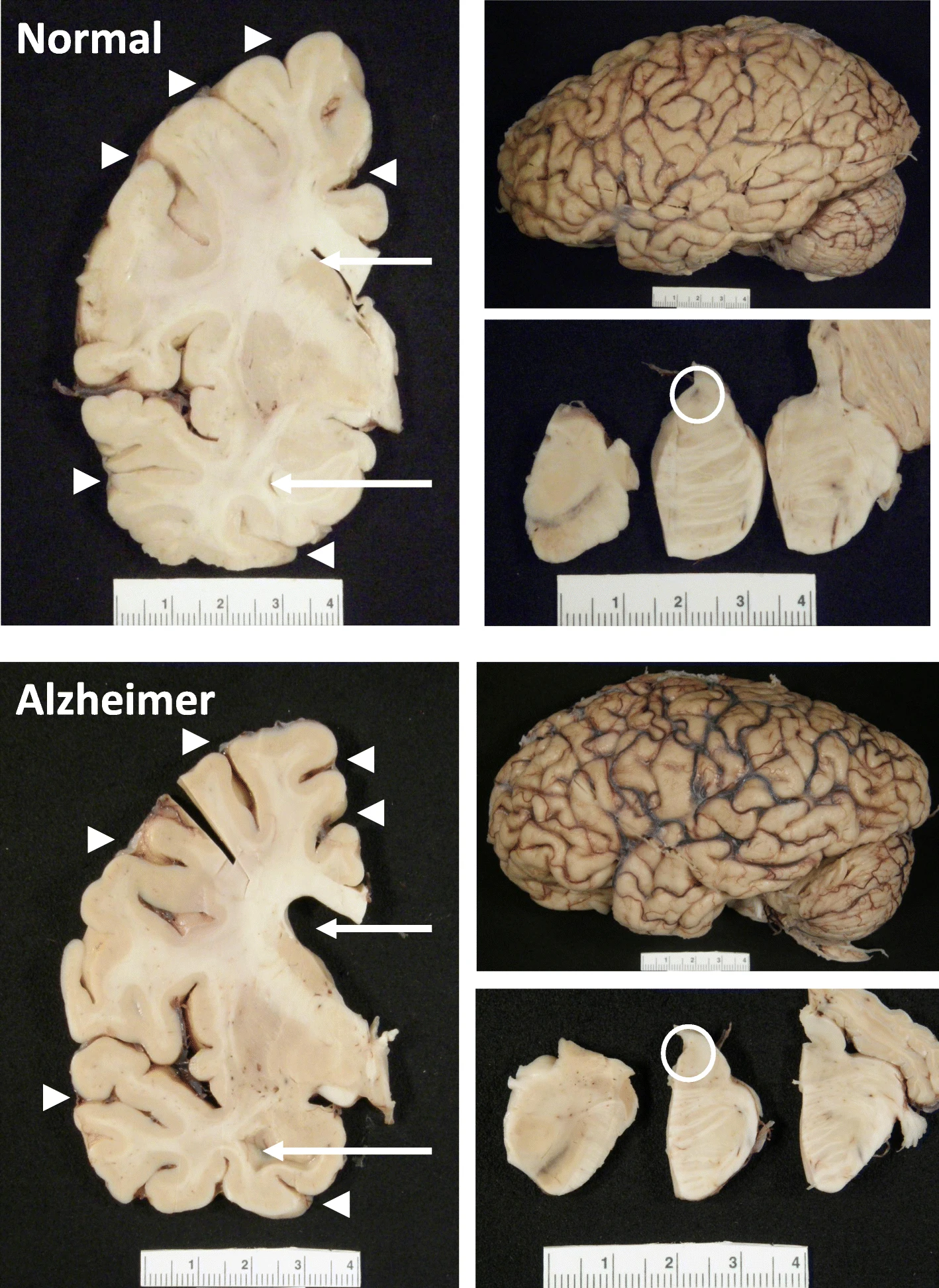
The hippocampus, a small, seahorse-shaped structure buried deep within the brain, is best known for its role in memory formation and learning. It is an exceptionally vulnerable structure, with perfusion deficits often observed in diseases related to learning and memory. However, a brain affected by Alzheimer's disease tends to exhibit at least moderate cortical atrophy, including in the precuneus and posterior cingulate gyrus. It should be noted that the posterior cingulate gyrus is adjacent to the hippocampus.

A recent study shows a relationship between blood flow and mechanical stiffness (an MRI concept) of the hippocampus. Researchers sought to understand how these physical properties interact in a healthy brain and what this might reveal about early brain changes in neurodegenerative diseases. The researchers used two advanced MRI techniques—magnetic resonance elastography (MRE) and arterial spin labeling (ASL).

Using these tools, the researchers measured: * Tissue stiffness (the resistance of an area to physical deformation) * Perfusion (blood flow at the tissue level)

Seventeen healthy adults were examined by the researchers at two different MRI intensities (3T and 7T), allowing for a cross-comparison between the two magnetic field strengths. They found that the hippocampus had the highest blood flow among the deep gray matter structures, followed closely by the caudate nucleus and putamen.

A strong positive correlation was observed between blood flow and stiffness in the hippocampus, but not in the caudate nucleus, although both regions are highly vascularized. This indicates that good brain health appears to be linked to good blood flow, manifested by the good stability (stiffness) of the tissue.

In a subgroup of ten subjects, it was found that higher blood flow resulted in larger tissue pulsations, suggesting that the dynamics of blood supply physically influence the hippocampus.

These results suggest a previously underestimated link between the physical characteristics of well-perfused brain tissue and its metabolic needs. This connection is not entirely surprising, as the macroscopic characteristics of tissue depend on the well-being of its constituent cells.

Although not mentioned in the article, one might wonder about the relationship between this mechanical property—rigidity—and beta-amyloid (Aβ), the signature protein implicated in Alzheimer's disease.

Beta-amyloid plaques begin to accumulate in the brain well before the onset of symptoms. These plaques can form: * Extracellularly: outside neurons, interfering with cell-to-cell communication and nutrient supply; * Intracellularly: inside neurons and other nerve cells, disrupting protein production.

Based on current knowledge, beta-amyloid likely appears before physical changes in tissues. It emerges early, sometimes decades before cognitive symptoms, triggering a cascade of tissue changes. Mechanical stiffness, as measured by MRE, is more likely a result of these changes than a cause.

Interestingly, numerous MRE studies have observed brain softening, particularly in the hippocampus and cortex, in Alzheimer's patients and those with mild cognitive impairment. This supports the perspective that stiffness decreases due to beta-amyloid pathology and its effects on brain tissue structure.

As fascinating as these findings are, it is important to acknowledge the limitations of the study, which suggest future research directions.

With only 17 participants, the study lacks statistical power, making it vulnerable to false positives or exaggerated effect sizes.

All subjects were young adults (22–35 years old) who were generally healthy, limiting the relevance of the results to aging populations or those at risk for Alzheimer's disease.

The sample did not include key groups, either clinical or high-risk individuals, such as APOE4 gene carriers or those with mild cognitive impairment (MCI).

The study was cross-sectional, capturing a single snapshot in time. We do not yet know how stiffness or perfusion might change over time or in response to pathology.

No cognitive data were collected; therefore, the relationship between hippocampal mechanics and actual memory performance remains unexplored.

There are considerable interpretation challenges: Stiffness, measured by magnetic resonance elastography (MRE), reflects a complex array of biological factors: neuronal density, inflammation, vascular integrity, etc. It is a valuable signal, but not biologically specific. Indeed, perfusion can vary depending on common physiological factors (e.g., hydration, stress).

Because the study did not include beta-amyloid PET scans or fluid biomarkers, the link between mechanical findings and Alzheimer's pathology remains hypothetical.

The analysis focused on the hippocampus and a few other deep gray matter structures. Key cortical regions involved in Alzheimer's disease (such as the entorhinal cortex or precuneus) were not examined.

The emerging link between perfusion and mechanics, and how this relationship deteriorates in the presence of beta-amyloid, could help us uncover subtle clues that precede cognitive decline. Ultimately, measuring something as simple as brain "firmness" could help us identify those at risk and determine when to act.

Skin pathology in ALS

- Posted by admin in English

Many ALS patients have noticed that their patients seem to share a particular skin type. Studies have shown that ALS patients often exhibit small fiber neuropathy in the skin, contributing to symptoms such as impaired thermoregulation, abnormal sweating, and sensory disturbances (e.g., numbness, and pain). Similar skin changes have been observed in diseases such as Parkinson's disease and Alzheimer's disease, suggesting that skin biomarkers could contribute to the early diagnosis and monitoring of ALS.

The article reviewed here is a review of this phenomenon, which rarely receives scientific attention. While the focus of the article is on early diagnosis of ALS, scientists, and physicians are not necessarily pleased that ALS is a disease far more complex than motor neuron disease, as this makes it difficult to conceptualize and makes the design of therapeutic strategies more challenging.

One factor that may explain this is that the skin and the nervous system share a common embryonic origin. The skin is composed of the epidermis, dermis, subcutaneous tissue, and appendages (such as sweat and sebaceous glands). In patients with ALS, the skin exhibits a soft, leathery texture, as well as a phenomenon called delayed return (DRP). enter image description here In healthy individuals, after a deformation or pinching, the skin quickly returns to its original shape. In patients with ALS, this return is slower. This is called the delayed return phenomenon (DRP).

In the context of ALS, DRP has been associated with abnormalities in the dermal connective tissue, such as altered collagen composition. Microscopic examination reveals fewer and less organized collagen bundles and increasing gaps in the connective tissue. Electron microscopy shows the progressive deposition of fine materials in the dermal matrix, disrupting collagen fibers and connective tissue integrity. These changes reduce the skin's resilience and elasticity, making it softer and slower to regenerate.

ALS patients also exhibit decreased sweat gland nerve fiber density (SGND) and pilomotor nerve fiber density (PNF).

Histological studies show thickening of the walls of small dermal blood vessels, particularly in sporadic ALS (sALS). Electron microscopy reveals onion-like structures formed by β-amyloid deposits and basement membrane duplications, reducing the surface area of ​​the vascular bed. This vascular remodeling, particularly in the papillary layer, may be linked to changes in autonomic innervation and contribute to preventing pressure ulcers.

One of the culprits for this state of affairs could be MMP-9, which belongs to the matrix metalloproteinase (MMP) family. Metalloproteinases degrade extracellular matrix components such as collagen. Proteins of the matrix metalloproteinase (MMP) family are involved in the restructuring of the extracellular matrix in processes such as embryonic development, wound healing, learning, and memory, as well as in pathological processes such as asthma, arthritis, intracerebral hemorrhage, and metastases.


Please, help us continue to provide valuable information: