How the gut microbiome influences insulin resistance.

- Posted by admin in English

Alzheimer's disease, Parkinson's disease, ALS, and FT Dementia share many biomarkers and comorbidities. One of them is insulin resistance which is found in half of patients.

Indeed insulin resistance is also a consequence of diabetes and aging. Scientists from Japan explore in a recent article how the gut microbiome influences insulin resistance. enter image description here 
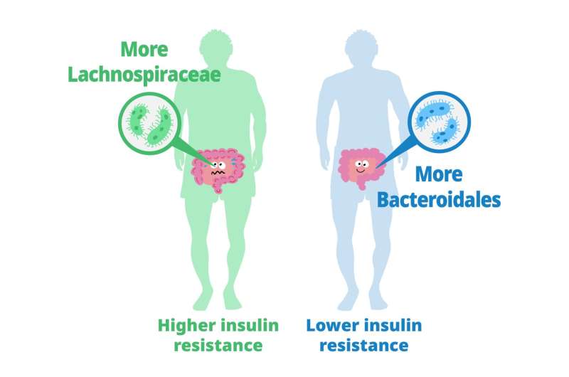
Previous studies have explored the role of gut microbiota in metabolizing nutrients in insulin resistance. This research aims to uncover the mechanisms underlying this relationship using a multi-omics approach. The study analyzes data from 306 individuals without diabetes, focusing on insulin resistance as defined by HOMA-insulin resistance scores.

The researchers used various techniques, including metabolomics, metagenomics, transcriptomics, and clinical data, to profile how the gut microbiome contributes to insulin resistance.

They found that certain carbohydrates in the feces, particularly those accessible to the host, are elevated in individuals with insulin resistance. These carbohydrates are linked to microbial carbohydrate metabolism and host inflammatory cytokines. Specific gut bacteria are associated with insulin resistance and insulin sensitivity, each displaying distinct carbohydrate metabolism patterns. In a mouse model, bacteria linked to insulin sensitivity demonstrate the potential to improve insulin resistance traits.

The study also involves analyzing metabolic syndrome (MetS) and its associations with fecal and plasma metabolites. Using human fecal cultures, the researchers discovered that Bacteroidales, a type of gut bacteria linked to insulin sensitivity, have a unique metabolic profile. These bacteria are efficient consumers of certain carbohydrates, affecting the production of fermentation products.

To explore causality, the researchers test the effects of seven candidate bacteria associated with insulin sensitivity on mice fed a high-fat diet. Several strains, notably Alistipes indistinctus, show promising results in reducing postprandial blood glucose levels and improving insulin resistance. These strains also impact body mass, lipid accumulation, and glucose intolerance.

Mechanistically, the researchers find that A. indistinctus administration reduces carbohydrate oxidation in mice, possibly due to decreased host-accessible carbohydrates in the intestine. This is supported by altered caecal metabolites, including reduced monosaccharides like fructose.

In conclusion, the study employs a comprehensive multi-omics strategy to investigate the relationship between gut microbiota and insulin resistance. It identifies specific bacteria associated with insulin resistance and sensitivity and highlights the potential of A. indistinctus in ameliorating insulin resistance in mice. However, further research is needed to understand the precise mechanisms and potential therapeutic implications of these findings.

Longevity factor klotho enhances cognition in aged Rhesus primates

- Posted by admin in English

Klotho is a protein that has been of interest in scientific research due to its potential role in aging and age-related diseases. It is believed to have several beneficial effects on various organ systems, including the brain, kidneys, and cardiovascular system. enter image description here Until now, studies on Klotho have primarily focused on its effects on animal models such as mice, rather than directly increasing its levels in primates.

This new study by scientists from the University of California, explores the potential of using the protein Klotho to enhance cognition in nonhuman primates, specifically rhesus macaques. Klotho declines with aging, and previous research has shown that increasing Klotho levels can improve cognitive function in mice. So the scientists wanted to verify whether this effect could be replicated in nonhuman primates, which have a higher genetic and functional complexity more similar to humans.

The researchers first validated the activity of the rhesus form of the Klotho protein (a variant of the Klotho protein that is found in rhesus macaques) in mice, demonstrating that it increased synaptic plasticity and cognition. They then tested different doses of Klotho in aged rhesus macaques and found that a single low-dose administration enhanced memory performance. Interestingly, higher doses did not produce the same cognitive benefits. The results suggest that systemic low-dose Klotho treatment may have therapeutic potential for aging humans.

The study emphasizes the importance of studying animal models with greater complexity, such as nonhuman primates, in the development of cognitive treatments for humans. By demonstrating the cognitive benefits of Klotho in rhesus macaques, the researchers bridge a knowledge gap and provide evidence for the potential translation of this treatment to humans.

There are several known ways to potentially increase Klotho levels in the body. Here are a few approaches that have shown promise:

  • Exercise: Regular physical exercise has been associated with increased Klotho levels.

  • Caloric restriction.

  • Pharmaceutical interventions: Certain drugs and compounds have been explored for their potential to increase Klotho levels.

  • Dietary factors: Some studies have suggested that certain dietary factors may influence Klotho levels. For example, diets rich in fruits, vegetables (rhubarb), and whole grains have been associated with higher Klotho levels.

  • Hormonal interventions: Hormonal factors, such as vitamin D and sex hormones (e.g., estrogen), have been implicated in Klotho regulation.

Yet it's not clear what is the mechanism of action of klotho on cognition. It must be stated also that there be nasty side effects to elevated levels of klotho like risks of weight gain, cancer or insulin resistance.

Improving sleep quality in neurodegeneration

- Posted by admin in English

A new publication in the Journal of Neuroscience, by Mount Sinai researchers, discusses a study conducted by researchers at Mount Sinai, which identifies a new form of treatment for rapid eye movement (REM) sleep behavior disorder. enter image description here This condition affects individuals, mostly adults over the age of 50, who physically act out their dreams during sleep, resulting in injuries to themselves or their bed partners. It's also suspected to be involved in premisses of Parkinson's disease. The study, published in the Journal of Neuroscience, presents a novel model that characterizes how REM sleep behavior disorder develops due to neurodegeneration associated with the accumulation of tau protein.

Tauopathies are a group of neurodegenerative disorders characterized by the abnormal accumulation of tau protein in the brain. DORAs have shown promise in preclinical studies as potential treatments for tauopathies, including diseases like progressive supranuclear palsy (PSP) and frontotemporal dementia with tau pathology.

DORAs have been found to modulate the activity of the orexin system, which is involved in regulating tau pathology. By targeting the orexin system, DORAs may potentially influence tau-related neurodegeneration and associated symptoms. However, further research is needed to fully understand the mechanisms and clinical implications of DORAs in tauopathies.

Using a mouse model, the researchers examined the effects of abnormal tau deposits on brain function and sleep. They analyzed various sleep-related factors, including wakefulness, REM sleep, non-REM sleep, sleep duration, and transitions between sleep stages. The study found that a significant number of older mice exhibited dream enactment behaviors similar to REM sleep behavior disorder, such as chewing and limb extension.

The researchers then administered a type of sleep medication called dual orexin receptor antagonists (DORAs) to evaluate their effects on sleep in the mice. They discovered that the medication not only improved sleep quality and duration but also significantly reduced dream enactment behaviors.

Orexin receptor antagonists (ORAs) are a class of medications that target the orexin system in the brain. Orexins, are neuropeptides that play a crucial role in regulating wakefulness and arousal. They promote wakefulness by activating orexin receptors in cells of the brain.

When orexin receptors are blocked or antagonized by medications, such as dual orexin receptor antagonists (DORAs), it leads to a decrease in wakefulness and an increase in sleepiness. DORAs are primarily used to treat insomnia, as they promote sleep initiation and maintenance.

In terms of sleep architecture, orexin receptor antagonists have been found to affect different sleep stages and sleep characteristics. Here are some of the effects observed:

  • Increased total sleep time, allowing individuals to obtain a longer and more restful sleep.

  • Increased non-rapid eye movement (NREM) sleep), which is deep and restorative sleep, and lighter stages of NREM sleep.

  • Decreased rapid eye movement (REM) sleep: DORAs have been found to suppress REM sleep, the stage associated with vivid dreaming and increased brain activity. This reduction in REM sleep may be beneficial in certain sleep disorders, such as REM sleep behavior disorder.

  • Improved sleep continuity: DORAs can enhance sleep continuity by reducing the number of awakenings during the night and decreasing the time spent awake after sleep onset. This leads to more consolidated and uninterrupted sleep.

It's important to note that the effects of orexin receptor antagonists on sleep architecture may vary depending on the specific medication, dosage, and individual factors.

These findings suggest that DORAs, which are already FDA-approved for treating insomnia, could serve as a promising new treatment for REM sleep behavior disorder. The researchers hope that their study will encourage further clinical trials of DORAs in humans with this disorder.

Overall, the study provides insights into the effects of neurodegeneration and tau protein accumulation on sleep and offers a potential therapeutic approach for managing REM sleep behavior disorder.

RAGE Inhibitors in Neurodegenerative Diseases

- Posted by admin in English

This post is about a review of the contribution of Advanced Glycation Endproducts to Neurodegenerative Diseases.

Advanced Glycation Endproducts (AGEs) are formed through non-enzymatic reactions between proteins, aminoglycosides, amino-terminal lipids, and reducing sugars like D-glucose. This process involves Amadori rearrangements and oxidative modifications. The accumulation of AGEs, especially under conditions of elevated oxidative stress, leads to various diseases. enter image description here 
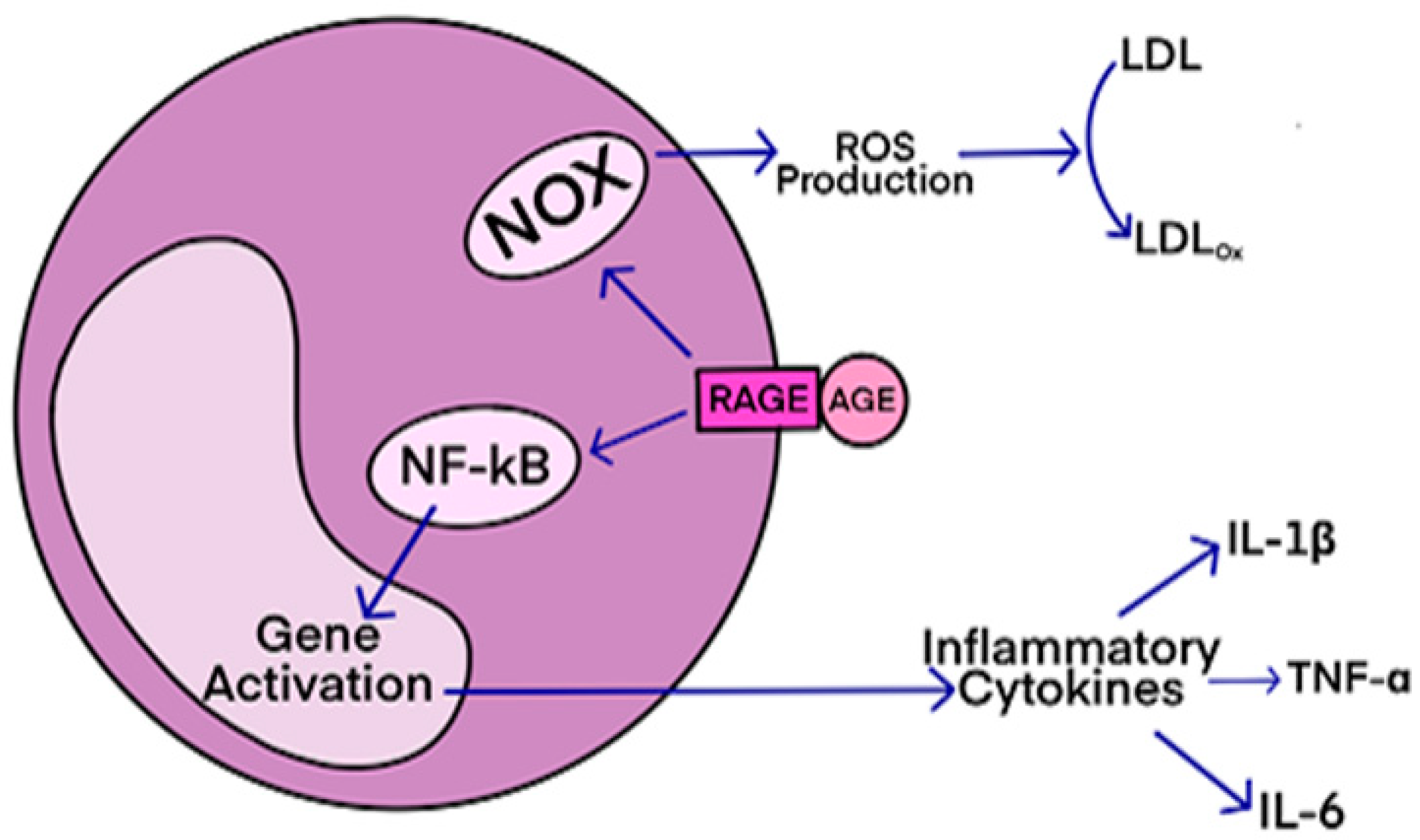
AGEs have diverse structures, but only a limited number have been characterized. Some AGEs are small molecules formed through proteolytic degradation of protein-crosslinked or protein-modified AGEs. Imbalance between the formation and destruction of AGEs, particularly under conditions of oxidative stress, results in excessive accumulation and disease progression.

Some of the well-characterized AGEs include pentosidine, glucosepane, Argpyrimidine, and Nε-(carboxymethyl)lysine (CML). The imbalance between the formation and destruction of AGEs, triggers a cascade of signaling events, inflammation, and oxidative stress. This inflammatory signaling cascade is associated with various neurological diseases, including Alzheimer’s disease (AD), amyotrophic lateral sclerosis (ALS), diabetic neuropathy, diabetes, and atherosclerosis.

Other endogenous ligands are also involved, such as high mobility group box1 (MGB1) proteins. Additionally, exogenously ingested AGEs contribute to disease onset. The formation and accumulation of AGEs overwhelm the body's detoxification mechanisms under conditions of enhanced oxidative stress, exacerbating neurodegenerative diseases and other inflammatory-associated conditions.

RAGE (receptor for advanced glycation end-products) is a receptor that interacts with AGE (advanced glycation end-products)-derived ligands, such as CML, CEL, and MG-H1. The ligands bind to specific residues on the receptor, initiating signal transduction and pro-inflammatory signaling events.

RAGE antagonists, either endogenous or exogenous compounds, bind to RAGE and attenuate the binding interactions between AGE and RAGE, thereby preventing disease progression. These antagonists have shown potential for treating neurodegenerative diseases, diabetes, atherosclerosis, and cancers. Some small molecule-based RAGE inhibitors, like FPS-ZM1 and Azeliragon, have entered clinical trials, but none have been FDA-approved yet.

FPS-ZM1 has shown selective binding to RAGE and inhibits the formation of Aβ peptides, which are associated with brain damage in diseases like Alzheimer's. RAGE inhibitors also have potential therapeutic applications in diabetic nephropathy, cancer cell metastasis, and Parkinson's disease.

Azeliragon, currently in phase 3 clinical trials, has demonstrated decreased levels of Aβ plaques in the brain, reduces inflammation, and slower cognitive decline in Alzheimer's patients. Other compounds, such as urolithin and its analogs, have shown comparable RAGE inhibition to Azeliragon.

RAGE antagonists are also considered as therapeutics for diabetic neuropathy and retinopathy. Inhibitors of the cytoplasmic tail of RAGE (ct-RAGE) have been investigated and shown to block AGE/RAGE-mediated signaling events effectively. Some of these small molecule antagonists have structural similarities to FPS-ZM1 and have demonstrated potential for treating neurological disorders and diabetic complications.

Dietary AGEs, especially from animal-derived foods cooked at high temperatures, can also contribute to AGE accumulation and the development of diseases. RAGE antagonists, AGE inhibitors, and soluble RAGE (sRAGE) have shown promise in the treatment of age-related pathologies. Polyphenolic compounds can attenuate AGE formation and its toxic effects by reducing oxidative stress.

Therapeutic interventions targeting the AGE-RAGE axis may provide effective treatment for neurodegenerative diseases, diabetes, atherosclerosis, and cancers.

In summary, RAGE antagonists show promise as therapeutics for various diseases by attenuating the binding interactions between AGE and RAGE and preventing downstream pro-inflammatory signaling events. Clinical trials are underway for several RAGE inhibitors, and they have shown potential for treating Alzheimer's, diabetes, and other AGE-related diseases.

Overall, understanding the formation, toxicity, and interactions of AGEs with RAGE is crucial for developing therapies to combat age-related diseases.

Le sommeil est important pour maintenir la santé du cerveau. Pendant le sommeil, le cerveau élimine les déchets métaboliques, les protéines et les débris cellulaires qui s'accumulent pendant l'activité cérébrale diurne. Ceet élimination se fait via grâce à des systèmes interconnectés d'activité neuronale et de flux de liquide interstitiel et céphalo-rachidien. enter image description here Des études précliniques ont montré que pendant le sommeil, l'infiltration du liquide interstitiel et céphalo-rachidien le long des espaces périvasculaires augmente, augmentant ainsi la clairance des solutés interstitiels. Lorsqu'ils sont agrandis, les espaces périvasculaires sont visibles par imagerie par résonance magnétique cérébrale (IRM). Le volume et le nombre de espaces périvasculaires augmentent avec l'âge. Ce phénomène est associés à des facteurs de risque vasculaire tels que l'hypertension, des marqueurs de microangiopathie telle que la leucoaraiose et entraine des effets néfastes sur la santé. Une question importante est de savoir si le manque de sommeil est la cause de ces risques vasculaires, ou simplement leur conséquence.

La microangiopathie est une angiopathie qui atteint des vaisseaux sanguins de petits calibres. Elle est souvent une complication du diabète et de l'hypertension artérielle. La présence de leucoaraiose sur l'IRM a également été associée à un déclin fonctionnel, à des troubles de la marche et à la dépression, peut-être parce qu'ils perturbent les réseaux neuronaux. enter image description here

Souvent, les leucoaraiose sont dus à un processus appelé infarctus incomplet, c'est à dire une réduction chronique du flux sanguin dans les zones profondes du cerveau causée par l'artériolosclérose, la lipohyalinose ou la nécrose fibrinoïde des petites artères et artérioles cérébrales. Un tel flux sanguin réduit conduit à l'hypoxie, altère les mécanismes d'autorégulation cérébrale et favorise la transcription des gènes inflammatoires, la rupture de la barrière hémato-encéphalique et l'entrée de protéines pro-inflammatoires dans les parois des vaisseaux et le parenchyme cérébral. Même en l'absence d'infarctus franc, ces processus entraînent une démyélinisation, une perte axonale, une réduction de la densité gliale, une vacuolisation et une atrophie du cortex sus-jacent.

En outre, d'autres processus pouvant être impliqués dans la genèse des leucoaraiose comprennent le dysfonctionnement des cellules précurseurs des oligodendrocytes, la défaillance du système glymphatique, la collagénose veineuse. Les études IRM qui utilisent des techniques telles que l'imagerie du tenseur de diffusion et le transfert d'aimantation pour examiner la diffusivité de l'eau et l'intégrité de la substance blanche montrent que certains des changements physiopathologiques précoces se produisent également dans les zones de la substance blanche qui semblent normales sur l'IRM conventionnelle. Ces études IRM des études suggèrent que les leucoaraiose visibles ne sont «que la pointe de l'iceberg» et que la physiopathologie sous-jacente est un processus diffus affectant les petits vaisseaux sanguins dans une grande partie de la substance blanche et d'autres parties du cerveau.

Les marqueurs de la qualité du sommeil ont été associés à plusieurs indicateurs structurels de la détérioration de la santé cérébrale, à savoir les hyperintensités de la substance blanche (Leucoaraiose) d'origine vasculaire présumée, les microhémorragies et la perte de tissu cérébral d'apparence normale, tous évalués à l'aide IRM. Cependant, les études démontrant des associations causales définitives entre le sommeil et la santé des tissus cérébraux au fil du temps chez les personnes âgées vivant dans la communauté font défaut.

Des scientifiques Ecossais essayent de répondre à cette question dans une nouvelle publication, pour cela ils utilisent les données longitudinales des personnes âgées vivant dans la communauté de la Lothian Birth Cohort 1936. Les participants à l'étude sont donc des personnes âgées toutes nées en 1936 à Édimbourg et dans les environs (Lothian). Le Lothian est une région d'Écosse où la langue des Lothiens et de l'ancien royaume de Northumbria est encore pratiquée. La Lothian Birth Cohort 1936 est une vaste étude basée sur la population du vieillissement cognitif, cérébral et général. Les auteurs se sont intéressés à toute association entre la durée, la qualité et l'efficacité du sommeil autodéclarées, la charge espaces périvasculaires au début de la 8e décennie de la vie et la détérioration des volumes cérébraux normaux et anormaux au cours de la décennie, à l'aide de l'IRM structurelle.

Là où des associations existent, les scientifiques ont cherché à savoir si la charge espaces périvasculaires influence ou non l'association entre le sommeil et les changements structurels du cerveau sur une période de temps. Ils ont testé trois hypothèses: (1) les mesures du sommeil serait associées indépendamment à une détérioration ultérieure du tissu cérébral, (2) la charge espaces périvasculaires serait associée à une détérioration du tissu cérébral, (3) l'association entre le sommeil et les changements structurels du cerveau serait en partie médiée par le espaces périvasculaires fardeau.

Dans ce grand échantillon d'individus, le fardeau des espaces périvasculaires élargies visibles dans l'examen IRM du cerveau à l'âge d'environ 73 ans était associé à une aggravation ultérieure de la santé de la substance blanche. Celle-ci était reflétée par une diminution plus rapide du volume de substance blanche d'apparence normale, une plus grande augmentation de leucoaraiose et de la «mesure des dommages à la substance blanche» des années 73 à 79.

Les personnes qui ont signalé une moins bonne efficacité du sommeil et plus de sommeil diurne étaient celles qui ont également connu une baisse significativement plus importante de la santé de leur substance blanche entre 73 et 79 ans, et leurs résultats montrent que cela pourrait être en partie médié par la charge espaces périvasculaires.

Ni la charge espaces périvasculaires, ni la qualité du sommeil ou les indicateurs d'efficacité n'ont été associés à une détérioration du volume de tissue cérébral ou des changements de matière grise au cours de la même période.

Une analyse longitudinale récente à 5 points dans le temps sur 28 ans dans la même cohorte avait auparavant identifié quatre groupes de trajectoires différents avec des durées moyennes de sommeil différentes allant d'environ 5 à 8 h, mais n'avait trouvé aucune différence significative dans la microstructure de la matière blanche ou les volumes de matière grise entre les groupes.

Une autre étude à la fin de l'âge adulte [60–82 ans] avait déjà révélé que le nombre de fois où les participants ont signalé une mauvaise qualité de sommeil sur cinq points temporels couvrant une période de 16 ans avant l'analyse, n'était pas associé à ces mesures de la substance blanche, suggérant ainsi que seule la qualité actuelle du sommeil affectait la substance blanche.

Avec cette étude, les résultats peuvent indiquer soit un effet à court terme du manque d'efficacité du sommeil sur la santé de la substance blanche à la fin de l'âge adulte, soit que l'altération des habitudes de sommeil chez les personnes âgées peut être liée à des changements cérébraux sous-jacents mais ne provoque pas nécessairement les changements cérébraux.

Bien que l'association entre les espaces périvasculaires élargi et le leucoaraiose ait été rapportée, l'étude actuelle suggère qu'en outre, une augmentation des espaces périvasculaires visible (en volume, en nombre ou en scores visuels) prédit également le taux d'atrophie de la substance blanche et la structure de la substance blanche. dommages à la fin de l'âge adulte, mais pas de changements de volume total du cerveau et de la matière grise.

Les troubles du sommeil sont associés à une détérioration plus rapide de la santé de la substance blanche chez les septuagénaires. Une charge plus élevée des espaces périvasculaires est associée à une détérioration plus rapide de la santé de la substance blanche chez les septuagénaires. Les espaces périvasculaires interviennent en partie dans l'effet du sommeil sur la santé du cerveau.

Le principal apport de cette étude semble donc être que c'est la détérioration de la santé vasculaire, plutôt que le manque de sommeil, qui amène la détérioration de la matière blanche.

Cependant, il faut faire preuve de prudence en ce qui concerne le manque apparent d'association avec la matière grise et les taux d'atrophie totale du tissu cérébral. Le logiciel utilisé pour générer des volumes de matière grise, FSL-FAST, bien que considéré comme un étalon-or pour la segmentation des tissus d'apparence normale, ne discrimine pas la matière grise saine de la matière grise malsaine.

L'absence d'association entre les paramètres du sommeil et l'atrophie de la matière grise est moins surprenante.

Une étude précédante examinant la qualité et la quantité du sommeil en relation avec les changements corticaux avait indiqué que la qualité du sommeil et les troubles du sommeil autodéclarés étaient liés à l'amincissement du cortex temporal latéral droit.

Aucune information sur l'apnée du sommeil n'a été recueillie, mais dans les dossiers médicaux, elle n'a été signalée que chez un seul participant.

Quadruple Misfolded Proteins in Older Adults

- Posted by admin in English

In this cohort study, Shama Karanth and colleagues from University of Kentucky, Lexington, investigated quadruple misfolded proteins and other proteinopathy combinations in a cohort of 375 deceased individuals with autopsy data in the University of Kentucky Alzheimer Disease Center (UK-ADC).

At least 3 proteinopathies were observed in 50% of brains.

In addition, quadruple misfolded proteins were associated with severe cognitive impairment at least 12 years before death.

Participants with 3 proteinopathies tended to have poorer global cognition earlier than with the presence of only tau and Aβ and were likely to have higher Braak stages.

Previous studies have reported cognitive decline associated with the presence of mixed pathologies, with study to study differences in methods and proteinopathies, the assessment and inclusion of cerebrovascular pathologies, and hippocampal sclerosis. In the present sample, as in other community based cohorts, FTLD in old age was rare (with an estimated incidence of 8.9 of 100 000 in individuals aged 60 to 69 years; no incidence data are available for older age groups) and was not found in brains of individuals who began follow up with normal cognition.

Individuals with FTLD-TDP-43 with data in the UK-ADC Brain Bank were recruited from a dementia clinic. The scientists excluded 6 individuals with FTLD-TDP-43 in the study; none had the quadruple misfolded proteins phenotype. No discernible overlap in any FTLD feature was observed in these individuals other than presence of TDP-43 proteinopathy, which is now detected in many different neurological diseases outside of the amyotrophic lateral sclerosis–FTLD spectrum.

Cognitive impairment was associated with quadruple misfolded proteins at autopsy, with 89.1% of participants developing dementia and some experiencing profound impairment up to 12 years before death. This finding suggests that quadruple misfolded proteins occur before end stage Alzheimer disease neuropathological change (ie, before high Braak stage).

Consistent with this hypothesis, the mild cognitive impairment to dementia transition was, on average, fastest in the quadruple misfolded proteins group. Estimation of the group cognitive trajectories was aided by the relatively long followup (mean duration of 10.4 years).

These data provide the basis for a novel hypothesis that quadruple misfolded proteins have a more aggressive phenotype from the early stages of the disease rather than accruing additional pathologies only after Alzheimer disease neuropathological change has progressed to high levels. About 10% of these participants died with normal cognition, and previous research has shown quadruple misfolded proteins were present in persons with apparently normal cognition. In the present study, all individuals with quadruple misfolded proteins who had normal cognition at the last visit before death had lower Braak NFT stages (I to III), had no APOEε4 allele, and were predominantly male (4 of 5 participants).

These individuals may represent an early stage of quadruple misfolded proteins, but there are complexities: clinical presentation of proteinopathy combinations may be cohort specific and depend on other currently unknown factors. Older cohorts that survive into advanced old age, like those in the UK-ADC study, may be more likely to experience multiple proteinopathies than younger cohorts. As previously described, APOE appeared to be associated with multiple proteinopathies in this study, particularly those proteinopathy combinations including Aβ plaques. Carriers of APOEε allele not only had increased odds of tau and Aβ, an expected result, but also had higher odds of tau, Aβ, and αsynuclein; tau, Aβ, and TDP-43; and quadruple misfolded proteins.

Unlike previous studies, this study did not find evidence that the ε4 allele was associated with tau or TDP-43 in the absence of Aβ, but the sample size was relatively small. The temporality of protein misfolding may play a clinically important and differentiating role in disease progression.

Autopsy data, although cross-sectional by nature, are compatible with the hypothesis that Aβ aggregates precede, and perhaps stimulate or exacerbate, the widespread misfolding of tau, TDP-43, and α-synuclein. These results suggest that TDP-43 pathology may be associated with poor global cognition.

The presence of multiple proteinopathies, particularly the quadruple misfolded proteins phenotype, appeared to have been associated with the cognitive decline in deceased individuals who participated in a longitudinal community based study at the UK-ADC.

These observations have potentially significant implications for clinical practice and public health, given that strategies to prevent or manage AD dementia may be complicated by the unrecognized presence of multiple additional neuropathologies.

Two epigenetic regulators interfere with healthy aging

- Posted by admin in English

It has long been assumed that lifespan and health are strongly correlated, but although there has been an overall increase in human life expectancy in recent decades, it is too often accompanied by deterioration of health.

A new study published on February 26 in Nature shows the influence of two epigenetic regulators on aging. Scientists led by Jie Yuan from the Chinese Academy of Sciences in Shanghai have studied the BAZ-2 and SET-6 proteins in Caenorhabditis elegans worms, which are orthologs of the human proteins BAZ2B and EHMT1.

Through genome-wide RNA-interference-based screening of genes that regulate behavioral deterioration in aging C. elegans, the researchers identified 59 genes as potential health modulators during aging. Essentially the proteins expressed by these genes, read and write epigenetic signals.

Among these modulators, they found that a neuronal epigenetic reader, BAZ-2, and a neuronal histone SET-6, accelerate the deterioration of the behavior of C. elegans by reducing the mitochondrial function, and repressing the expression of the encoded mitochondrial proteins. in the cell nucleus.

The researchers found that the levels of the two proteins increase with age in C. elegans and mice, which in turn attenuates the expression of genes involved in mitochondrial function.

BAZ-2 and SET-6 are complementary epigenetic mechanisms. SET-6 is an "epigenetic writer" and BAZ-2 is an "epigenetic reader" which recognizes modified histones and recruits transcriptional regulators.

Histones are proteins located in the nucleus of eukaryotic cells. They are the main protein components of chromosomes. They are closely associated with DNA and allow their compaction, but they also modify the expression of proteins by various epigenetic mechanisms known as the "histone code".

enter image description here Source Wikipedia.

How do BAZ-2 and SET-6 accelerate aging? The researchers found that the two proteins bind together to the promoter regions of more than 2,000 genes, and decrease their expression via methylation of histones. Among these target genes are many mitochondrial genes encoded nuclear. By suppressing the expression of these genes, BAZ-2 and SET-6 reduce oxygen consumption and ATP production, and decrease the critical stress responses that maintain mitochondrial proteostasis. The resulting metabolic slowdown discourages the worms from assimilating their food and they mate less.

This mechanism is conserved in the neurons of cultured mice and human cells. What about the orthologs of these epigenetic proteins in humans? A review of the databases shows that expression by human orthologs of the two proteins mentioned above, BAZ2B and EHMT1, increases with age and is positively correlated with the progression of Alzheimer's disease. Researchers have verified that ablation of BAZ-2 mouse ortholog Baz2b attenuates age-dependent body weight gain and prevents cognitive decline in aging mice.

enter image description here While wild-type mice grew fat with age, animals lacking both copies of the epigenetic reader Baz2b stayed trim, indicating improved mitochondrial function. [Yuan et al., Nature, 2020.]

However, it must be asked whether BAZ-2 and SET-6 would rather mediate age-related physiological adaptation, rather than the agents of aging itself. Indeed their action could reflect a mechanism of adaptation to a progressively more hostile biological environment.

The scientists here investigated the feasibility of using wearable actigraphy in agitated late-stage dementia patients.

Read the original article on Pubmed

The aim of this study was to investigate the role of support from the social environment for the life expectancy in people with dementia beyond well-established individual demographic and clinical predictors over a period of up to 8 years.

Read the original article on Pubmed

This study aimed to develop and validate clinical prediction models using machine learning algorithms for reliable prediction of subsequent hip fractures in older individuals, who had previously sustained a first hip fracture, and facilitate early prevention and diagnosis, therefore effectively managing rapidly rising healthcare costs in China.

Read the original article on Pubmed


Please, help us continue to provide valuable information: