About another ridiculous claim in Alzheimer research

- Posted by admin in English

Another article from a university's public relations department claims that a major breakthrough has been made in Alzheimer's disease. There is one of these crazy articles every day. enter image description here Source: Peta

This time, the bar is set very high by the public relations department: "Our research demonstrates that by targeting synaptic activity early, we may be able to prevent or slow the progression of Alzheimer's. This could revolutionize the way we approach treatment for this disease" noted Drs. Miranda Reed and Michael Gramlich.

Since Alzheimer's is characterized by significant loss of brain matter, we expect at least an article showing the genesis of new neurons in several model animals of different species. Showing a change in very different species gives hope that the action of a drug will be as effective on humans.

Alzheimer's disease scientists usually work on mouse models of the disease 3xTg mice that declare the disease at the age of 8 months. This age is convenient for academics to use the free labor of students, but in a human, it is about 25 years old. This is very young to model Alzheimer's disease.

But what makes me jump is that all that is measured by these scientists is the change in synapses in an in vitro culture of 3xTg mice!

In fact, using in-vitro culture of cells from an animal model is very convenient, no need to raise mice! But the odds this is translatable to humans are minuscule. Nobody does this in preclinical studies. The claims of a revolution in the treatment of human Alzheimer's patients are extremely ridiculous.

In addition, the names of authors Michael Gramlich and Miranda Reed appear in all sorts of publications, most of which have nothing to do with neurodegenerative diseases.

Additionally, one of the authors of this article is the CEO of Biohaven Pharmaceuticals. Another author is the Chief Medical Officer of this company, and while he is a medical doctor, he is also not an Alzheimer's specialist.

Running induces neurogenesis in mice

- Posted by admin in English

A great dream in neurodegenerative diseases is to regenerate the brain's cells. Alas, half of the brain's cells are neurons and they do not divide, except for a tiny portion of them which are located in a small structure deep in the brain.

Neurogenesis is most active during embryonic development and is responsible for producing all the various types of neurons of the organism, but it continues throughout adult life in a variety of organisms. Once born, neurons do not divide (mitosis), and many will live the lifespan, except under pathogenic circumstances.

In humans, adult neurogenesis has been shown to occur at low levels compared with development, and in only three regions of the brain: the adult subventricular zone (olfactory sense) of the lateral ventricles, the amygdala emotion, socialization) and the dentate gyrus of the hippocampus (inhibition of impulses, episodic memory, and spatial cognition). Neurogenesis in the human hippocampus decreases sharply after the first years. The hippocampus is a neural structure located deep inside the medial temporal lobe of the brain. It has a distinctive, curved shape that has been likened to the sea horse.

Prior research has demonstrated that there are conditions which increase the rate of neurogenesis, such as voluntary running or pharmacological treatment with memantine. On the contrary, conditions that decrease neurogenesis, such as natural aging or pharmacological treatment, have been shown to increase the expression density of perineuronal nets in the hippocampus's CA1 area. Perineuronal nets (PNNs) are extracellular matrix structures that regulate the excitability and potential for plasticity of the cells they surround. So it seems (but everything is complicated in biology) that the more perineuronal nets means the less neurogenesis.

Aerobic exercise has been shown to modulate short-term cognitive performance and long-term cognitive outcomes in many ways. In particular, running has been found to impact learning and memory ability in a timing-dependent manner. enter image description here Running before learning aids in the formation of new memories, yet, running after learning promotes the forgetting of recently acquired information!

One of the mechanisms that seems to contribute to this relationship between running and cognition is adult hippocampal neurogenesis, which increases with running. With increased neurogenesis, the excitability of the dentate gyrus changes, which has knock-on effects across the hippocampus.

Aerobic exercise has many effects on brain function, particularly in the hippocampus. Exercise has been shown to increase the rate of adult neurogenesis within the dentate gyrus and decrease the density of perineuronal nets in the hippocampus's CA1 area. The relationship between the rate of neurogenesis and the density of perineuronal nets in CA1 is robust; however, these studies only ever examined these effects across longer time scales, with running manipulations of 4 weeks or longer. With such long periods of manipulation, the precise temporal nature of the relationship between running-induced neurogenesis and reduced perineuronal net density in CA1 is unknown.

Here, the authors provided male and female mice with home cage access to running wheels for 0, 1, 2, or 4 weeks and quantified hippocampal neurogenesis and CA1 perineuronal net density. In doing so, the authors observed a 2-week delay before the increase in neurogenesis, coinciding with the delay before the decreased CA1 perineuronal net density. These results highlight the closely linked temporal relationship between running-induced neurogenesis and decreased perineuronal net expression in CA1.

If this article is about mice, one reader of this blog could speculate if running could improve the conditions of Alzheimer's or Parkinson's disease sufferers. Obviously it would be complicated to make those patients to run periodically. Most of them are disabled and some of them have nearly no physical activity. Maybe some adapted device like an exercise bike that is usable in a recumbent or lying position and under supervision by health professionals could be useful.

This is an interesting study on Alzheimer’s disease. Unlike the multitude of low-quality academic studies, here the authors explore the complexity of the central nervous system, which is not limited to neurons, and show that this population of neuronal and non-neuronal cells interact and change over time. enter image description here Currently, the diagnosis of Alzheimer’s disease includes cognitive decline leading to dementia, associated with the observation of two major proteinopathies in the brain tissue. These two proteinopathies are plaques, formed by the aggregation of beta-amyloid proteins, and tangles, which are composed of hyperphosphorylated tau proteins.

However, the interaction between amyloid, tau, and cognitive decline is complex.

These proteinopathies follow a stereotypical propagation throughout the brain during the disease. This has led to staging paradigms such as CERAD, Thal, and Braak.

In this new study, scientists reconstructed the changing dynamics of the brain’s cellular environment and identified a pathway to Alzheimer’s disease that is distinct from other aging-related effects.

The authors suggest that two different types of non-neuronal cells initiate the process of amyloid and tau accumulation that defines Alzheimer’s disease.

Once pathology has accumulated, different cells called astrocytes play a key role in altering the brain’s electrical connectivity leading to cognitive impairment. The cells communicate with each other and bring in additional cell types that lead to a profound disruption in human brain function.

To achieve this, the authors constructed a comprehensive cellular atlas of the aged prefrontal cortex from 1.65 million single-nuclear RNA sequencing profiles sampled from 437 older adults and identified specific glial and neuronal subpopulations associated with Alzheimer’s disease-related traits.

Modeling identified two distinct lipid-associated microglial subpopulations: - one drives amyloid-β proteinopathy - the other mediates the effect of amyloid-β on tau proteinopathy In addition, a subpopulation of astrocytes mediates the effect of tau on cognitive decline.

They designed the BEYOND methodology to model the temporal change in cellular environments. It identified two distinct trajectories of brain aging, each defined by coordinated progressive changes in certain cellular communities that lead to either Alzheimer's dementia or classical brain aging.

La restauration du métabolisme du glucose dans la maladie d’Alzheimer

- Posted by admin in Français

Pour ce blog, je recherche plutôt des articles scientifiques récents concernant des résultats sur des humains, les essais sur des animaux modèles n'étant quasiment jamais transposables aux humains. Dans les maladies neurodégénératives il n'y a quasiment aucun essai clinique ayant montré une régression des symptômes. Actuellement il y a un fort lobbying des acteurs de l'industrie pharmaceutique et des milieux universitaires pour faire dépendre la définition des maladies de marqueurs techniques plutôt que des symptômes cliniques.

La maladie d'Alzheimer est souvent liée à l'âge, et caractérisée par une perte progressive et irréversible des neurones, à la fois des corps (soma) des neurones et des appendices comme l 'axone et les synapses. La cause de cette dégénérescence est encore inconnue, quoique l'industrie pharmaceutique et le nombre d'académiques pensent que cette destruction est due à des peptides comme les bêta-amyloïdes, ou/et la protéine Tau ou encore TDP-43, comme dans la SLA et la FTD ou encore l'alpha-synucléine comme dans la maladie de Parkinson. La majorité des études n'étudie aussi que les neurones comme si nous étions toujours en 1980. Personnellement je crois plutôt, comme l'affirme un certain nombre d'études, qu'en vieillissant nous développons un ensemble de commorbidités. Quand nous sommes arrivés à 70 ans, nous avons tous un peu de maladies d'Alzheimer et de Parkinson.

Une nouvelle étude académique semble aller dans ce sens, mais évidemment elle est faite sur des modèles de souris de la maladie et pas sur des humains.

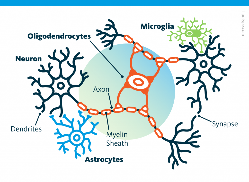
Pour les auteurs, une certaine molécule, IDO1, provoque un cercle vicieux de déclin du soutien du métabolisme cérébral du glucose par la production de marqueurs, amyloïdes β et tau chez les sujets atteints de la maladie d'Alzheimer. Les auteurs pensent que ces marqueurs amyloïdes β et tau perturbent à leur tour le métabolisme des astrocytes et de la microglie, c'est-à-dire des cellules non neuronales qui représentent 20 % du volume total, en accroissant la production d'IDO1. enter image description here Ces cellules, contrairement aux neurones, ressemblent plus à leurs consoeurs du reste du corps à la fois par la morphologie et la durée de vie. A l'inverse un neurone est caractérisé par un ou plusieurs appendices dendritiques et axonaux et ne se reproduisent pas. Les neurones ont besoin des astrocytes et de nombreux autres types de cellules pour survivre.

Les astrocytes produisent notamment du lactate qui est exporté vers les neurones pour alimenter la respiration mitochondriale et soutenir l’activité synaptique. Ce processus est connu sous le nom de navette lactate astrocytes-neurones (ANLS). Il s'agit d'une voie métabolique par laquelle les astrocytes absorbent le glucose de la circulation sanguine, le métabolisent en lactate, puis le libèrent dans l'espace extracellulaire. Les neurones peuvent alors absorber ce lactate et l'utiliser comme source de carburant pour leurs besoins énergétiques.

Ce processus est particulièrement important pendant les périodes de forte activité neuronale, lorsque les neurones nécessitent un apport d'énergie rapide et soutenu. La monnaie énergétique des cellules est l'ATP. L'ATP est produit par les mitochondries des cellules à partir du glucose. Le glucose pénètre dans la membrane cellulaire lorsque les récepteurs cellulaires détectent l'insuline. Les neurones absorbent le glucose du sang, mais ils le font moins efficacement que les astrocytes. La navette lactate astrocyte-neurone contribue à garantir que les neurones disposent du carburant nécessaire pour maintenir leur fonction.

Les scientifiques suggèrent que l'indoleamine-2, 3-dioxygénase 1 (IDO1), une enzyme exprimée dans les astrocytes et dans de nombreux troubles neurodégénératifs, dont la maladie d'Alzheimer, est une molécule clé dans ce processus.

  • L'IDO1 peut favoriser la neuroinflammation qui augmente la production de bêta-amyloïde
  • L'IDO1 est une enzyme qui catalyse la dégradation de l'acide aminé tryptophane. Le tryptophane produit la sérotonine qui est un neurotransmetteur. En tant que neurotransmetteur, la sérotonine peut influencer l'activité des synapses, les jonctions entre les neurones, car les neurones libèrent davantage de neurotransmetteurs lors de la transmission synaptique, ce qui peut déclencher des voies de signalisation favorisant la production de bêta-amyloïde.
  • IDO1 peut contribuer à la neuroinflammation chronique. La neuroinflammation a été associée à une augmentation de la phosphorylation de la protéine tau.

Dans le cerveau, l'IDO1 est exprimé dans les astrocytes et la microglie, ces cellules en support des neurones, mais pas dans les neurones. Bien que l'IDO1 ne participe pas directement à la voie ANLS, il joue un rôle crucial dans la régulation de l'environnement métabolique global du cerveau. La dégradation du tryptophane produit également de la kynurénine. La kynurénine peut stimuler la production de facteurs neuroprotecteurs, tels que le BDNF (facteur neurotrophique dérivé du cerveau). Ces facteurs peuvent aider à protéger les neurones contre les dommages et favoriser leur survie. En régulant les niveaux de tryptophane et de ses métabolites, IDO1 peut influencer indirectement le métabolisme équilibre du cerveau, y compris la disponibilité du glucose et du lactate.

Les auteurs rapportent que l'inhibition de l'IDO1 et la production de kynurénine sauvent la plasticité synaptique dans l'hippocampe et la fonction de mémoire dans les animaux modèles de pathologie amyloïde et tau en rétablissant le soutien métabolique astrocytaire des neurones. Au contraire l'activation de l'IDO1 dans les astrocytes par les oligomères amyloïdes β et tau, augmenterait le kynurénine et supprime la glycolyse (la transformation du glucose en ATP) d'une manière dépendante de l'AhR.

L’inhibition pharmacologique de l’IDO1 restaurerait la glycolyse astrocytaire et la production de lactate. Chez les souris APPSwe-PS1∆E9 et 5XFAD productrices d’amyloïde et chez les souris P301S productrices de tau, l’inhibition de l’IDO1 améliore le glucose de l’hippocampe et restaure la mémoire spatiale. Cette affirmation est assez surprenante : Lors de la maladie d'Alzheimer, une partie conséquente du cerveau meurt et disparaît, comment la mémoire pourrait-elle être restituée ?

Le blocage de l’IDO1 sauve également la potentialisation hippocampique à long terme d’une manière dépendante du transporteur de monocarboxylate, ce qui suggère que l’activité de l’IDO1 perturbe le soutien métabolique astrocytaire des neurones. En effet, l’IDO1 régule la production de lactate par les astrocytes qui est ensuite absorbé par les neurones humains. Dans les cocultures d'astrocytes et de neurones issus de sujets atteints de la maladie d'Alzheimer, la production déficiente de lactate des astrocytes et son transfert vers les neurones ont été corrigés par l'inhibition de l'IDO1, ce qui a permis d'améliorer le métabolisme neuronal du glucose.

Il se trouve que des inhibiteurs de l’IDO1 pénétrants dans le cerveau ont déjà été développés comme traitement d’appoint contre le cancer, ils pourraient donc être réutilisés pour traiter des maladies neurodégénératives telles que la maladie d’Alzheimer. A tout le moins cela veut dire qu'une phase II pourrait être démarrée rapidement si un financement est trouvé.

Cette étude suggère également qu'en plus de la maladie d'Alzheimer, la manipulation de l'IDO1 peut être pertinente pour la démence de la maladie de Parkinson, qui est caractérisée par une accumulation d'amyloïdes en plus de l'α-synucléine, ainsi que pour le grand spectre des tauopathies. Il est possible qu'un métabolisme astrocytaire déficient du glucose puisse également être à l'origine d'autres maladies neurodégénératives caractérisées par l'accumulation d'autres protéines mal répondues où des augmentations des métabolites de la voie de la kynurénine ont été observées.

En effet, des états dysfonctionnels d’étapes distinctes de la voie de la kynurénine ont été décrits dans de nombreux troubles neurologiques.

Weight-loss drug may slow Alzheimer’s decline

- Posted by admin in English

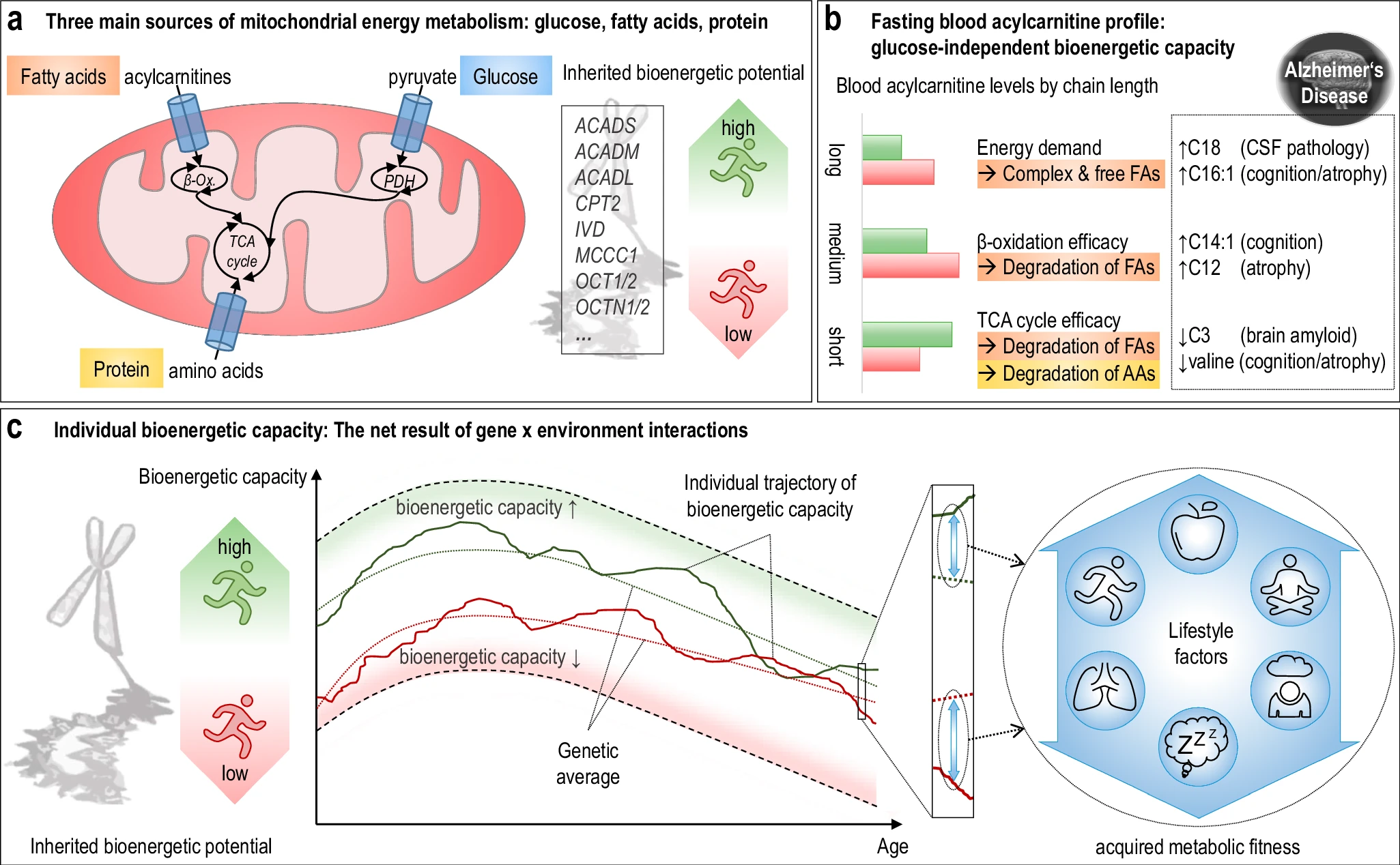
A growing body of evidence suggests that cardiometabolic risk factors play a significant role in Alzheimer’s disease (AD). Diabetes and hypertension, the famous silent killers, are highly prevalent and can accelerate neurodegeneration and perpetuate the burden of Alzheimer’s disease. Insulin resistance and enzymes including insulin-degrading enzymes are implicated in Alzheimer’s disease where the breakdown of insulin is prioritized over the breakdown of amyloid-β. enter image description here Studies have shown that immune cells in the brain, particularly microglia, and astrocytes, are key regulators of the inflammatory response in the central nervous system and are involved in the pathogenesis of AD. These cells have various roles, including supporting neurons.

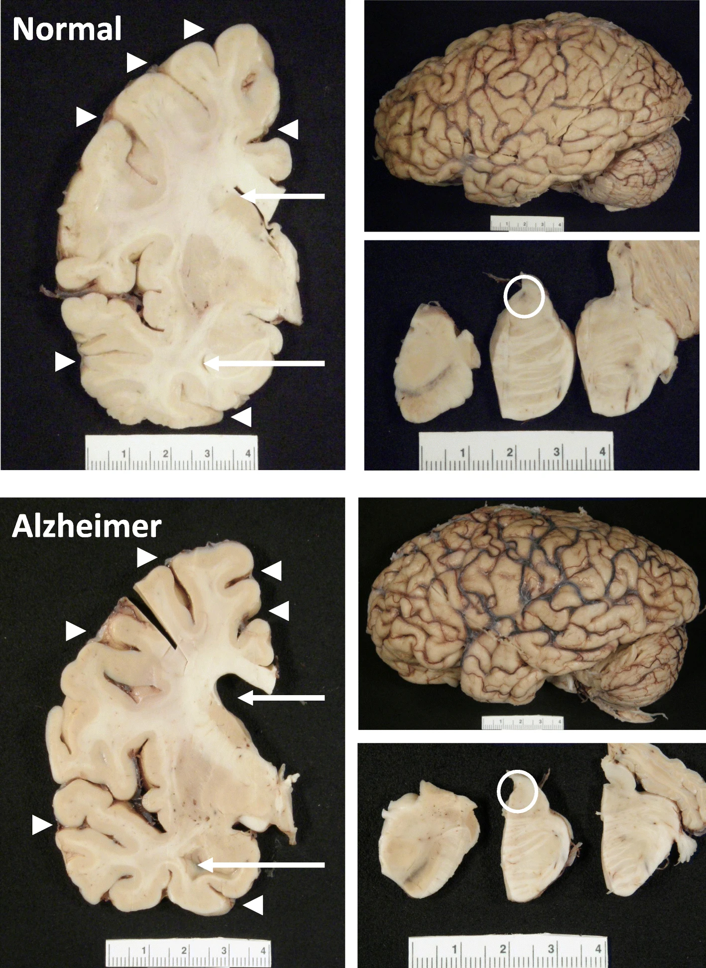
Alzheimer’s disease (AD) is characterized by progressive neurodegeneration associated with synaptic dysfunction and neuronal death, which culminates in brain atrophy. It can also be characterized by the deposition of senile plaques composed of β-amyloid (Aβ) and neurofibrillary tangles (NFTs), formed of hyperphosphorylated Tau protein.

Glucagon-like peptide-1 receptor agonists (GLP-1 RAs), currently marketed for type 2 diabetes and obesity, may offer novel mechanisms to delay or prevent neurotoxicity associated with Alzheimer's disease. The impact of semaglutide in amyloid positivity (ISAP) trial is investigating whether the GLP-1 RA semaglutide reduces accumulation in the brain of cortical tau protein and neuroinflammation in individuals with preclinical/prodromal AD.

Several clinical trials are testing the effect of Glucagon-like peptide-1 receptor agonists on Alzheimer's patients. For example ISAP clinical trial tests oral semaglutide (Ozempic). Two independent phase 3 trials are already underway, with findings due at the end of 2025. Liraglutide is another glucagon-like peptide-1 (GLP-1) analog currently approved for type 2 diabetes and obesity.

The results of a clinical trial were presented at the Alzheimer’s Association International Conference in the United States. It suggests that liraglutide may protect the brains of people with mild Alzheimer’s disease and reduce cognitive decline by as much as 18% after one year of treatment.

ELAD was a 12-month, multi-center, phase IIb trial of liraglutide in participants with mild Alzheimer's dementia. A total of 204 participants were randomized to receive either liraglutide or a placebo in a daily injection for a year. The patients with mild Alzheimer’s disease were seen at 24 clinics throughout the United Kingdom. Each received a daily subcutaneous injection for one year: half received up to 1.8 mg of liraglutide and half received a placebo. Before the study began, all patients had magnetic resonance imaging (MRI) to evaluate brain structure and volumes, glucose metabolism PET scans, and detailed memory testing. These were repeated at the end of the study with regular safety visits.

The primary outcome was the change in cerebral glucose metabolic rate in the cortical regions (hippocampus, medial temporal lobe, and posterior cingulate) from baseline to follow-up in the treatment group compared with the placebo group. The key secondary outcomes were the change from baseline to 12 months in scores for clinical and cognitive measures and the incidence and severity of treatment-emergent adverse events or clinically important changes in safety assessments. Other secondary outcomes were a 12-month change in magnetic resonance imaging volume, diffusion tensor imaging parameters, reduction in microglial activation in a subgroup of participants, reduction in tau formation, and change in amyloid levels in a subgroup of participants measured by tau and amyloid imaging, and changes in composite scores using support machine vector analysis in the treatment group compared with the placebo group.

ELAD clinical trial was led by Prof. Paul Edison, M.D., Ph.D., professor of science from Imperial College London and probably the most cited author in the field of Alzheimer's research.

While the primary endpoint (change in the cerebral glucose metabolic rate) was not met, scores for clinical and cognitive measures and the exploratory endpoint of brain volume showed statistically significant benefits. The patients who received liraglutide had nearly 50% less volume loss in several areas of the brain, including frontal, temporal, parietal, and total gray matter, as measured by MRI. Patients who received liraglutide had an 18% slower decline in cognitive function in a year compared to those who got the placebo.

Uncomfortable side effects often result from administration of GLP-1 agonists. Here gastrointestinal problems such as nausea were the most common side effects, which totaled 25.5% of all adverse events in the liraglutide group. Twenty-five serious side effects occurred in 18 participants (17.6%) in the placebo arm and seven participants (6.9%) in the treatment arm. The most serious side effects were considered unlikely to be related to the treatment of the study.

Clinical trials are costly, this study was funded by the UK Alzheimer’s Society, Alzheimer’s Drug Discovery Foundation, Novo Nordisk, John and Lucille Van Geest Foundation, and the National Institute for Health and Care Research (NIHR) Biomedical Research Centre.

In conclusion, if these results are confirmed in a phase III clinical trial, they are revolutionary. Indeed they are much better than the recently authorized but controversial drugs that use antibodies to target amyloid plaques.

Yet, this is not a cure, it will slow the disease, not cure it. However no drugs currently slow the disease, and the US-authorized medicines have severe side effects (ARIA).

Biomarkers of Neurodegeneration in Young Adults with Diabetes

- Posted by admin in English

Adult-onset diabetes is a known risk factor for cognitive impairment and dementia, yet the study of diabetes in young people has been neglected until now.

The average age of onset of diabetes in adults is ~46 years, and roughly thirty years later neurodegenerative diseases may appear. People with early-onset diabetes when they reach the age of 46 years, have lived with their disease for at least 30 years. It is therefore possible that early-onset diabetes leads to early-onset dementia.

A recent prospective population-based cohort in the United Kingdom did find that a younger age at the onset of diabetes corresponded to a younger age at the onset of dementia, but this study did not specifically look at people with early diabetes. A new study on this subject indicates that people with early-onset diabetes are at significant risk of prematurely developing cognitive impairment and dementia with possible neuropathology. enter image description here This study aimed to explore neurodegenerative disease biomarkers in cohort-derived biomarker banks as changes in key plasma biomarkers between the time of diabetes diagnosis and early adulthood have been correlated with worsening cognitive function in young adults with early-onset diabetes.

Participants with youth-onset diabetes (age of onset less than 20 years) were found in the SEARCH for Diabetes in Youth study, a multicenter population-based registry and cohort. A randomly selected subset of 50 SEARCH participants (n=25 type 1 diabetics, n=25 type 2 diabetics) was identified for inclusion in the plasma biomarker analysis.

Among SEARCH participants eligible for plasma biomarker analyses, the authors recruited and enrolled a subset of the Colorado SEARCH clinic site to complete positron emission tomography (PET) imaging to measure plasma accumulation amyloid and tau density in brain regions susceptible to Alzheimer's disease.

For their study of plasma biomarkers of neurodegeneration, scientists identified age-matched controls without diabetes from two cohorts with plasma samples stored to include adolescent controls from the Exploring Perinatal Study Outcomes in Children (EPOCH) (n = 25) and young adult controls from the CROCODILE (Control of Renal Oxygen Consumption, Mitochondrial Dysfunction and Insulin Resistance) study (n = 21).

The authors also recruited and enrolled a group of young adult controls from the University of Colorado Anschutz Medical Campus to complete PET imaging for amyloid density and tau in brain regions susceptible to Alzheimer's disease.

By studying these two types of biomarkers (plasma and molecular imaging), scientists found evidence of potentially greater neuropathology of neurodegenerative diseases in young adults with early-onset diabetes, where plasma pTau181 was significantly higher and Aβ40 and Aβ42 were significantly lower, compared to controls, and over time from diabetes diagnosis from adolescence to young adulthood.

Furthermore, changes in key plasma biomarkers of neurodegeneration from diabetes diagnosis to early adulthood have been correlated with worsening cognitive function in young adults with early-onset diabetes. These preliminary data suggest the possibility of an early risk trajectory for Alzheimer's disease among individuals diagnosed with diabetes during childhood or adolescence.

It is important to emphasize that the participants with youth-onset diabetes had lower plasma Aβ42 and Aβ40 concentrations than age-matched controls both in adolescence and early adulthood, suggesting amyloid dysregulation potentially early and sustained in diabetes beginning in young people. Lower levels of Aβ40, Aβ42 and their ratio, especially in plasma and cerebrospinal fluid, correspond to monomer sequestration and amyloid plaque formation. Overall, the lower plasma concentrations of Aβ42 and Aβ40 in their sample suggest the development of Alzheimer's disease neuropathology, but could also indicate disrupted neurodevelopment in those with early-onset diabetes.

NfL was not different from controls in adolescence, but was higher on average in the group of young adults with youth-onset diabetes, compared to young adult controls. These results are consistent with other larger studies in adults with diabetes. However, the scientists are cautious in interpreting their NfL results, given that NfL is a non-specific marker of neuronal damage and could also indicate involvement of peripheral neuropathy in people with diabetes. diabetes

The moral integrity of the authors of this study is reflected in the number of limitations highlighted by the authors. Too often, scientists (and their university's public relations department) make overblown, ridiculous, and deliberately misleading claims.

  • The young age of their sample limits confounding by age, such that changes in plasma biomarkers are more likely to be attributed to diabetes pathophysiology and not typical aging-related processes.

  • Their age-matched control groups were sampled from different cohorts. Thus, scientists cannot interpret biomarker differences between adolescent controls and young adult controls as a typical developmental change in these biomarkers. It should be noted that the shelf life of plasma samples from each group differed, which imposed yet another limitation on their study, with diabetes samples appearing in young people having on average a longer shelf life than control samples. .

Longer storage duration could impact the observed protein concentrations of the measured plasma biomarkers. However, if protein levels were differentially impacted between groups given variability in storage duration, we might expect to see lower concentrations in the youth diabetes group compared to control groups for all proteins measured. This was not the case in their study.

  • The scientists did not have APOE4 status among the subjects of their study.

  • The SEARCH study did not measure cognitive function at the initial visit so scientists could not study cognitive changes over time in relation to changing plasma biomarker levels.

  • Scientists do not have corresponding biomarkers measured in the CSF.

  • The sample of young adults with early-onset diabetes who participated in the PET imaging study was small.

In conclusion, although neurodegenerative diseases are conceptualized as a disease of the elderly, increasing evidence suggests that factors linked to early life may have an impact on risk trajectories.

Such life-span course disease models will contribute to a better understanding of how currently used neurodegeneartion biomarkers evolve during critical periods of development across the lifespan, and how they can be used to predict the risk of neurodegenerative diseases early onset and cognitive impairment in high-risk clinical populations such as early-onset diabetes.

Sleep and the Price of Plasticity

- Posted by admin in English

The reason we need sleep seems clear: without sleep, we become tired, and irritable and our brain functions less well. Conversely, after a good night's sleep, the brain and body feel refreshed. But why does the brain need to disconnect from the environment for hours every day? What is restored by sleep has proven difficult to explain. enter image description here Sleep efficiency in older adults. Sleep deprivation increases amyloid-β (Aβ) concentrations in the interstitial fluid of experimental animal models and in cerebrospinal fluid in humans, while increased sleep decreases Aβ. Sleep abnormalities may therefore represent a risk factor for neurodegeneration.

It has recently been shown that sleep is likely a time for clearing waste in the brain or repairing damaged cells. Circadian sleep-wake rhythm disorders are strong predictors of institutionalization.

During a period of wakefulness, coping with the environment requires increasing the number and strength of connections at the synapses between neurons in the brain. This increased activity increases cellular requirements for energy and materials, leading to cellular stress, a major factor in neurodegenerative diseases, and forcing changes in supporting cells such as glial cells, while hindering learning. During sleep, this synaptic activity is decreased which helps restore cellular health and increase plasticity through negative selection of synapses. This may also explain the benefits of sleep for memory acquisition, consolidation, and integration.

In other words, for the theory of synaptic homeostasis, sleep is “the price we pay for our learning and memory abilities”. Increased synaptic activity reduces the selectivity of neuronal responses and limits the ability to learn. By renormalizing synaptic activity, sleep reduces the plasticity burden of neurons and other cells while restoring neuronal selectivity and the ability to learn, and the consolidation and integration of memories.

A new study has just confirmed this, at least in translucent zebrafish larvae. However, these results obtained with zebrafish larvae should only be extrapolated with great caution to humans, but it is nevertheless an interesting discovery for fundamental neuroscience. The study authors used in vivo synaptic labeling tools in larval zebrafish to image the same neurons and their synapses repeatedly over long periods, allowing them to map the synapse changes of a single neuron in states of sleep and wakefulness. In effect, this meant genetically modifying these neurons to allow fluorescence upon firing.

By tracking the synapses of single tectal neurons across sleep-wake states and circadian time, scientists resolve several outstanding questions about the magnitude, universality, and mechanisms of sleep-related plasticity.

They show that synaptic dynamics are present in many cells on average, but when examined neuron by neuron, more diverse patterns of synaptic changes are revealed. These observations may explain some discrepancies between previous studies on the synaptic homeostasis hypothesis, as such single-neuron synaptic dynamics were not captured by one-time snapshots of synapse number or function at the population level.

The authors also found that sleep-related synapse loss depends on molecular signals related to elevated sleep pressure and, notably, also reflects slow-wave activity by occurring primarily at the beginning of the sleep period. This finding raises the question of whether sleep periods associated with low sleep pressure, such as in the second half of the night, play an additional role in non-synaptic remodeling.

Despite all this work was carried out on zebrafish larvae equipped with genetically modified neurons. Thus, any extrapolation to mammals, without even thinking about humans, is highly speculative. Still, it's another clue that sleep is an integral part of good neuronal health, probably along with vascular health and physical activity.

Alzheimer disease and RT inhibitors

- Posted by admin in English

Two very interesting new articles have been published on the (supposed) action of a drug against Alzheimer's disease. enter image description here These articles provide the reader with several points for further reflection. - One of them is that the clinical trial cannot be the only method of validating the marketing of a drug. Indeed, for diseases that develop over decades, often asymptomatically, how can the effectiveness of a drug be tested? We can always dream of a pill that will cure in a few months a patient, as this is true for diseases where there is an identified pathogen, but it is very unlikely with chronic diseases. Even for communicable diseases, this type of medication does not always exist; a patient can be considered permanently cured, even though they suffer very serious after-effects.

HIV and many other viruses rely on an enzyme, called reverse transcriptase (RT), to copy their RNA molecules and change them into complementary DNA duplicates that can then be inserted back into the host cell's DNA, producing permanent sequence changes.

As RT hijacks a host's cells to establish a chronic infection, so drugs that block the RT enzyme's activity have become a common part of treatment cocktails for keeping HIV at bay.

The team of the first article analyzed anonymized medical records with prescription claims from more than 225,000 controls and patients and found that RT inhibitor exposure was associated with a statistically significant reduced incidence and prevalence of Alzheimer's disease. There were 2.46 Alzheimer's disease diagnoses per 1,000 persons taking these inhibitors, versus 6.15 for the general population. And this with a drug that is not optimized for Alzheimer's disease. Indeed the drugs patients took in this retrospective study were designed to counter a specific form of RT used by HIV and it may only have a limited effect on many different possible forms of the enzyme in the brain. So there is significant room for progress to perfect this drug.

  • Another point of reflection concerns the etiology of Alzheimer's disease. This is the subject of numerous debates and gigantic financial bets. Clearly, the pharmaceutical industry ecosystem considers that the appearance of intra and extra-cellular beta-amyloid plaques is the cause of this disease. Few scientists question why these plaques appear, or whether competing hypotheses have been properly studied. In this new article, it is shown that an anti-viral drug significantly reduces the risk of developing Alzheimer's disease.

In a second article, an explanation is proposed as to why clinical trials for this disease most often fail. The amyloid hypothesis, or the theory that the accumulation of a protein called beta-amyloid in the brain causes Alzheimer's disease, has driven Alzheimer's research to date. However, treatments that target beta-amyloid have notoriously failed in clinical trials. The authors of the second article found that most Alzheimer's disease brain samples contained an over-abundance of distinct APP gene variants, compared to samples from normal brains. Among these Alzheimer's-enriched variations, the scientists identified 11 single-nucleotide changes identical to known mutations in familial Alzheimer's disease—a very rare inherited form of the disorder. Although found in a mosaic pattern, identical APP variants were observed in the most common form of Alzheimer's disease, further linking gene recombination in neurons to disease.

“The thousands of APP gene variations in Alzheimer's disease provide a possible explanation for the failures of more than 400 clinical trials targeting single forms of beta-amyloid or involved enzymes,” says Chun. "APP gene recombination in Alzheimer's disease may be producing many other genotoxic changes as well as disease-related proteins that were therapeutically missed in prior clinical trials. The functions of APP and beta-amyloid that are central to the amyloid hypothesis can now be re-evaluated in light of our gene recombination discovery."

  • We have all learned that all the cells of an organism have the same genetic heritage since they come from the same original cell. This vision, which dates from the middle of the last century, is beginning to be called into question in different cases: cancers, cases of mosaicism, and aging. These two articles, which come from the same laboratory, raise the question of how to detect these cases of mosaicism. Indeed, if a virus modifies the genome of a cell, but only in certain tissues (such as the brain) and only for a fraction of the cells, a genetic analysis of an easily accessible tissue will not necessarily highlight an alteration of the genome. of a significant fraction of the cell population of another tissue.

The researchers note that: "It is important to note that none of this work would have been possible without the altruistic generosity of brain donors and their loving families, to whom we are most grateful. Their generosity is yielding fundamental insights into the brain and is leading us toward developing new and effective ways of treating Alzheimer's disease and possibly other brain disorders—potentially helping millions of people.

As usual, it is not the first time that RT inhibitors have been proposed against Alzheimer's disease animal models, but it is the first time the effect has been shown in humans.

Atrial fibrillation, stroke and dementia

- Posted by admin in English

Atrial fibrillation is a risk factor for stroke and multiple forms of dementia, including Alzheimer’s disease. enter image description here The exact reasons why atrial fibrillation (a common heart disease) increases dementia risk aren't fully understood, but there are likely several factors involved. Some of these factors directly cause damage, while others increase the chances of getting dementia over time. These factors include tiny blood blockages and bleeding in the brain's small blood vessels. Small strokes you might not even notice, reduce blood flow to the brain, increase long-term inflammation, and lead to shrinkage of brain tissue.

"Silent strokes" seem to be especially important because they're found in up to a quarter of atrial fibrillation patients. In these cases, dementia develops because of damage to the brain's tissues caused by a stroke or a temporary lack of blood flow (transient ischemic attack). So, by preventing these silent strokes, we can also reduce the risk of getting dementia or slow it down. This is why using blood thinners (anticoagulants or anticoagulants) is so important for people with atrial fibrillation.

Catheter ablation of atrial fibrillation is a minimally invasive procedure used to treat atrial fibrillation, a heart condition that causes irregular heart rhythm. Catheter ablation is typically an option for people with atrial fibrillation that is not controlled by medications or who experience significant side effects from medications. It is not a cure for atrial fibrillation, but it can significantly improve symptoms and quality of life for many people.

During the procedure, a thin, flexible tube called a catheter is inserted into a blood vessel in the groin and threaded up to the heart. The catheter uses radiofrequency energy or extreme cold (cryoablation) to create tiny scars on the heart tissue in the areas where the abnormal electrical signals originate. These scars block the faulty electrical signals, preventing them from spreading through the atria and causing irregular heartbeats.

However, studies of atrial fibrillation did not consistently report on the influence of periprocedural anticoagulation and long-term use of anticoagulants on dementia risk.

Swedish scientists evaluated the protective effect of atrial fibrillation ablation in a large cohort who received optimized anticoagulation and compared them with patients whose cases were recorded in the Swedish Patient Register.

They studied the cases of 5,912 patients who underwent first-time catheter ablation for atrial fibrillation between 2008 and 2018 and compared them with 52,681 control individuals from the Swedish Patient Register. The majority of patients were on anticoagulants. The mean follow-up time of those patients in the Swedish Patient Register was 5 years.

The Swedish authors found that catheter ablation and anticoagulation treatment were associated with a lower risk of dementia diagnosis compared with the control group. The result was similar when including patients with a stroke diagnosis, which tends to confirm the vascular origin of these cases of dementia.

These results are in line with similar findings in other countries.

Brain's health, increased lactate and decreased pH levels

- Posted by admin in English

The body's pH level is often stated to become slightly more acidic (below pH 7) as we age, but studies are inconsistent. Ideally, blood pH remains tightly regulated within a narrow range (around 7.4). This is crucial for many bodily functions. It's less clear how the brain's PH is modified with aging and disease. A collaborative research group comprising 131 researchers from 105 laboratories mostly from Japan has published an impressive paper in eLife on the influence of brain pH and lactate levels in neurological disorders. The research group's previous findings in a 2018 small study, suggest that the brain's PH alteration is a consequence of diseases. This conclusion was drawn from five animal models of schizophrenia/developmental disorders, bipolar disorder, and autism.

The recent study, suggests that changes in brain pH and lactate levels are a common feature in a diverse range of animal models of diseases, including schizophrenia/developmental disorders, bipolar disorder, autism, as well as models of depression, epilepsy, and Alzheimer's disease. enter image description here What could be the mechanism between increased lactate and decreased pH? Lactate is a byproduct of metabolism and it is an acid, hence the acidification of pH. Neuronal activity relies heavily on glucose for energy. Under normal conditions, oxygen is readily available, and glucose is broken down through aerobic respiration, producing ATP (energy) with minimal lactate as a byproduct. However, during periods of high neuronal activity or limited oxygen supply, cells shift to anaerobic metabolism, relying more on glycolysis. This process produces more lactate as a byproduct.

These changes were observed in the postmortem brains of patients deceased of several neurological diseases, including Alzheimer's disease. However, the observed phenomena are potentially confounded by secondary factors such as the administration of antipsychotic drugs. In addition, the brains of deceased patients have undergone multiple changes during the transition between life and death, and the conservation process, and these postmortem brains may not reflect the condition of living patients.

This time the authors included autism, epilepsy, bipolar disorder, autism, models of depression, epilepsy, and Alzheimer's disease, as well as peripheral diseases or conditions comorbid with psychiatric disorders (e.g. diabetes mellitus [DM], colitis, and peripheral nerve injury). These animal models included 109 mice, rats, and chick animal models with genetic modifications, drug treatments, and other experimental manipulations such as exposure to physical and psychological stressors. More than 2200 animals were used in a hundred laboratories.

First, this large-scale animal model study revealed that alterations in brain pH/lactate levels can be found in approximately 30% of the animal models examined. 30% is not 100% so more studies are needed to understand this result.

The authors observed no significant correlation between brain pH and age in the wild-type/control rodents. In human studies, inconsistent results have been obtained regarding the correlation between brain pH and age. There isn't necessarily a direct cause-and-effect relationship between aging and altered pH, but rather a decline in the body's efficiency to maintain homeostasis (stable internal environment).

  • Reduced Kidney Function: Kidneys play a key role in balancing blood pH by filtering out acids. As kidney function declines with age, the body may struggle to eliminate excess acid: Drink more, and quality water with low minerals!
  • Decreased Metabolic Activity: A lower metabolic rate can lead to the buildup of acidic byproducts in the body. Decreased Metabolic Activity manifests itself in slower digestion, heart rate, and breathing, lower body temperature, and reduced strength.

So the authors conclude that if this correlation between brain pH and age, is a feature of disease and not aging, then brain diseases might be instrumental in the appearance of these changes in pH in the brain, but the mechanism of action is far from clear.

Their analysis found that poorer working memory performance in animal models of neuropsychiatric disorders may be predominantly associated with higher lactate levels, which was confirmed in an independent cohort. They found the same result for anxiety, probably because it causes a large increase in brain activity.

In conclusion, this is an excellent study, there are too few studies of that caliber. In the future research will improve the brain's ability to remove lactate. This might involve stimulating transporters that clear lactate from the CNS or potentially using enzyme therapies to convert lactate into other metabolites. Another possibility is with therapies that encourage the brain to rely more on aerobic respiration (oxygen-dependent energy production) as they could minimize lactate production as a byproduct. This could involve optimizing blood flow to the brain or exploring potential metabolic drugs.


Please, help us continue to provide valuable information: