Alzheimer's disease is a severe neurodegenerative disease characterized by progressive damage to synaptic bonds and neuronal cells mainly in the cerebral region of the hippocampus.
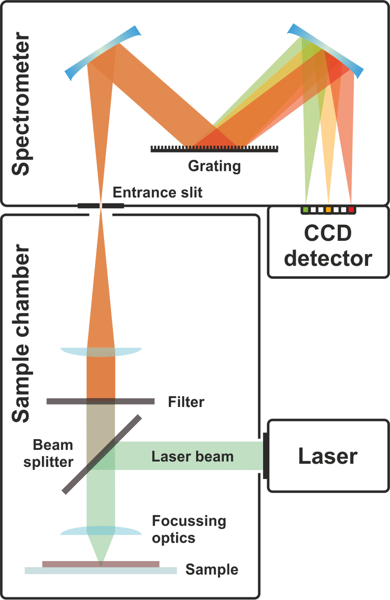
Optical spectroscopy methods can provide a unique means of analyzing body fluids, enabling rapid, sensitive, non-invasive and reliable diagnosis.
 * Source: Toommm via Wikipedia. *
The development of Alzheimer's disease has been associated with a dysfunctional blood-brain barrier with a reduced ability to clear Aβ and tau proteins from the brain. These two substances are biomarkers of the progression of Alzheimer's disease and are a reliable sign of a neurodegenerative process even at an early stage. These biomarkers can be transported in the vascular system across the blood-brain barrier. The blood-brain barrier plays an important role in the selective passage of several substances between the brain and the blood system and in the maintenance and integrity of the brain.
Traces of these biomarkers are present in the systemic bloodstream and can be found in the vasculature of other areas of the body. The relevance of changes occurring in the eye as a consequence of Alzheimer's disease has been reported by Lim et al. Therefore, tears are an interesting way to study the changes induced by neurodegenerative pathologies. They are easily accessible and can be collected using minimally invasive methods.
Over the past decades, a large number of studies have been conducted to analyze the composition of tears using conventional biochemical methods.These conventional techniques have been largely replaced by optical spectroscopy methods, such as those based on Raman scattering, which take less time and require only little preparation of the samples.
Raman spectroscopy has already been used to identify biomarkers of Alzheimer's disease in blood serum and saliva and to study the basic aggregation mechanisms of amyloids. However, it has some limitations regarding sensitivity, noise / signal level and repeatability.
The objective of the study which concerns us today was to demonstrate the spectral differences in the SERS spectroscopic response of human tears in subjects suffering from Alzheimer's disease, subjects suffering from mild cognitive and healthy control subjects. Human tears were characterized by SERS coupled with multivariate data analysis.
Thirty-one informed subjects (Ctr, MCI and AD) were considered. Eighteen subjects with Alzheimer's disease (7 women, 11 men and mean age 71 ± 10 years) and seven subjects with mild cognitive impairment (3 women, 4 men and mean age 73 ± 9 years) were included In this study.
While the Raman spectrum does not provide any valuable information apart from very weak characteristics, conversely, the SERS spectrum clearly demonstrated the various contributions of human tears components. SERS measurements were performed with standard microscope glasses coated with a homemade colloid of gold nanoparticles (GNP).
The data processing was implemented using software routines (wavelet toolbox).
The fully processed dataset of all mean spectra was analyzed by PCA and i-PCA to describe it using a set of orthogonal eigenvectors separately accounting for different sources of variance in the original data.

The differences between the SERS spectra of tears from subjects at different clinical states are sometimes very subtle and are mainly localized in certain specific spectral ranges. Notably, the authors found that PCA performed over the entire spectral range did not differentiate between the subtle changes that occurred in the spectra examined. To better highlight these differences and take advantage of them to classify the spectra, the authors therefore performed the PCA in intervals (interval partial component analysis (i-PCA)).
The average SERS spectra of the Ctr, MCI and AD subjects showed differences related to the protein components of lactoferrin and lysozyme. Quantitative changes were also observed by determining the intensity ratio between the selected bands. Scientists also built a classification model that discriminates between AD, MCI and Ctr subjects. This model was built using the scores obtained by performing a interval principal component analysis on specific spectral regions (i-PCA).

Although it has not been possible to discriminate specific biomarkers of Alzheimer's disease, the overall response of SERS reflects small but interesting changes in tear composition that can be attributed to altered levels of specific pathological substances. and stimulated by disease.
The analysis of the partial interval components (i-PCA) of the spectra by the authors made it possible to distinguish subjects with Alzheimer's disease from healthy subjects with MCI. The precision of classification of the constructed method was very encouraging with interesting prospects for medical applications as a support for clinical diagnosis and discrimination of Alzheimer's disease from other forms of dementia.

