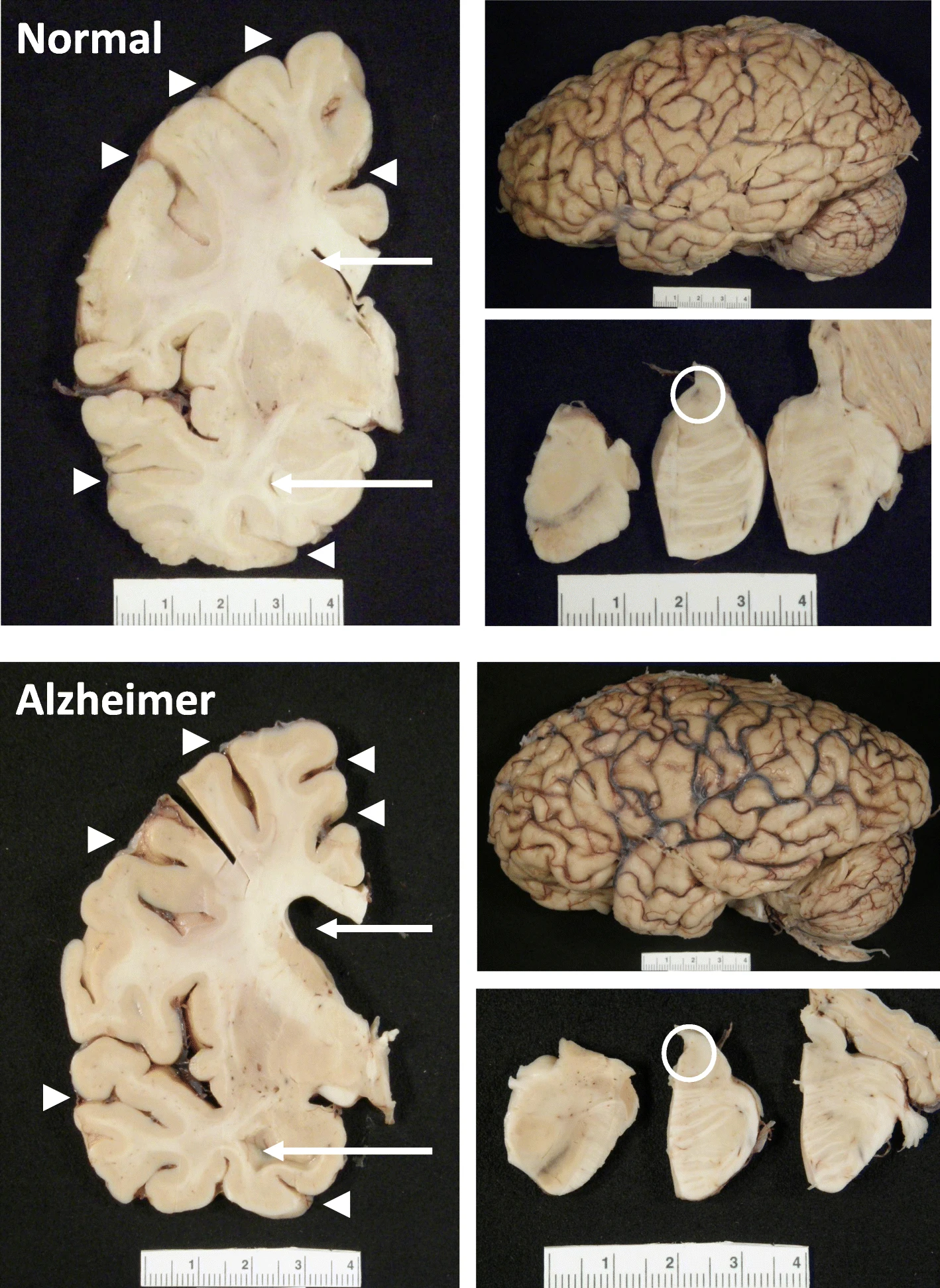
Researchers are currently very interested in finding biomarkers for the early detection of many neurodegenerative diseases. The market for these technologies is likely to be quite large. Detecting weakened cerebral rigidity before the onset of irreversible damage could pave the way for early intervention in Alzheimer's disease and related disorders. This could be achieved using a device that has now become relatively common: MRI. enter image description here The hippocampus, a small, seahorse-shaped structure buried deep within the brain, is best known for its role in memory formation and learning. It is an exceptionally vulnerable structure, with perfusion deficits often observed in diseases related to learning and memory. However, a brain affected by Alzheimer's disease tends to exhibit at least moderate cortical atrophy, including in the precuneus and posterior cingulate gyrus. It should be noted that the posterior cingulate gyrus is adjacent to the hippocampus.
A recent study shows a relationship between blood flow and mechanical stiffness (an MRI concept) of the hippocampus. Researchers sought to understand how these physical properties interact in a healthy brain and what this might reveal about early brain changes in neurodegenerative diseases. The researchers used two advanced MRI techniques—magnetic resonance elastography (MRE) and arterial spin labeling (ASL).

Using these tools, the researchers measured: * Tissue stiffness (the resistance of an area to physical deformation) * Perfusion (blood flow at the tissue level)

Seventeen healthy adults were examined by the researchers at two different MRI intensities (3T and 7T), allowing for a cross-comparison between the two magnetic field strengths. They found that the hippocampus had the highest blood flow among the deep gray matter structures, followed closely by the caudate nucleus and putamen.

A strong positive correlation was observed between blood flow and stiffness in the hippocampus, but not in the caudate nucleus, although both regions are highly vascularized. This indicates that good brain health appears to be linked to good blood flow, manifested by the good stability (stiffness) of the tissue.

In a subgroup of ten subjects, it was found that higher blood flow resulted in larger tissue pulsations, suggesting that the dynamics of blood supply physically influence the hippocampus.

These results suggest a previously underestimated link between the physical characteristics of well-perfused brain tissue and its metabolic needs. This connection is not entirely surprising, as the macroscopic characteristics of tissue depend on the well-being of its constituent cells.

Although not mentioned in the article, one might wonder about the relationship between this mechanical property—rigidity—and beta-amyloid (Aβ), the signature protein implicated in Alzheimer's disease.

Beta-amyloid plaques begin to accumulate in the brain well before the onset of symptoms. These plaques can form: * Extracellularly: outside neurons, interfering with cell-to-cell communication and nutrient supply; * Intracellularly: inside neurons and other nerve cells, disrupting protein production.

Based on current knowledge, beta-amyloid likely appears before physical changes in tissues. It emerges early, sometimes decades before cognitive symptoms, triggering a cascade of tissue changes. Mechanical stiffness, as measured by MRE, is more likely a result of these changes than a cause.

Interestingly, numerous MRE studies have observed brain softening, particularly in the hippocampus and cortex, in Alzheimer's patients and those with mild cognitive impairment. This supports the perspective that stiffness decreases due to beta-amyloid pathology and its effects on brain tissue structure.

As fascinating as these findings are, it is important to acknowledge the limitations of the study, which suggest future research directions.

With only 17 participants, the study lacks statistical power, making it vulnerable to false positives or exaggerated effect sizes.

All subjects were young adults (22–35 years old) who were generally healthy, limiting the relevance of the results to aging populations or those at risk for Alzheimer's disease.

The sample did not include key groups, either clinical or high-risk individuals, such as APOE4 gene carriers or those with mild cognitive impairment (MCI).

The study was cross-sectional, capturing a single snapshot in time. We do not yet know how stiffness or perfusion might change over time or in response to pathology.

No cognitive data were collected; therefore, the relationship between hippocampal mechanics and actual memory performance remains unexplored.

There are considerable interpretation challenges: Stiffness, measured by magnetic resonance elastography (MRE), reflects a complex array of biological factors: neuronal density, inflammation, vascular integrity, etc. It is a valuable signal, but not biologically specific. Indeed, perfusion can vary depending on common physiological factors (e.g., hydration, stress).

Because the study did not include beta-amyloid PET scans or fluid biomarkers, the link between mechanical findings and Alzheimer's pathology remains hypothetical.

The analysis focused on the hippocampus and a few other deep gray matter structures. Key cortical regions involved in Alzheimer's disease (such as the entorhinal cortex or precuneus) were not examined.

The emerging link between perfusion and mechanics, and how this relationship deteriorates in the presence of beta-amyloid, could help us uncover subtle clues that precede cognitive decline. Ultimately, measuring something as simple as brain "firmness" could help us identify those at risk and determine when to act.

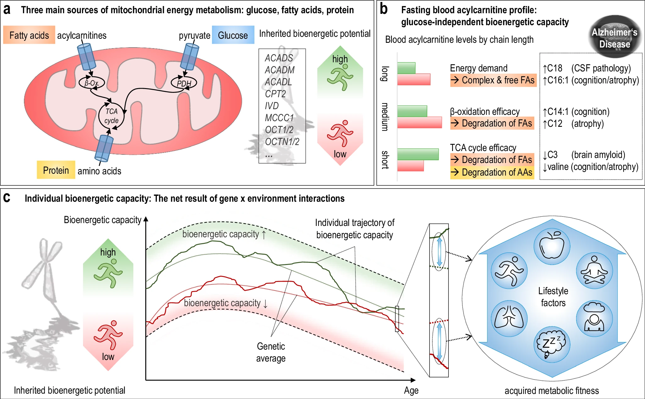
An interesting research article was recently published on bioenergetic subgroups in Alzheimer's Disease. The study found a connection between acylcarnitines, bioenergetic age, and Alzheimer's progression. It opens up interesting possibilities for how we might approach brain health from a metabolic perspective as the study suggests brain health to be largely modifiable rather than genetically determined. Focusing on general metabolic health through evidence-based approaches like regular exercise, quality sleep, and dietary patterns that support mitochondrial function could potentially be beneficial. enter image description here The researchers used acylcarnitine profiles from blood samples to identify distinct bioenergetic subgroups in Alzheimer's Disease (AD) patients and evaluate how bioenergetic capacity relates to disease progression. They used data from 1,531 participants in the Alzheimer's Disease Neuroimaging Initiative (ADNI), and identified several bioenergetic subgroups with significant differences in AD biomarkers, cognitive function, and brain glucose metabolism. These subgroups were primarily determined by modifiable factors (40-60%) related to beta-oxidation function, rather than genetic factors, suggesting potential for intervention.

The researchers developed a "bioenergetic age" metric based on acylcarnitine levels that strongly correlated with AD pathology. Individuals with "younger" bioenergetic ages showed less severe disease markers. Baseline bioenergetic age predicted cognitive decline over time in multiple studies, independent of APOE ε4 status in most cases. Specific genetic variants (SNPs rs17806888 and rs924135) influenced cognitive decline trajectories, but their protective effect appeared limited to individuals with younger bioenergetic ages. A simulated clinical trial showed that individuals with younger bioenergetic ages had significantly better outcomes on multiple clinical measures, with effect sizes comparable to those seen in the lecanemab anti-amyloid antibody trial.

The research suggests that targeting bioenergetic capacity could be a promising intervention approach for AD, particularly for the approximately 30% of individuals with protective genotypes but older bioenergetic ages.

In addition to the usual recommendations (Exercise/physical activity, dietary approaches, sleep optimization, stress reduction) supplementation (with medical supervision) might be an option:

  • L-carnitine/acetyl-L-carnitine - directly involved in fatty acid transport for beta-oxidation
  • Omega-3 fatty acids - support mitochondrial membrane health
  • Coenzyme Q10 - important for mitochondrial energy production

Today there isn't a widely available, inexpensive rapid test specifically for comprehensive acylcarnitine profiling that consumers can easily access. Yet your doctor could order acylcarnitine profiling, though it's not a routine test. Some companies offer more comprehensive metabolic panels that include some acylcarnitine measurements, though these typically cost $300-500+ and aren't widely validated. There's no equivalent to something like a glucose meter or rapid cholesterol test for measuring acylcarnitines at home or in point-of-care settings.

Yet another theory about the Alzheimer's disease

- Posted by admin in English

Here is a paper that outlines a new theory on the cause of Alzheimer's disease with implications for Parkinson's disease as well as ALS.

This is just speculation, based on almost only one fact: The expression of many genes is involved in this disease, so it would imply a global deregulation of the cellular machinery. Unfortunately, as usual in biology, this is a purely qualitative theory and, therefore, susceptible to many possibly contradictory interpretations. However, it is a theory that sees many neurodegenerative diseases as belonging to a spectrum rather than as distinct diseases. I endorse this point of view. enter image description here Alzheimer's disease research has produced many hypotheses over the years, including cholinergic, inflammatory, viral, mitochondrial, tau, and amyloid. However, none of these hypotheses have led to treatments that can stop or reverse the disease. This leads to a search for new theories to explain these failures. But this may be because interventions occur too late in the disease progression, with brain damage irreparable and compensatory mechanisms saturated.

Most publications ignore physiology, such as the importance of drainage in the cerebral lymphatic channels that have been discovered in recent years. This publication is no exception to this unfortunate trend, it is a discussion of the functioning of a cell in general, not even a brain cell like a neuron or an astrocyte, and the theory is even mostly not specific to humans or mammals, which still leaves one very skeptical. In this publication, the authors suggest that a disrupted nucleocytoplasmic transport system, linked to the formation of stress granules (SG), plays a central role. Cellular stress itself can have multiple causes independent of each other. There is no clear explanation why a general blockage of the cell would specifically lead to the appearance of beta-amyloid in Alzheimer's disease, nor that of alpha-synuclein in Parkinson's disease or TDP-43 in ALS.

In this model, cellular stress triggers SGs, which disrupt the movement of molecules between the nucleus and the cytoplasm, affecting RNA transport, chromatin accessibility, and alternative splicing. These changes lead to synaptic dysfunction, metabolic disorders, protein processing defects, and ultimately cell death. When this process propagates to brain regions, it results in clinical Alzheimer's disease.

The authors present a multistep mechanism linking SGs, NCT dysfunction, and amyloid propagation: * SG formation disrupts nucleocytoplasmic transport, altering gene expression and RNA localization. * Aβ clearance is decreased due to impaired lysosomal function, reduced proteostasis, and disrupted Aβ export. * Aβ production may increase via impaired APP processing. * Seeding and spreading of Aβ aggregates are facilitated by exosome dysregulation and chaperone sequestration. * Glial activation and BBB dysfunction further enhance Aβ diffusion in the brain.

The mention of ALS, FTD, and other conditions with similar transport disruptions strengthens the model's plausibility by showing how dysfunction of nucleocytoplasmic transport is implicated in multiple diseases.

Eukaryotic cells regulate the movement of molecules between the nucleus and the cytoplasm through nuclear pore complexes (NPCs), which are composed of nucleoporins. This transport is controlled by importins, exportins, and the protein Ran, which provides the energy for molecular movement.

Stress granules (SGs) are nonmembranous cytoplasmic structures that form in response to cellular stress, typically through phosphorylation of eukaryotic initiation factor 2 (eIF2α). During transient stress, SGs help cells recover, but during chronic stress, such as in Alzheimer's disease (AD), SGs abnormally persist and sequester key molecules, disrupting transcription and nucleocytoplasmic transport.

Disruption of nucleocytoplasmic transport in Alzheimer's disease (AD) was first reported in 2006, when cytoplasmic accumulation of nuclear transport factor 2 (NTF2) was observed in hippocampal neurons, even before the formation of neurofibrillary tangles (NFTs). This suggests that dysfunction of the transport system occurs early in the progression of AD. Analysis of gene expression data shows similar transport-related disruptions in tangle-bearing and non-tangle-bearing neurons.

Similar disruptions are observed in ALS, FTD, Huntington's disease, and even in non-neurological diseases such as cancer and heart failure. However, the specific transport disruptions vary by disease, likely due to different patterns of SG sequestration.

Some neurons maintain normal expression of the transport system and show enrichment in translational and neuronal function pathways, while others, with altered expression of the transport system, display stress-related pathways and deficits in mitochondrial function and metabolism. These findings are consistent with in vitro studies, suggesting that AD progresses along a continuum at the cellular level, ultimately leading to widespread neuronal dysfunction and clinical symptoms.

Conclusion The text suggests a causal role for SGs and transport dysfunction in AD, but much of the available supporting evidence comes from in vitro studies or studies of related diseases (e.g., ALS, FTD). The available direct in vivo evidence demonstrating SG-mediated pathology in AD patients is still limited.

Although the text discusses tau tangles and Aβ, their role appears secondary to SGs. Since amyloid and tau pathology remain at the core of AD research and therapeutic efforts, their relative downplaying constitutes a potential weakness.

The proposed model is primarily based on molecular and cellular studies, with little reference to clinical data.

In recent days, there has been a lot of talk on social networks and mainstream press about a surgical procedure that is being carried out in a Shanghai hospital by a team led by Li Xia from the Faculty of Medicine of Shanghai Jiao Tong University, which could slow the progression of Alzheimer's disease, or even allow a temporary regression, in half of the participants.

Five weeks after the operation, clinical assessments revealed an improvement in cognitive function: the Mini-Mental Status Examination score went from 5 to 7, and the Clinical Dementia Rating-sum of boxes test score went from 10 to 8. The Geriatric Depression Scale score went from 9 to 0. The PET scan examination provided objective proof of this improvement. enter image description here

This recent increase of interest is quite curious because a publication was made in June of this year by the team that innovated with this technique.

This study presents a new surgical procedure, neck shunting to unclog the cerebral lymphatic systems, aimed at improving the elimination of waste accumulated in the glymphatic system of the brain to manage Alzheimer's disease. The glymphatic system facilitates the removal of harmful proteins such as beta-amyloid and tau, the accumulation of which is linked to Alzheimer's disease.

The surgical procedure uses lymphatic-venous anastomosis (LVA) to decompress the cervical lymphatic trunk, allowing cerebrospinal fluid (CSF) and residual proteins to flow more efficiently from the brain into the venous system.

It is an extracranial procedure and therefore less invasive and potentially safer than intracranial methods. The surgical treatment, which involves four small incisions in the patient's neck, has been performed on at least 30 patients in two public hospitals in China. Other much larger numbers of patients treated are circulating on the Internet.

Early results indicate that the procedure holds promise as an innovative strategy for the prevention and treatment of Alzheimer's disease.

This surgical technique has not been considered for the treatment of Alzheimer's disease, or even the brain in general, but has been used for some time for other diseases such as lymphedema.

The field of lymphedema surgery has seen enormous progress over the years and has been associated with the rapid growth of super microsurgery techniques. A lymphovenous bypass or lymphaticovenular anastomosis requires the identification of residual lymphatic channels and the creation of an anastomosis on a recipient venule, thus allowing the flow of lymphatic fluid and the improvement of a patient's lymphedema.

A technique similar to that of Chinese doctors has long been used in the context of hydrocephalus. The cause of this disease is a blockage of cerebral-spinal fluids which leads to an accumulation of these fluids in the brain. Treatment for hydrocephalus then involves creating a way to drain the excess fluid from the brain.

In the long term, some hydrocephalus patients require a permanent cerebral shunt. This involves placing a ventricular catheter (a silastic tube) into the brain's ventricles to bypass the flow obstruction and drain the excess fluid into other body cavities, where it can be reabsorbed. Most shunts drain fluid into the peritoneal cavity (in the abdomen), but other sites include the right atrium (heart), pleural cavity, or gallbladder.

Hydrocephalus is known to cause Alzheimer's disease. Normal pressure hydrocephalus is common in elderly patients. Curiously, as Western physicians consider this hydrocephalus to be independent of Alzheimer's disease they are reluctant to operate on these patients because they believe the benefits are small.

The Chinese approach is the opposite. She believes that this accumulation of cerebrospinal fluid in Alzheimer's disease should be treated because, at the very least, it can temporarily relieve about two-thirds of patients. This pragmatic approach considers the patients and does not seek to demonstrate whether or not this accumulation causes Alzheimer's disease.

In a series of videos posted last month on social media, Cheng Chongjie, a doctor in Chongqing who adopted the technique following his colleagues, said the operation was effective in more than half of his patients.

"Two national medical centers have performed hundreds of LVA operations, and their results show that this procedure is effective in 60 to 80 percent of patients.

Astrocytes conversion into interneurons

- Posted by admin in English

In the case of neurodegenerative diseases, it is certainly more useful to try to develop new neuronal cells than to "cure" cells that we are told are dying or even dead for months. This is the purpose of regenerative medicine.

One of the difficulties is that neurons are cells that do not reproduce, we live with the stock that we had at the end of our puberty. These cells come from stem cells.

In our brain and the rest of the central nervous system, half of the cells are not neurons, but more classic cells that nourish and maintain neurons.

These cells reproduce by fission like most cells. In particular, astrocytes, although tiny, have many points in common with neurons. It is therefore natural to want to transform astrocytes into neuronal stem cells. enter image description here Curiously, scientists are rather looking to directly convert astrocytes into neurons, despite the enormous morphological difference between these two types of cells.

A few years ago, following the discovery of Yamanaka factors, it was believed that this goal could be achieved quickly. However, the results obtained have been contested, in particular, because of methodological flaws. Despite the promising results of previous studies, the lack of robust lineage tracing methods has led to uncertainty as to whether the induced neurons originated from glial cells or whether native neurons were mistakenly labeled.

A new publication describes recent advances in converting glial cells into induced neurons in the central nervous system, particularly in regions where stem cell activity is limited. The previous “lineage reprogramming” approach used transcription factors to change the identity of terminally differentiated cells, such as glial cells, to other cell types.

The current study focuses on the transcription factor Ascl1, known for its role in neuronal fate decisions and its potential to reprogram various cell types into neurons. While Ascl1 can efficiently convert glial cells into induced neurons in vitro, its efficacy in vivo varies widely, often leading to limited neurogenic effects or, in some cases, inducing glial proliferation rather than neuronal conversion.

This inconsistency suggests that regulatory challenges affect Ascl1’s performance as a reprogramming factor, particularly due to context-specific modifications such as phosphorylation, which modulate its activity. Previous research has found that a mutant variant of Ascl1, known as Ascl1SA6, engineered to resist phosphorylation, enhances neurogenic activity, prompting this study to determine whether Ascl1SA6 could enhance glia-to-neuron reprogramming in vivo.

The researchers tested Ascl1SA6 in the early postnatal mouse cerebral cortex, which showed superior neuronal induction capacity compared to wild-type Ascl1. Furthermore, the combination of Ascl1SA6 with the survival factor Bcl2 further enhanced the efficiency of iN conversion. The study’s lineage tracing confirmed that the newly formed induced neurons were primarily from astrocytes. Interestingly, many of these induced neurons exhibited characteristics of fast-spiking parvalbumin-positive (PV+) interneurons.

In summary, the study demonstrates that Ascl1SA6, particularly in combination with Bcl2, could improve the efficiency and fidelity of glia-to-neuron reprogramming in vivo, with potential implications for therapies targeting neuronal loss and brain circuit restoration.

It should be kept in mind, however, that these cell type conversions a priori promote the appearance of cancers, that the new neurons are hardly useful, that the method used is gene therapy and therefore has very low efficacy and elevates cancer risks, that is is a report on experiences done in mice and they usually do not translate in humans and that it is not a question of conversion to neuronal stem cells which would be free of many of these problems.

Nevertheless, it's a step in the right direction.

Here is a somewhat interesting article about Alzheimer's disease, but I guess it has also value for studies of ALS and Parkinson's disease.

Until the 1980' all cells in the brain that were big enough to be studied routinely were the neurons. Neurons are impressive cells because they are extremely long. The other cells were called the glue "glia" because they had no obvious functions. Neurons are also impressive because they can convey electrical and chemical signals over long distances. Neurons once mature, mostly do not divide, especially the ones with long axons. Other cells more conventionally divide every few days. enter image description here Those other cells, which compose half of the brain's cells, are receiving more attention. There are multiple types but normally they are there to assist neurons in their task. A simplified view tells that neurons are a sort of plumbing system and the glial cells are the real actors in the brain.

One of the glial cells, the astrocytes, seems to play important roles, and these roles may change depending on biological events such as stress or pathogen intrusion. When they enter the state called reactive state (M1), they kill neurons, in a similar manner that macrophages (white cells) kill infected cells.

In Alzheimer's disease, the shrinking of the brain which is the result of dying neurons and glia, is associated with aggregates of amyloïd proteins (Aβ). The study found that Aβ protein triggers autophagy in astrocytes. Autophagy is a cellular process involved in waste removal and recycling. Cells need new proteins every passing minute to function correctly, and the diet would never be able to provide protein building blocks at the required speed, so it is of the utmost importance for cells to recycle used proteins. There are several kinds of autophagy mechanisms in cells. Neurons being quite passive giant cells, autophagy is mostly assured by astrocytes.

Recycling proteins is not a clean job, it involves breaking proteins into smaller components (the metabolites or even amino acids) in a series of steps. Some of these intermediate components are toxic.

This article investigates the role of autophagy in astrocytes in the context of Alzheimer's disease (AD). Autophagy plays a crucial role in clearing Aβ so disrupting autophagy in astrocytes leads to increased Aβ plaques and cognitive decline in AD mouse models. Conversely, enhancing autophagy by over-expressing the LC3B pathway with a genetic therapy in astrocytes can reduce Aβ plaques and improve cognitive function.

It is believed by the authors that Aβ is toxic because it induces urea cycle activation in astrocyte as a compensatory mechanism to deal with the toxic effects of Aβ.. The urea cycle is a metabolic pathway that primarily occurs in the liver, but it can also be active in astrocytes. This pathway is involved in detoxifying ammonia, a toxic byproduct of amino acid metabolism.

Overall, this study provides evidence that targeting autophagy in astrocytes may be a promising therapeutic approach for AD. Understanding the relationship between Aβ, autophagy, and the urea cycle could potentially lead to new therapeutic strategies for AD. For example, targeting the urea cycle or autophagy pathways might be explored as potential approaches to reduce Aβ toxicity.

**A new study claims that it observed that higher levels of soluble amyloid-β42, one of the main forms of amyloid-β associated with Alzheimer's disease, is associated with better cognition. Yet there is no description of a mechanism of action, this is just a correlation study. enter image description here Source: Nephron via Wikipedia It is known that reduced CSF Aβ42 levels in Parkinson's disease and dementia with Lewy bodies predict cognitive impairment and a more aggressive disease course and correlate with postmortem β-amyloid plaques in the brain. Recently it was shown the same phenomenon also appears in Alzheimer's disease.

This article brings new insight in that it suggests that increasing the levels of CSF's Aβ42 with anti-Aβ monoclonal antibodies benefits Alzheimer's disease.

The authors analyzed data from 25,966 AD patients in 24 clinical trials of anti-Aβ drugs that either lowered or increased CSF Aβ42 levels. They focused on long-term (12 months or more) placebo-controlled trials of anti-Aβ drugs published up to November 2023. The scientists examined the effects of new anti-amyloid-β (Aβ) monoclonal antibodies on Alzheimer’s disease (AD). These antibodies are designed to reduce amyloid plaques in the brain, which are associated with AD. While their primary effect is to reduce amyloid in the brain, some also increase levels of the 42-amino acid isoform (Aβ42) in cerebrospinal fluid (CSF).

The study evaluated changes in cognitive function and clinical status and compared these changes with alterations in CSF Aβ42 and brain amyloid measured by PET imaging. Increased levels of CSF Aβ42 were associated with slower cognitive and clinical decline, as measured by ADAS-Cog and CDR-SB.

The authors hypothesized that normal, soluble Aβ42 in the brain is crucial for neuron health and that the loss of Aβ42, rather than the buildup of plaques, drives Alzheimer's, but it's not apparent in the article if this is verified.

Yet, as stated above there is no mechanism of action described, in my opinion, scientists focus too much on drugs using brute force. They seem to think only in terms of removing or more rarely increasing the level of some molecules, based on correlations, but not on complex descriptions involving multiple biological systems.

About another ridiculous claim in Alzheimer research

- Posted by admin in English

Another article from a university's public relations department claims that a major breakthrough has been made in Alzheimer's disease. There is one of these crazy articles every day. enter image description here Source: Peta

This time, the bar is set very high by the public relations department: "Our research demonstrates that by targeting synaptic activity early, we may be able to prevent or slow the progression of Alzheimer's. This could revolutionize the way we approach treatment for this disease" noted Drs. Miranda Reed and Michael Gramlich.

Since Alzheimer's is characterized by significant loss of brain matter, we expect at least an article showing the genesis of new neurons in several model animals of different species. Showing a change in very different species gives hope that the action of a drug will be as effective on humans.

Alzheimer's disease scientists usually work on mouse models of the disease 3xTg mice that declare the disease at the age of 8 months. This age is convenient for academics to use the free labor of students, but in a human, it is about 25 years old. This is very young to model Alzheimer's disease.

But what makes me jump is that all that is measured by these scientists is the change in synapses in an in vitro culture of 3xTg mice!

In fact, using in-vitro culture of cells from an animal model is very convenient, no need to raise mice! But the odds this is translatable to humans are minuscule. Nobody does this in preclinical studies. The claims of a revolution in the treatment of human Alzheimer's patients are extremely ridiculous.

In addition, the names of authors Michael Gramlich and Miranda Reed appear in all sorts of publications, most of which have nothing to do with neurodegenerative diseases.

Additionally, one of the authors of this article is the CEO of Biohaven Pharmaceuticals. Another author is the Chief Medical Officer of this company, and while he is a medical doctor, he is also not an Alzheimer's specialist.

Running induces neurogenesis in mice

- Posted by admin in English

A great dream in neurodegenerative diseases is to regenerate the brain's cells. Alas, half of the brain's cells are neurons and they do not divide, except for a tiny portion of them which are located in a small structure deep in the brain.

Neurogenesis is most active during embryonic development and is responsible for producing all the various types of neurons of the organism, but it continues throughout adult life in a variety of organisms. Once born, neurons do not divide (mitosis), and many will live the lifespan, except under pathogenic circumstances.

In humans, adult neurogenesis has been shown to occur at low levels compared with development, and in only three regions of the brain: the adult subventricular zone (olfactory sense) of the lateral ventricles, the amygdala emotion, socialization) and the dentate gyrus of the hippocampus (inhibition of impulses, episodic memory, and spatial cognition). Neurogenesis in the human hippocampus decreases sharply after the first years. The hippocampus is a neural structure located deep inside the medial temporal lobe of the brain. It has a distinctive, curved shape that has been likened to the sea horse.

Prior research has demonstrated that there are conditions which increase the rate of neurogenesis, such as voluntary running or pharmacological treatment with memantine. On the contrary, conditions that decrease neurogenesis, such as natural aging or pharmacological treatment, have been shown to increase the expression density of perineuronal nets in the hippocampus's CA1 area. Perineuronal nets (PNNs) are extracellular matrix structures that regulate the excitability and potential for plasticity of the cells they surround. So it seems (but everything is complicated in biology) that the more perineuronal nets means the less neurogenesis.

Aerobic exercise has been shown to modulate short-term cognitive performance and long-term cognitive outcomes in many ways. In particular, running has been found to impact learning and memory ability in a timing-dependent manner. enter image description here Running before learning aids in the formation of new memories, yet, running after learning promotes the forgetting of recently acquired information!

One of the mechanisms that seems to contribute to this relationship between running and cognition is adult hippocampal neurogenesis, which increases with running. With increased neurogenesis, the excitability of the dentate gyrus changes, which has knock-on effects across the hippocampus.

Aerobic exercise has many effects on brain function, particularly in the hippocampus. Exercise has been shown to increase the rate of adult neurogenesis within the dentate gyrus and decrease the density of perineuronal nets in the hippocampus's CA1 area. The relationship between the rate of neurogenesis and the density of perineuronal nets in CA1 is robust; however, these studies only ever examined these effects across longer time scales, with running manipulations of 4 weeks or longer. With such long periods of manipulation, the precise temporal nature of the relationship between running-induced neurogenesis and reduced perineuronal net density in CA1 is unknown.

Here, the authors provided male and female mice with home cage access to running wheels for 0, 1, 2, or 4 weeks and quantified hippocampal neurogenesis and CA1 perineuronal net density. In doing so, the authors observed a 2-week delay before the increase in neurogenesis, coinciding with the delay before the decreased CA1 perineuronal net density. These results highlight the closely linked temporal relationship between running-induced neurogenesis and decreased perineuronal net expression in CA1.

If this article is about mice, one reader of this blog could speculate if running could improve the conditions of Alzheimer's or Parkinson's disease sufferers. Obviously it would be complicated to make those patients to run periodically. Most of them are disabled and some of them have nearly no physical activity. Maybe some adapted device like an exercise bike that is usable in a recumbent or lying position and under supervision by health professionals could be useful.

This is an interesting study on Alzheimer’s disease. Unlike the multitude of low-quality academic studies, here the authors explore the complexity of the central nervous system, which is not limited to neurons, and show that this population of neuronal and non-neuronal cells interact and change over time. enter image description here Currently, the diagnosis of Alzheimer’s disease includes cognitive decline leading to dementia, associated with the observation of two major proteinopathies in the brain tissue. These two proteinopathies are plaques, formed by the aggregation of beta-amyloid proteins, and tangles, which are composed of hyperphosphorylated tau proteins.

However, the interaction between amyloid, tau, and cognitive decline is complex.

These proteinopathies follow a stereotypical propagation throughout the brain during the disease. This has led to staging paradigms such as CERAD, Thal, and Braak.

In this new study, scientists reconstructed the changing dynamics of the brain’s cellular environment and identified a pathway to Alzheimer’s disease that is distinct from other aging-related effects.

The authors suggest that two different types of non-neuronal cells initiate the process of amyloid and tau accumulation that defines Alzheimer’s disease.

Once pathology has accumulated, different cells called astrocytes play a key role in altering the brain’s electrical connectivity leading to cognitive impairment. The cells communicate with each other and bring in additional cell types that lead to a profound disruption in human brain function.

To achieve this, the authors constructed a comprehensive cellular atlas of the aged prefrontal cortex from 1.65 million single-nuclear RNA sequencing profiles sampled from 437 older adults and identified specific glial and neuronal subpopulations associated with Alzheimer’s disease-related traits.

Modeling identified two distinct lipid-associated microglial subpopulations: - one drives amyloid-β proteinopathy - the other mediates the effect of amyloid-β on tau proteinopathy In addition, a subpopulation of astrocytes mediates the effect of tau on cognitive decline.

They designed the BEYOND methodology to model the temporal change in cellular environments. It identified two distinct trajectories of brain aging, each defined by coordinated progressive changes in certain cellular communities that lead to either Alzheimer's dementia or classical brain aging.


Please, help us continue to provide valuable information: