Novocure's Tumor treating fields

- Posted by admin in English

Tumor treating fields (TTF), is a type of electromagnetic field therapy using low-intensity, intermediate frequency electrical fields to treat cancer. Novocure's TTF is approved in the US and EU for the treatment of glioblastoma (Brain cancer).

About a year and a half after receiving approval from the FDA as a first-line treatment for mesothelioma, Novocure has secured a CE mark for its Tumor Treating Fields therapy in Europe.

The NovoTTF-100L system, known as Optune Lua in the U.S., will similarly be offered in combination with pemetrexed and platinum-based chemotherapy for the treatment of inoperable, advanced or metastatic malignant pleural mesothelioma, a rare lung cancer linked to asbestos exposure. enter image description here

Worn on the chest as a large pad, the device delivers electromagnetic energy to the tumor to disrupt the division and replication of cells of a certain size. The technology has also been approved for glioblastoma.

Mesothelioma is a type of cancer that develops from mesothelium, the thin layer of tissue that covers many of the internal organs. The most common area affected is the lining of the lungs and chest wall.

“We believe recent financing transactions create financial flexibility in our capital structure to support ongoing investments intended to drive near-term growth and unlock future value at an extremely favorable cost of capital,” said Novocure’s chief financial officer, Ashley Cordova.

Merck’s has signed up a few months ago to test its Keytruda PD-1 antibody alongside Novocure’s bioelectric treatment as well.

The two companies plan to launch a phase 2 study in advanced or metastatic non-small cell lung cancer, putting the drug-plus-device regimen forward as a potential first-line treatment.

Despite earning regulatory approval, the efficacy of this technology remains controversial among medical experts.

Metastasis as a metabolic disease

- Posted by admin in English

Metastase as a metabolic disease

The risk of cancer and associated mortality increases substantially in humans from the age of 65 years onward. Nonetheless, our understanding of the complex relationship between age and cancer is still in its infancy. For decades, this link has largely been attributed to increased exposure time to mutagens in older individuals. However, more and more publications point toward metabolic aging as an important factor in cancer etiology and Ana Gomes, John Blenis, at Weill Cornell Medicine in New York, and their colleagues have made significant progress in this direction.

It is well known that many physiological processes are degraded or even severely altered in aging:
* lack of normal hepatic synthesis, excess of ammonia in the blood is a dangerous condition that may lead to brain injury and death. * gut microbiome dysbiosis, * development of insulin resistance, * impaired immune processes with persistent chronic neuro-inflammation, and persistent infectious.

Metabolic deregulation of the aged host may play a central role in the acquisition of aggressive properties that contribute to tumor progression.

Considering the growing body of evidence that cancer cell-extrinsic factors are key in modulating tumor progression, the scientists hypothesized that aging might produce a systemic environment that supports tumor progression and aggressiveness. To test this hypothesis, they cultured human cancer cells from 30 young and 30 old healthy donors.

Cells into young serum

Whereas the majority (25 out of 30) of cells treated with young donor serum (plasma from which the clotting proteins have been removed) maintained their epithelial morphology, cells treated with 25 out of the 30 old donors sera became mesenchymal, losing their polarity and displaying a spindle-shaped morphology. These phenotypes were independent of donor ethnicity, and resembled the epithelial-to-mesenchymal transition (EMT), a developmental process that is hijacked by cancer cells to acquire pro-metastatic properties.

Cells into old blood serum

Cells cultured with aged-donor serum displayed a pronounced loss of the epithelial marker E-cadherin and gain of the mesenchymal markers fibronectin and vimentin, in addition to increased expression of serpine1 and MMP2 (proteins associated with aggressive phenotypes). Moreover, the aged sera promoted resistance to two distinct and widely used chemotherapeutic drugs, carboplatin and paclitaxel.

Cancer cells into mice

To determine whether the cells treated with the old donor sera would also show heightened metastatic potential, the scientists treated breast cancer cells with human serum before injecting them into the tail veins of athymic mice. In contrast to the young sera, the aged sera potentiated the ability of the cells to colonize the lungs and form metastatic lesions.

Assessment of metabolites

Out of the 179 circulatory metabolites detected by targeted metabolomics, only 10 were altered at a statistically significant level. A pronounced decline in levels of glutathione, spermidine, glutamine and α-ketoglutarate was expected, considering their known or suggested roles in the ageing process. Notably, only three metabolites were consistently increased in the sera of aged donors: phosphoenolpyruvate, quinolinate and methylmalonic acid (MMA). * phosphoenolpyruvate is involved in glycolysis and gluconeogenesis * Quinolinic acid has a potent neurotoxic effect. * Methylmalonic acid (MMA), is converted into succinyl-CoA by methylmalonyl-CoA mutase, in a reaction that requires vitamin B12 as a cofactor. In this way, it enters the Krebs cycle (a series of chemical reactions used by all aerobic organisms to release stored energy through the oxidation of acetyl-CoA derived from carbohydrates, fats, and proteins). 20–25% of patients over the age of 70 have elevated levels of MMA, but 25–33% of them do not have B12 deficiency. For this reason, MMA test is not routinely recommended in the elderly.

Which metabolite is responsible for the cells metastase-like behavior?

To test whether any of these three metabolites was responsible for inducing the pro-aggressive effects, the scientists treated cancer cells with each metabolite. Only MMA induced a complete pro-aggressive EMT-like phenotype with a decline in E-cadherin and a concurrent increase in fibronectin and vimentin. Loss of E-cadherin function or expression has been implicated in cancer progression and metastasis. Fibronectin may promote lung tumor growth/survival and resistance to therapy

Focus on MMA

The scientists measured the absolute concentration of MMA in the sera from all 60 donors. This analysis revealed that MMA levels were higher in the sera of the old donors (15–80 μM) than in that of the young donors (0.1–1.5 μM). Moreover, in the case of the ten outlier samples (five samples from old donors that did not induce EMT and five samples from young donors that did induce EMT), MMA levels consistently correlated with the phenotypes observed in cancer cells, supporting the idea that MMA is, at least in part, responsible for the observed age-related aggressive phenotypes.

Confirming that MMA is implicated in metastasis

To better understand the pro-aggressive properties of MMA, the scientists treated cells model for EMT studies with MMA. Concentrations of 1 mM and above were sufficient to induce an EMT-like phenotype and the expression of pro-aggressive proteins. Notably, the pro-aggressive effects of MMA were specific, as different acids with similar structures and pK a values did not induce the same complete phenotype under the specific conditions used.

MMA also induced resistance to carboplatin and paclitaxel, two common chemotherapy medication, and increased the migratory and invasive capacity of the cells, and promoted stem-like properties, as shown by an upregulation of CD44 and a decline in CD24.

Treatment of MDA-MB-231 cells in vitro with MMA was sufficient to robustly increase the ability of the cells to colonize the lungs of athymic mice in a concentration-dependent manner

MMA is not enough to induce metastasis

To assess whether another component of the serum could facilitate the entrance of MMA into cancer cells, the scientists depleted the old blood serum of lipids or of molecules larger than 3 kDa—two manipulations that should not affect the levels of polar metabolites such as MMA.

In both cases, the ability of the depleted old blood serum to induce pro-aggressive properties was abolished. Strikingly, both manipulations also caused a pronounced decrease in serum MMA levels.

MMA complexed with large lipidic structures

This suggests that the MMA has to be complexed with lipidic structures larger than 3 kDa in the serum in order to facilitate its entry into cancer cells.

To test this hypothesis, the scientists first complexed MMA with synthetic lipidic structures (lipofectamine) or with lipidic structures purified from fetal bovine serum (FBS). With both approaches, the concentration of MMA necessary to induce pro-aggressive properties was reduced to the levels similar to that of the old donor serum. Moreover, MMA complexed with lipidic structures from FBS produced a similar intracellular concentration of MMA within the same time frame as treatment with old donor serum.

MMA complexed with lipidic structures has similar properties to old blood serum

In support of this idea, treatment of cancer cells with lipidic structures isolated from old blood serum, but not from young serum, or isolated from young serum and loaded with MMA at concentrations similar to the ones found in the old blood serum, was sufficient to drive pro-aggressive properties. Conversely, depletion of lipidic structures from old blood serum resulted in a reduction in total serum MMA levels and was sufficient to abrogate the pro-aggressive phenotype. Orthotopic injections of MDA-MB-231 cells into the mammary fat pads of athymic mice with elevated circulatory MMA levels further demonstrated that circulatory MMA has a substantial role in tumor progression by promoting tumor growth and metastatic spread.

Conclusion

Aging promotes an increase in circulatory MMA, which in turn endows cancer cells with the properties necessary to migrate, invade, survive and thrive as metastatic lesions, which results in decreased cancer-associated survival. Although more in-depth studies are necessary to fully determine the scope of age-driven changes that contribute to the tumorigenic process, this study adds metabolic reprogramming to the complex relationship between aging and cancer.

An innovative hypothesis on cancer metastasis

- Posted by admin in English

A scientist from the Universitätsklinikum Erlangen, Heiko Bruns, pursues an innovative hypothesis on cancer metastases. He wrote his doctoral thesis on tuberculosis bacteria hidding in macrophages, and his postdoctoral thesis focused on the importance of macrophages in the context of cancer metastasis.

Macrophages are a type of white blood cell, which gobbles up and digests cell debris, foreign substances, microbes and cancer cells. Macrophages enter damaged tissues by the endothelium of blood vessels, a process known as extravasation. This process is very similar to what is currently thought to be the metastasis process.

enter image description here Classical conception of the metastasis process Source: doi: 10.1038 / nri3789
It is well known that macrophages can contribute to the growth and progression of cancers. Macrophages can also positively and negatively influence the results of anti-cancer treatments. Unfortunately some pathogens managed to lives inside those powerful macrophages. This allows them to escape from the immune system. This is the case with Mycobacterium tuberculosis or HIV.

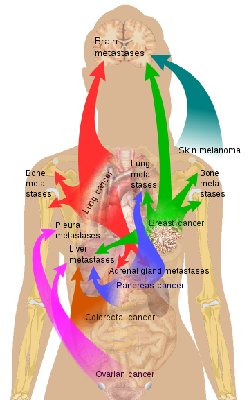
Metastases are responsible for most of the deaths caused by cancer. Dr. Bruns believes that it is currently unclear how metastases form. So far, it has been assumed that they spread throughout the body via the lymphatic vessels and the bloodstream. But this mechanistic hypothesis cannot explain why some organs are preferably targeted in metastasis while other are relatively preserved. This was first discussed as the "seed and soil" theory by Stephen Paget in 1889. enter image description here Source Mikael Häggström via Wikipedia

Paradoxically, cancer patients with a high number of macrophages have a reduced life expectancy. In a mouse model, tumor growth almost stops when the macrophages where removed.

Heiko Bruns assumes that individual tumor cells are consumed by phagocytes, but are not necessarily eliminated by them. Instead, he suspects that tumor cells are using macrophages as "Trojans horses". They could thus escape detection and travel through the body to colonize other organs.

Dr. Heiko Bruns' idea was accepted into the 'Experiment! In search of bold research ideas' based on this unusual question. This idea received funding of 120,000 euros from the Volkswagen Foundation until the end of 2021.

Les phagocytes peuvent-ils transporter des cellules tumorales?

- Posted by admin in Français

Un scientifique de l'Universitätsklinikum Erlangen, Heiko Bruns, poursuit une hypothèse innovante sur les métastases cancéreuses. Il a rédigé sa thèse de doctorat sur la survie des bactéries de la tuberculose dans les macrophages, et sa thèse postdoctorale porte sur l'importance des macrophages dans le contexte de la métastase cancéreuse.

Les macrophages sont un type de globules blancs du système immunitaire, qui engloutit et digère les débris cellulaires, les substances étrangères, les microbes, les cellules cancéreuses. Les macrophages pénètrent dans les tissus endommagés par l'endothélium d'un vaisseau sanguin, un processus connu sous le nom d'extravasation de leucocytes. Ce processus ressemble beaucoup à celui qu’on pense être celui de la métastase.

enter image description here Classical conception of the metastasis process Source: doi: 10.1038 / nri3789

Il est bien connu que les macrophages peuvent contribuer à la croissance et à la progression des cancers. Les macrophages peuvent également influencer positivement et négativement les résultats des traitements anti-cancéreux. Cependant certains agents pathogènes réussissent à vivre même à l'intérieur des macrophages. Cela permet alors à l'agent pathogène d’échapper au système immunitaire. C'est le cas de Mycobacterium tuberculosis ou du VIH.

Les métastases sont responsables de la plupart des décès causés par le cancer. Le Dr Bruns pense qu'à l'heure actuelle, on ne sait pas vraiment comment les métastases se forment. Jusqu'à présent, on a supposé qu'elles se propageaient dans tout le corps via les vaisseaux lymphatiques et la circulation sanguine.

Paradoxalement, les patients cancéreux avec un nombre élevé de macrophages ont une espérance de vie diminuée. Un modèle de souris a démontré que la croissance tumorale s'arrête pratiquement lorsque les macrophages ont été retirés.

Heiko Bruns suppose que les cellules tumorales individuelles sont consommées par les phagocytes, mais ne sont pas nécessairement éliminées par ceux-ci. Au lieu de cela, il soupçonne que les cellules tumorales utilisent les macrophages comme «chevaux de Troie». Elles pourraient ainsi échapper à la détection et voyager à travers le corps pour coloniser d'autres organes.

L’idée du docteur Heiko Bruns a été acceptée au programme de financement «Expérience! A la recherche d'idées de recherche audacieuses 'sur la base de cette question inhabituelle. Il a reçu un financement de 120 000 euros de la Fondation Volkswagen jusqu'à fin 2021. Le projet devrait démarrer au printemps.

Evolutionary Connection Between Pregnancy and Cancer Metastasis

- Posted by admin in English

Placental invasion into the maternal endometrium of the uterus has substantial similarities with the early spread of cancer in the stroma (the part of a tissue or organ with a structural or connective role). These similarities have inspired the hypothesis that trophoblasts (the continuous cell layer of fibroblasts that limit the egg, which became blastocyst at day 6 after fertilization) have developed the ability to invade the endometrium, leading to invasive placentation. Invasion of a specific type of trophoblast (extravillous trophoblast) in the maternal uterus is a vital step in the establishment of pregnancy. * Zephyris Source CC BY-SA 3.0, * https://commons.wikimedia.org/w/index.php?curid=10811330

These mechanisms can be reactivated in cancer cells, leading to a predisposition to metastasis. It had been hypothesized (the ELI hypothesis) that cancer malignancy should be limited to placental mammals where invasive placentation first evolved. But there are several counterexamples.

In a recent article, the authors explore an alternative scenario in which stromal cells of the uterus evolved to resist or allow invasion, determining the outcome of placental invasiveness. The likelihood that changes in the stromal environment will lead to changes in cancer malignancy is reinforced by the fact that the molecular mechanisms used by cancer cells to metastasize are shared with other biological processes.

For example, mechanisms regulating gastrulation, wound healing, leukocyte extravasation, etc., are shared with both trophoblast and cancer invasion. This implies that invasive cancer cells use mechanisms that have evolved much earlier than placental invasion and, therefore, the evolution of invasive placentation per se can not be responsible for the origin of malignant cancer.

It is important to note, however, that the invasive nature of the placenta continued to evolve after its origin. Mammalian species differ in their tumorigenesis potential, as well as their vulnerability to cancer metastasis.

While the evolution resulted in an even higher degree of invasiveness in great apes, which includes humans, a complete loss of placental invasion has evolved in hoofed mammals, such as cows and horses and their parents, and these animals have lower malignancy rates for a variety of cancers.

In a recent review, Constanzo et al. presented compelling arguments for a model in which cancer progression in humans includes reactivation of the expression of embryonic genes normally controlling placental development and the development of the placenta. immune evasion.

For example, melanoma occurs in cattle and equines but remains largely benign; while it is very malign in the human. This correlates with the phenotype of the fetal-maternal interface (the degree of placental invasion during pregnancy). In particular, these results support that TGF-β secretion and high non-canonical WNT signaling in stromal cells are causal factors accounting for the high vulnerability of human stromal tissues to cancer invasion, at least in the case of melanoma .

Their data support the ELI hypothesis, suggesting that differences in stromal gene expression between species are critical in determining the degree of embryo implantation as well as stromal resistance to early cancer dissemination.

Advertisement


This book retraces the main achievements of ALS research over the last 30 years, presents the drugs under clinical trial, as well as ongoing research on future treatments likely to be able stop the disease in a few years and to provide a complete cure in a decade or two.

L'invasion placentaire dans l'endomètre maternel de l'utérus présente des similitudes substantielles avec la dissémination précoce du cancer dans le stroma (la partie d'un tissu ou d'un organe ayant un rôle structurel ou conjonctif).

Ces similitudes ont inspiré l'hypothèse que les trophoblastes (la couche cellulaire continue formée de fibroblastes qui limite l'œuf, devenu blastocyste au 6e jour après la fécondation) ont développé la capacité d'envahir l'endomètre, conduisant à une placentation invasive. L'invasion d'un type spécifique de trophoblaste (trophoblaste extravilleux) dans l'utérus maternel est une étape vitale dans l'établissement de la grossesse. Source Zephyris CC BY-SA 3.0, https://commons.wikimedia.org/w/index.php?curid=10811330

Ces mécanismes peuvent se réactiver dans les cellules cancéreuses, entraînant une prédisposition aux métastases. Il avait donc été fait l'hypothèse (nommée ELI) que la malignité cancéreuse devait être limitée aux mammifères placentaires où la placentation invasive a d'abord évolué. Mais il y a plusieurs contre-exemples.

Dans un article récent, les auteurs explorent un scénario alternatif dans lequel les cellules stromales de l'utérus ont évolué pour résister ou permettre l'invasion, déterminant le résultat de l'invasivité placentaire. La probabilité que l'évolution de l'environnement stromal entraîne l'évolution de la malignité cancéreuse est renforcée par le fait que les mécanismes moléculaires utilisés par les cellules cancéreuses pour métastaser sont partagés avec d'autres processus biologiques.

Par exemple, les mécanismes régulant la gastrulation, la cicatrisation des plaies, l'extravasation par les leucocytes, etc., sont partagés à la fois avec le trophoblaste et l'invasion du cancer. Cela implique que les cellules cancéreuses envahissantes utilisent des mécanismes qui ont évolué beaucoup plus tôt que l'invasion placentaire et, par conséquent, l'évolution de la placentation invasive en soi ne peut pas être responsable de l'origine du cancer malin.

Il est important de noter, cependant, que la nature invasive du placenta a continué d'évoluer après son origine. Les espèces de mammifères diffèrent par leur potentiel de tumorigenèse, ainsi que leur vulnérabilité aux métastases cancéreuses.

Alors que l'évolution a entraîné un degré d'envahissement encore plus élevé chez les grands singes, qui comprend les humains, une perte complète de l'invasion placentaire a évolué chez les mammifères à sabots, tels que les vaches et les chevaux et leurs parents, et ces animaux ont des taux de malignité inférieurs pour une variété des cancers.

Dans une revue récente, Constanzo et ses collaborateurs ont présenté des arguments convaincants pour un modèle où la progression du cancer chez l'homme comprend la réactivation de l'expression des gènes embryonnaires contrôlant normalement le développement du placenta et l'évasion immunitaire.

Par exemple, le mélanome survient chez les bovins et les équidés mais reste largement bénin; alors qu'il est très malin chez l'homme. Ceci est en corrélation avec le phénotype de l'interface fœtale-maternelle (le degré d'invasion placentaire pendant la grossesse).

En particulier, ces résultats soutiennent que la sécrétion de TGF-β et la signalisation WNT non canonique élevée dans les cellules stromales sont des facteurs causaux expliquant la forte vulnérabilité des tissus stromaux humains à l'invasion du cancer, au moins dans le cas du mélanome.

Leurs données soutiennent l'hypothèse ELI, suggérant que les différences d'expression génique stromale entre les espèces sont déterminantes pour déterminer le degré d'implantation de l'embryon ainsi que la résistance stromale à la dissémination précoce du cancer.

Publicité


Ce livre retrace les principales réalisations de la recherche sur la SLA au cours des 30 dernières années. Il présente les médicaments en cours d’essai clinique ainsi que les recherches en cours sur les futurs traitements susceptibles d’ici quelques années, d’arrêter la maladie et de fournir un traitement complet en une décennie ou deux.

Qu'est-ce que la chimioprévention

La chimioprévention (également la chimioprophylaxie) fait référence à l'administration d'un médicament dans le but de prévenir une maladie ou une infection. Des antibiotiques, par exemple, peuvent être administrés à des patients présentant des troubles du système immunitaire afin de prévenir les infections bactériennes (en particulier les infections opportunistes). Il peut également s'agir d'administrer de l'héparine pour prévenir la thrombose veineuse profonde chez les patients hospitalisés.

Comment l'incidence du cancer du poumon?

Le cancer du poumon est la principale cause de décès par cancer aux États-Unis et en Europe. Parmi les autres agents cancérigènes, citons l'amiante et les matières radioactives. Par conséquent, la prévention du tabagisme est primordiale pour la prévention du cancer du poumon. Les stratégies efficaces pour réduire l'incidence du cancer du poumon (au-delà du sevrage tabagique) font actuellement défaut.

Par exemple, le développement du cancer du poumon à cellules squameuses commence par un épithélium normal et progresse par l'hyperplasie, la métaplasie, la dysplasie (légère, modérée et grave) et le carcinome in situ. À ce jour, aucun biomarqueur intermédiaire n'a été validé pour l'interception du cancer du poumon, en partie en raison de l'absence de traitement éprouvé, et l'histologie est actuellement considérée comme le meilleur marqueur.

Quelles pistes pour la chimioprévention du cancer du poumon?

Les produits de la voie de l'acide arachidonique, en particulier les prostaglandines (PG), jouent un rôle essentiel dans la carcinogenèse pulmonaire et la chimioprévention. De grandes études épidémiologiques ont montré un lien entre l'utilisation régulière d'aspirine et la diminution du taux de certains cancers. Ces résultats ont conduit à un essai clinique montrant que l'iloprost oral (un analogue de la prostacycline) améliorait de manière significative la dysplasie endobronchique chez les anciens fumeurs. Des études supplémentaires portant sur le mécanisme chimio-préventif ont montré que les effets de la prostacycline étaient indépendants du récepteur de la PGI2 à la surface d'une cellule et pouvaient s'appuyer sur la capacité de la PGI2 à agir en tant qu'agoniste de la PPARγ.

En quoi les thiazolidinediones sont-elles intéressantes?

Les thiazolidinediones (TZD) sont des agonistes de PPARγ couramment utilisés dans le traitement du diabète et la pioglitazone TZD a été étudiée dans de nombreux modèles de cancer précliniques. Des études précliniques sur des surexpresseurs de PPARγ génétiquement modifiés et des agonistes de PPARγ par voie orale ont confirmé que l'activation de PPARγ favorise la différenciatio, inhibe la croissance tumorale et empêche la progression des lésions préinvasives dans les modèles murins. Dans des modèles précliniques, la pioglitazone par voie orale, et plus récemment inhalée, prévient à la fois les carcinomes à cellules adéno et épidermoïdes, en monothérapie ou en association avec des corticostéroïdes et de la metformine inhalés. Les raisons d'un rôle dans la chimioprévention du cancer du poumon ont également été étayées par une vaste étude portant sur les taux de cancer du poumon, de la prostate et du côlon chez des vétérans diabétiques traités avec TZD. Govindarajan et ses collègues ont rapporté une diminution de 33% de l'incidence du cancer du poumon par rapport aux utilisateurs non-TZD, suggérant que l'activation de PPARγ pourrait prévenir le cancer du poumon.

Pourquoi un essai clinique?

Ceci a conduit à un essai à double insu, randomisé, de phase II et contrôlé par placebo de la pioglitazone orale chez des fumeurs à haut risque ou d'anciens fumeurs atteints d'atypie cytologique des expectorations ou de dysplasie endobronchique connue. Cet essai a été répertorié et enregistré sur ClinicalTrials.gov (identifiant: NCT00780234).

Cet essai de chimioprévention de phase II, monocentrique, à double insu, à évalué la pioglitazone par voie orale chez des fumeurs actuels ou anciens à haut risque présentant une atypie cytologique des expectorations ou une dysplasie endobronchique connue.

Quel en est le résultat?

Après six mois de traitement, la pioglitazone n’a pas significativement amélioré l’histologie endobronchique par rapport au placebo; Cependant, le traitement par la pioglitazone a entraîné une amélioration histologique de certaines de ces lésions. Les auteurs concluent que de futures études visant à mieux caractériser la dysplasie réactive dans ce contexte sont justifiées.

To study immune cells, we must take into account their environment

- Posted by admin in English

Living organisms use energy in a radically different way than immune cells in vitro.

For years, scientists have used cells grown in Petri dishes to study the metabolic processes that fuel the immune system. But a new article suggests that living organisms use energy in a radically different way from immune cells in vitro.

The scientific consensus since Warburg's work is that immune cells, called T cells, convert glucose into energy to fuel cellular function. In fact there are different mechanisms by which a cell can get energy, the so-called metabolism or also respiration.

How do the cells get energy?

On the one hand, we distinguish anabolism, which represents all the biosynthetic pathways of cellular constituents, and on the other hand, catabolism, which represents all the pathways of degradation of these cellular constituents into small molecules to release their energy by oxidation or to rebuild other cellular constituents.

Catabolism can be differentiated between aerobic and anaerobic respiration. Aerobic respiration includes glycolysis, oxidative decarboxylation of pyruvate, citric acid cycle, oxidative phosphorylation.

The main degradation pathway is glycolysis, where sugars such as glucose and fructose are converted to pyruvate and generate ATP. Pyruvate is an intermediate in several metabolic pathways, but the majority is converted to acetyl-CoA by aerobic glycolysis (with oxygen) and introduced into the citric acid cycle.

Lipids are catabolized by hydrolysis to free fatty acids and glycerol. Glycerol enters glycolysis and the fatty acids are decomposed by beta-oxidation to release acetyl-CoA, which is then introduced into the citric acid cycle.

There are two important microbial methane formation pathways, by carbonate reduction (respiration) and acetate fermentation.

Warburg hypothesized that cancer growth is caused by energy-generating tumor cells (such as, for example, adenosine triphosphate / ATP) primarily through anaerobic degradation of glucose (called fermentation or anaerobic respiration). This contrasts with healthy cells, which primarily generate energy from the oxidative decomposition of pyruvate. Pyruvate is a final product of glycolysis and is oxidized in mitochondria. Therefore, according to Warburg, cancer should be interpreted as mitochondrial dysfunction.

For multicellular organisms, during brief periods of intense activity, muscle cells use fermentation to supplement ATP production from slower aerobic respiration.

What was discovered?

Jones and colleagues found that T cells in a living system use glucose as a building block for DNA replication and other maintenance tasks, in addition to converting glucose into raw energy. They also discovered that the way T cells treat glucose evolves during an immune response. The metabolism of glucose in T cells changes dynamically during an immune response. Glucose-dependent serine biosynthesis promotes T-cell proliferation in vivo.

enter image description here

This suggests that T cells can use resources differently in the body when they are fighting a bacterial infection such as Listeria or a disease like cancer.

Naïve CD8+ T cells differentiating into effector T cells increase glucose uptake and transition from resting metabolism to anabolic metabolism. Although much is known about the metabolism of cultured T cells, the way in which T cells utilize nutrients during the in vivo immune response is less well defined. The researchers therefore combined the bioenergetic profiling and 13C glucose perfusion techniques to study the metabolism of CD8+ T cells responding to Listeria infection.

In contrast to the in vitro activated T cells, which exhibit Warburg metabolism, physiologically activated CD8+ T cells exhibited higher levels of oxidative metabolism, higher bioenergetic capacity, differential pyruvate utilization, and high 13C carbon flux. glucose to the anabolic pathways, including the biosynthesis of nucleotides and serine. The glucose-dependent serine biosynthesis induced by the Phgdh enzyme was essential for the expansion of CD8+ T cells in vivo.

Our immune cells do not work in isolation

"It's like watching animal behavior in a zoo or in the wild - our immune cells do not work in isolation - they work with a host of other cells and factors that influence how and when they are used. of energy, "said Russell Jones, Ph.D., lead author of the study and head of the Metabolic and Nutritional Programming Group at the Van Andel Institute. "Understanding cell metabolism is a crucial part of therapeutic development, and our results reinforce the need to study these cells in an environment as close as possible to nature."

The findings have profound implications for how scientists study the complex and interconnected systems that underlie health and disease and how they translate this information into new diagnostic and treatment strategies.

"Immune cells react much more dynamically to infections and diseases than we previously thought," Jones said. "For a while, we're at a stage of metabolism research, it's like we're in the dark under a lamppost, we could only see in front of us, and these results will help us better understand this. which immune cells need for optimal function. "

Which suite will be given?

The results were made possible by a new method developed by collaborator Ralph DeBerardinis, MD, Ph.D., which allowed Jones and his colleagues to map how T cells use nutrients in living organisms. They have developed an infusion method to study T cell metabolism in vivo

"In the future, this new mapping technique will be invaluable in pursuing studies of specific diseases," said Eric Ma, Ph.D., lead author of the study and a postdoctoral researcher in the field. Jones's laboratory.

In the future, the team plans to design human studies to measure how T cells use glucose and other nutrients when they respond to pathogens or other diseases such as injuries or diseases such as cancer.

Contact the author

Advertisement


This book retraces the main achievements of ALS research over the last 30 years, presents the drugs under clinical trial, as well as ongoing research on future treatments likely to be able stop the disease in a few years and to provide a complete cure in a decade or two.

les organismes vivants utilisent l'énergie de façon radicalement différente de la façon de celle des cellules immunitaires in-vitro.

Pendant des années, les scientifiques ont utilisé des cellules cultivées dans des boîtes de Pétri pour étudier les processus métaboliques qui alimentent le système immunitaire. Mais un nouvel article suggère que les organismes vivants utilisent l'énergie de façon radicalement différente de la façon de celle des cellules immunitaires in-vitro.

Le consensus scientifique depuis les travaux de Warburg, est que les cellules immunitaires, appelées cellules T, convertissent du glucose en énergie pour alimenter la fonction cellulaire. En fait il y a différents mécanismes par lequel une cellule peut obtenir de l'énergie, ce que l'on appelle métabolisme ou aussi respiration.

Comment les cellules acquièrent-elle de l'énergie?

on distingue d'une part l'anabolisme, qui représente l'ensemble des voies de biosynthèse des constituants cellulaires, et d'autre part le catabolisme, qui représente l'ensemble des voies de dégradation de ces constituants cellulaires en petites molécules pour en libérer l'énergie par oxydation ou pour rebâtir d'autres constituants cellulaires.

On peut différencier le catabolisme en respiration aérobie et anaérobie. La respiration aérobie comprend la glycolyse, la décarboxylation oxydante du pyruvate, le cycle de l'acide citrique, la phosphorylation oxydative.

La principale voie de dégradation est la glycolyse, où des sucres tels que le glucose et le fructose sont convertis en pyruvate et génèrent de l'ATP. Le pyruvate est un intermédiaire dans plusieurs voies métaboliques, mais la majorité est convertie en acétyl-CoA par glycolyse aérobie (avec oxygène) et introduite dans le cycle de l'acide citrique.

Les lipides sont catabolisés par hydrolyse en acides gras libres et en glycérol. Le glycérol entre dans la glycolyse et les acides gras sont décomposés par bêta-oxydation pour libérer de l'acétyl-CoA, qui est ensuite introduit dans le cycle de l'acide citrique.

Il existe deux voies de formation de méthane microbiennes importantes, par réduction du carbonate (respiration) et fermentation par l'acétate.

Warburg a émis l'hypothèse que la croissance du cancer est causée par des cellules tumorales générant de l'énergie (comme, par exemple, l'adénosine triphosphate / ATP) principalement par la dégradation anaérobie du glucose (appelée fermentation ou respiration anaérobie). Cela contraste avec les cellules saines, qui génèrent principalement de l'énergie à partir de la décomposition oxydante du pyruvate. Le pyruvate est un produit final de la glycolyse et est oxydé dans les mitochondries. Par conséquent, selon Warburg, le cancer devrait être interprété comme un dysfonctionnement mitochondrial.

Pour les organismes multicellulaires, lors de brèves périodes d'activité intense, les cellules musculaires utilisent la fermentation pour compléter la production d'ATP à partir de la respiration aérobie plus lente.

Qu'est-ce qui a été découvert?

Jones et ses collègues ont découvert que les cellules T dans un système vivant utilisent le glucose comme éléments de base pour la réplication de l'ADN et d'autres tâches de maintenance, en plus de la conversion du glucose en énergie brute. Ils ont également découvert que la façon dont les cellules T traitent le glucose évolue au cours d'une réponse immunitaire. Le métabolisme du glucose dans les cellules T change de façon dynamique au cours d'une réponse immunitaire. La biosynthèse de la sérine dépendante du glucose favorise la prolifération des lymphocytes T in vivo.

enter image description here

Ce qui suggère que les cellules T peuvent utiliser les ressources de manière différente dans le corps lorsqu’elles combattent une infection bactérienne telle que Listeria ou une maladie comme le cancer.

Les cellules T naïves CD8 + se différenciant en cellules T effectrices augmentent l'absorption du glucose et le passage du métabolisme au repos au métabolisme anabolique. Bien que l'on en sache beaucoup sur le métabolisme des cellules T en culture, la façon dont les cellules T utilisent les nutriments au cours de la réponse immunitaire in vivo est moins bien définie. Les chercheurs ont donc combiné les techniques de profil bioénergétique et de perfusion de glucose 13C pour étudier le métabolisme des lymphocytes T CD8 + répondant à une infection par Listeria.

Contrairement aux cellules T activées in vitro, qui présentent le métabolisme de Warburg, les cellules T CD8 + activées physiologiquement présentaient des taux plus élevés de métabolisme oxydatif, une capacité bioénergétique plus élevée, une utilisation différentielle du pyruvate et un flux important du carbone 13C-glucose vers les voies anaboliques, y compris la biosynthèse des nucléotides et de la sérine. La biosynthèse de la sérine dépendante du glucose induite par l’enzyme Phgdh était essentielle à l’expansion des cellules T CD8 + in vivo.

Nos cellules immunitaires ne fonctionnent pas en vase clos

"Cela revient à observer le comportement des animaux dans un zoo ou dans la nature. Nos cellules immunitaires ne fonctionnent pas en vase clos - elles travaillent de concert avec une foule d'autres cellules et facteurs qui influencent le mode et le moment d'utilisation de l'énergie, "a déclaré Russell Jones, Ph.D., auteur principal de l'étude et responsable du groupe de programmation métabolique et nutritionnelle de l'Institut Van Andel. "Comprendre le métabolisme cellulaire est un élément crucial du développement thérapeutique. Nos résultats renforcent la nécessité d'étudier ces cellules dans un environnement aussi proche que possible de la nature."

Les résultats ont des implications profondes sur la façon dont les scientifiques étudient les systèmes complexes et interconnectés qui sous-tendent la santé et la maladie et sur la manière dont ils traduisent ces informations en de nouvelles stratégies de diagnostic et de traitement.

"Les cellules immunitaires réagissent de manière beaucoup plus dynamique aux infections et aux maladies que nous ne le pensions auparavant", a déclaré Jones. "Pendant un certain temps, nous en sommes à un stade de la recherche sur le métabolisme, c'est comme si nous étions dans l'obscurité sous un réverbère. Nous ne pouvions voir que devant nous. Ces résultats nous aideront à mieux comprendre ce dont les cellules immunitaires ont besoin pour une fonction optimale ".

Quelle suite va être donnée?

Les résultats ont été rendus possibles grâce à une nouvelle méthode mise au point en consultation avec son collaborateur Ralph DeBerardinis, M.D., Ph.D., qui a permis à Jones et ses collègues de cartographier la manière dont les cellules T utilisent les nutriments dans des organismes vivants. Ils ont mis au point d'une méthode de perfusion 13C pour étudier le métabolisme des cellules T in vivo

"A l'avenir, cette nouvelle technique de cartographie sera d'une valeur inestimable dans la poursuite d'études sur des maladies spécifiques", a déclaré Eric Ma, Ph.D., premier auteur de l'étude et chercheur postdoctoral dans le laboratoire de Jones.

À l’avenir, l’équipe prévoit de concevoir des études sur l’homme afin de mesurer la manière dont les cellules T utilisent le glucose et d’autres nutriments lorsqu’elles réagissent à des agents pathogènes ou à d’autres atteintes telles que des blessures ou des maladies telles que le cancer.

Contact the author

Advertisement


This book retraces the main achievements of ALS research over the last 30 years, presents the drugs under clinical trial, as well as ongoing research on future treatments likely to be able stop the disease in a few years and to provide a complete cure in a decade or two.

A new kind of cancer vaccine

- Posted by admin in English

Immunotherapy on demand

Our frequent readers know that this site contains a peptide generator for designing simple, inexpensive and personalized cancer vaccines. Scientists believe that it is possible to use a system on the same principle, but much more effective.

Checkpoint inhibitors are not effective for many patients

Control point inhibitor immunotherapies are revolutionizing the treatment of cancer. However, even in the most sensitive cancers, a substantial proportion (50-80%) of patients have a low to no positive response. A surprising finding in the analysis of these patients was that one of the best correlates of response was the total number of neo-antigens in the tumor.

Basics of the new proposal

When information is transferred from DNA to RNA in cancer cells, errors occur frequently, resulting in the production of proteins that can be recognized by the immune system. Scientists at the Biodesign Institute at Arizona State University say these proteins, known as cancer-specific antigens, are important because they can be used to design vaccines that can treat or prevent tumor progression.

The common point of all cancer tumor mutations is the production of neo-antigens, or small fragments of proteins called peptides, formed inside a cancer cell, which the host's immune system had not yet never seen. These aberrant peptides, present only in cancer cells, can boost the immune system through a vaccine. They propose that mis-splicing and RNA transcription errors, particularly INDELs of MS in coding regions, in cancer cells could also be a source of neo-antigens.

These errors made by cancer cells can be used to develop a vaccine against some cancers

"In a cancer cell, it turns out that all levels of information transfer from DNA to RNA to protein become more prone to errors," said Johnston. "We proposed that these errors in cancer cells could also be the source of a cancer vaccine."

A "frame shift" or "splicing" error occurs when the DNA information of a gene is poorly processed during the fabrication of RNA, in a basic cellular process called transcription and splicing RNA.

For the most part, these alterations can be managed and cleaned by the cell, without ever being exposed to the immune system. As the cancer progresses, due to the increased number of errors, protein waste accumulates more rapidly, submerging the cell and aberrant proteins are exposed and recognized by the immune cell.

"These are overwhelming the quality control systems of a cell, generating errors in the RNA and proteins released by the cancer cell, which the immune system can respond to," said Johnston.

A new tool

To discover neo-antigens in tumors, Johnston's team has developed a new type of chip. They made fleas with the 200,000 possible neo-antigens for five common cancers, allowing them to simply search for antibodies present in the blood collected by patients. This is much simpler than the current practice of obtaining and sequencing tumor DNA, a starting point for the "personal cancer vaccines" that many companies are currently pursuing.

This set makes it possible to detect all possible predicted frame shift peptides that any tumor cell could potentially produce. They customized this chip, which contained nearly 400,000 peptides, and analyzed them with blood samples from cancer patients (and healthy control samples) to look for the peptides with the most reactive antibodies.

The basic idea is to make a vaccine containing multiple (up to 100) abnormal peptides.

Interest of this new type of immunotherapy

"Personal vaccines against cancer are complicated and expensive," said Johnston. "In addition, only about 40% of tumors have enough mutations in the DNA to make a vaccine. We have found that even cold tumors at the DNA level make many errors in the DNA. RNA: The peptides we generate are much more immunogenic than the point mutations used in personal cancer vaccines. More importantly, we can make standard vaccines for much cheaper therapeutic or even preventive vaccines. "

A vaccine that helps fight the development of resistance

It should be difficult for tumor cells to move away from the vaccine because these FSs are variants and not inherited mutations. In particular, if the FS antigen were produced in the RNA of an essential gene, the tumor cells would restrict the presentation of the MHC or create an immunosuppressive environment to evade an immune response.

In summary, scientists have discovered another class of neo-antigens that could be useful for developing different types of cancer vaccines. They have also created a matrix format for directly detecting immune responses to these tumor antigens.

What will happen to these works

Too often university laboratories stop their research when they can file a patent, in the very uncertain hope, that a large company will offer a golden bridge to buy that patent. This hope is often disappointed, simply because the patents resulting from academic research are not reproducible, but also because they do not respond to a large number of medical and regulatory issues. It is normally the role of biotech to solve these difficulties to make a process attractive for large companies. Our scientists are much more adept. They recently launched a large clinical trial of a pan-cancer prophylactic dog dog cancer vaccine funded by the Open Philanthropy Project. If that succeeds, Johnston is eager to move on to the first human clinical trials.

Regarding this web site, we could possibly design a peptide generator based on this principle. Please do not hesitate to contact us. "contact at padiracinnovation dot org"


Please, help us continue to provide valuable information: