Bats, although mammals, are not phylogenetically very close to humans, so we would not expect them to harbor many harmful viruses to our species.
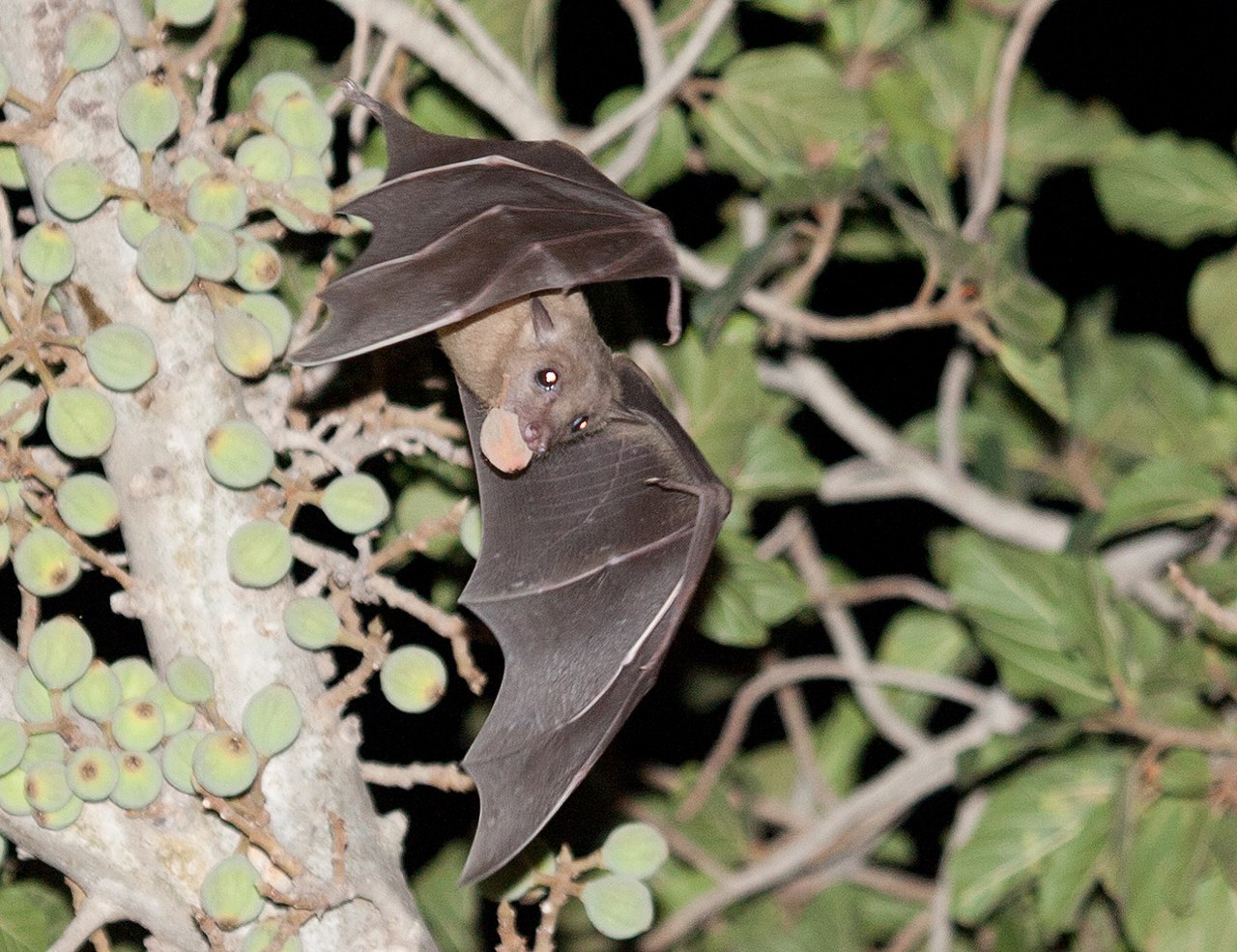 Source: Вых Пыхманн via Wikipedia
The order of bats contains more than 1,200 species. Because of this genetic diversity, information about one species may not apply to other species. In recent years, however, it has emerged that viruses have spread from bats to other mammals. For example, serological evidence and detection of viruses linked to ebolaviruses in bats suggest that bats are indeed one of the reservoirs of the virus.
However, although bats harbor many different viruses, their spread to other animals is extremely rare. One reason is that for such an event to occur, several factors should be conducive to transmission.
The ability of bats to host many viruses without showing pathology suggests that bats have developed immune mechanisms different from those of other mammals. This tolerance to viruses goes hand in hand with reduced inflammation.To explain this singular characteristic, it is necessary to compare it with another singular characteristic of bats: It is a mammal with the capacity to fly.
The metabolic rate of bats in flight is double that of running rodents of similar size. The increased metabolic rate that accompanies the flight would result in higher levels of oxygen-free radicals. To mount an immune response to the damage caused by this high metabolism, would be energetically expensive.
Bats have therefore developed mechanisms leading to reduced inflammation. It would also explain why bats of some species live longer than expected given their high metabolism and small size. Some bats can live up to 40 years, while a rodent of the same size can live only two years.
Small animals with a fast heart rate and metabolism generally have a shorter lifespan than larger animals with a slower heartbeat and a slower metabolism. But bats are unique because they have a much longer lifespan than other mammals of the same size.
But a reduced inflammatory response enables rapid replication of viruses, especially in stressful conditions that affect the immune system.
The awakening of hibernation is a stressful event for bats. Many large brown bats are latently infected and Gerowet and his colleagues have shown that the virus reactivates when it comes out of hibernation. This reactivation is also associated with a low level of antibodies to the virus. After hibernation, antibody levels rise, which puts the virus back to latency.
The virus also reactivates when bats are infected with a bacterial or fungal infection. Small brown bats are particularly susceptible to an often fatal fungal infection known as white nose syndrome. A study examining the effects of stress induced by this infection showed that bats infected with fungi had 60 times more coronavirus in their intestines than uninfected bats. The specific protection mechanisms of these bats results in a rapid response that blocks the virus outside the cells. Indeed, paradoxically a host that tolerates well the presence of viruses, because it controls inflammation, allows viruses to increase their rate of replication, by genetic selection, without damaging their host.
Disturbances in bat habitat also seem to stress these animals and cause them to spread even more viruses in their saliva, urine and feces which can infect other animals. "Increased environmental threats to bats can add to the threat of zoonosis," said Brook, who is also working with a Madagascar-based field project that explores the link between loss of bat habitat and spread of bat viruses to other animals and humans.
Such rapidly reproducing viruses generate extreme virulence when overflowing to hosts that do not have the same immune capabilities as bats.
When these virulent viruses travel from bats to animals without a rapid response immune system, they quickly overwhelm their new hosts.
Brook and Boots are developing a more formal model of disease progression in bats to better understand the spread of the virus to other animals and humans.

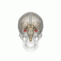 Source: Life Science Databases(LSDB) via Wikipedia.