Alzheimer's, FTD, bvAD and bvFTD
The signs of Alzheimer's disease, which is characterized by a selective amnesia, for example of his relatives, are well known. Fronto-Temporale Dementia (FTD), it is associated with significant changes in social and personal behavior, apathy, blunted emotions and language deficits. The FTD shares gene mutations with ALS. The behavioral variant of frontotemporal dementia (bvFTD) is characterized by changes in social behavior and behavior, with loss of social awareness and insufficient control of impulses.
How to differentiate bvAD from bvFTD?
Singleton and colleagues have shown that Alzheimer's disease, which typically leads to amnesic dementia, may also have behavioral variants (bvAD) and that the phenotype determinant is the anatomy of neurodegeneration.
In a small percentage of people with Alzheimer's disease, early behavioral changes, such as disregard for social norms or loss of empathy, may lead physicians to misdiagnose a behavioral variant of front-line dementia. temporal. How can they better distinguish this variant of Alzheimer's disease? The diagnosis between bvAD and bvFTD is clinically very difficult without imaging.
While structural MRI does not make a significant distinction between bvAD and typical Alzheimer's disease, metabolic PET shows a decrease in the activity of frontense and anterior default mode networks in bvAD, similar to bvFTD .
What is a default neurology network?
In neuroscience, the default network (DMN) is composed of interacting brain regions, whose activity is highly correlated with each other and distinct from other brain networks.
Initially, it was assumed that the default mode network was most often active when a person did not focus on the outside world and his brain was dormant, such as daydreaming or mental observation. However, it is now known that this can contribute to experience elements related to the performance of external tasks. He is also active when the individual thinks of others, thinks of himself, remembers the past and prepares himself for the future. Although the DMN was initially noted as disabled in some goal-oriented tasks and is sometimes referred to as a negative-task network, it may be active in other objective-oriented tasks, such as social work memory or autobiographical tasks. DMN is not correlated with other brain networks such as attention networks.
Studies on DMN have shown disturbances in the DMN of people with Alzheimer's disease and Autism Spectrum Disorder.
The value of neuroimaging for the diagnosis of bvAD
"This is the first study focused on bvAD to show such a variety of neuroimaging features," Singleton said. "This suggests that FDG-PET is more accurate in differentiating these diseases than MRI."
In a previous structural MRI study in 2015, Ossenkoppele and colleagues expected to find frontal cortical atrophy in people with bvAD, but were puzzled to find none.
Singleton and his colleagues examined the 150-person MRI and FDG-PET scans recruited at the University of California, San Francisco, and the University of Berkeley. BvAD was diagnosed in 29 patients, 28 in patients with typical Alzheimer's disease, 28 in patients with DVBt and 65 in cognitively normal subjects.
How does a TEP-FDG work?
Positron emission tomography (PET) scanners detect photons of light energy and construct three-dimensional images from the photons they receive. Scientists can use this ability to detect photons for medical diagnostic purposes if they know where these photons come from and what they represent.
The cells of the human body use glucose, a sugar, as the main source of energy to trigger all reactions and growth. FDG is a glucose molecule to which is attached a radioactive fluorine atom. It is radioactive, but it is not powerful enough to pose a significant risk to health.
When the FDG decomposes, it emits a particle called positron, which then divides into two photons. These are the photons produced by the FDG that the PET scanner detects. As FDG collects in highly active cells such as tumors, most photons come from these regions.
 Author Jens Maus http://jens-maus.de/
Metabolism and Alzheimer's
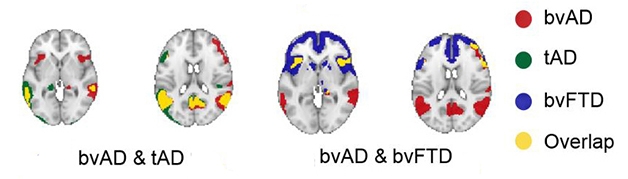
On PET-FDG scans, Singleton and colleagues found a pattern in patients with bvAD that was largely consistent with patients with typical Alzheimer's disease: hypometabolism in the posterior cingulate cortex, precuneus, and temporoparietal lateral. In addition, patients with bvAD had subtle metabolic deficits in fronto-insular areas, including the right lateral frontal lobe and bilateral insula, which did not appear in typical Alzheimer's disease (see image below). -above). This earlier pattern was more similar to that observed in sweeping examinations of patients with bvFTD.
The researchers also measured the uptake of FDG through the brain networks to look for changes in metabolic connectivity. Deficiency in the posterior default network (DMN) was lower in patients with bvAD and typical Alzheimer's disease than in controls, suggesting that these areas were affected in both types of AD. However, less absorption at the previous DMN distinguished bvAD from typical Alzheimer's disease and corresponded to the trend seen in bvFTD.
The researchers found no difference in terms of subcortical atrophy or white matter lesion between typical and bvAD.
These data suggest that common metabolic and connectivity deficits are at the basis of the behavioral phenotype shared by patients with bvAD and bvFTD.
